Review
. 2023 Aug 17:14:1255727.
doi: 10.3389/fphar.2023.1255727. eCollection 2023.

Trinity of inflammation, innate immune cells and cross-talk of signalling pathways in tumour microenvironment

Affiliations
Review

Trinity of inflammation, innate immune cells and cross-talk of signalling pathways in tumour microenvironment

Ali Attiq et al. Front Pharmacol. .

Abstract

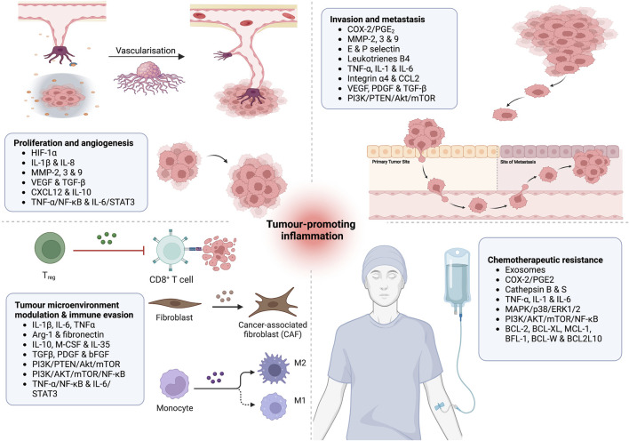
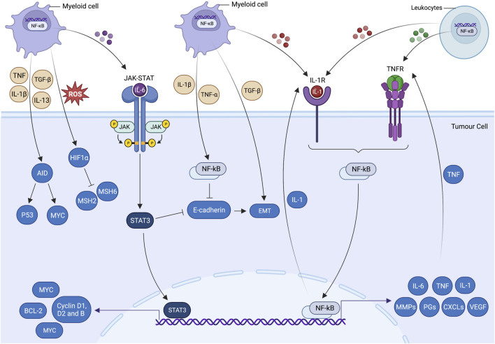
Unresolved inflammation is a pathological consequence of persistent inflammatory stimulus and perturbation in regulatory mechanisms. It increases the risk of tumour development and orchestrates all stages of tumorigenesis in selected organs. In certain cancers, inflammatory processes create the appropriate conditions for neoplastic transformation. While in other types, oncogenic changes pave the way for an inflammatory microenvironment that leads to tumour development. Of interest, hallmarks of tumour-promoting and cancer-associated inflammation are striking similar, sharing a complex network of stromal (fibroblasts and vascular cells) and inflammatory immune cells that collectively form the tumour microenvironment (TME). The cross-talks of signalling pathways initially developed to support homeostasis, change their role, and promote atypical proliferation, survival, angiogenesis, and subversion of adaptive immunity in TME. These transcriptional and regulatory pathways invariably contribute to cancer-promoting inflammation in chronic inflammatory disorders and foster "smouldering" inflammation in the microenvironment of various tumour types. Besides identifying common target sites of numerous cancer types, signalling programs and their cross-talks governing immune cells' plasticity and functional diversity can be used to develop new fate-mapping and lineage-tracing mechanisms. Here, we review the vital molecular mechanisms and pathways that establish the connection between inflammation and tumour development, progression, and metastasis. We also discussed the cross-talks between signalling pathways and devised strategies focusing on these interaction mechanisms to harness synthetic lethal drug combinations for targeted cancer therapy.

Keywords: cross-talks; inflammation; innate immunity; signalling pathways; tumorigenesis; tumour microenvironment; tumour-promoting inflammation.


Conflict of interest statement

The authors declare that the research was conducted in the absence of any commercial or financial relationships that could be construed as a potential conflict of interest.

Figures

FIGURE 1
FIGURE 1
Pathways involved in tumour-promoting inflammation. Insufficient mechanisms to resolve inflammation and perseverance in instigating stimuli result in non-resolving inflammation. Development of angiogenesis to support the tissue remodelling, infiltration of tumour-associated macrophages, immature myeloid and T cells, abundance of inflammatory mediators and imbalance of pro- and anti-inflammatory cytokines creates an ideal microenvironment for non-resolving inflammation. Extrinsic and intrinsic pathways bridge inflammation and cancer. In the extrinsic pathway, an inflammatory response is triggered by exogenous factors, including the PAMPs from pathogens or DAMPs from necrotic cells, which activate the inflammatory cells and establish a microenvironment that potentiates cancer risk. On the contrary, the intrinsic pathway is induced by a mutation in cancer-associated genetic factors such as activating proto-oncogenes, tumour suppressor and DNA repair genes inactivation and chromosomal aberration. These events pave the way for inflammatory microenvironment and neoplastic transformation by upregulating the expression of inflammation-related programs. These two pathways congregate and activate the transcriptional factors (e.g., NF-κB, STAT3, HIFα), which propagate the production of inflammatory mediators, including cytokine, chemokines, COX2/PGE2 and ROS, in inflammatory, stromal, and tumour cells. Perpetually activated transcriptional factors orchestrate pro-tumorigenic inflammatory microenvironment, which is well-known for its tumour-promoting effects.
FIGURE 2
FIGURE 2
Putative role of inflammatory effector molecules in (1) tumour proliferation and angiogenesis; (2) tumour metastasis and invasion; (3) modulation of tumour microenvironment and immune evasion; and (4) induction of chemotherapeutic resistance.
FIGURE 3
FIGURE 3
Inflammatory cell signalling pathways and tumorigenesis. Cross talk of cell-centric and -extrinsic interactions pave the way for tumorigenesis. These processes propagate cancer-associated mutations, which give rise to genetic volatility, hyperproliferation, restructured stromal milieu and poorly differentiated states of epithelial and mesenchymal cells. Inflammatory cells have a unique potential to trigger essential molecular processes that are prerequisites of tumorigenesis. Inflammatory cytokine produced by tumour-infiltrating myeloid cells allows cancer cells to evade apoptosis and promote cell growth via activating transcriptional factors, including signal transducer and activator of transcription 3 (STAT3) and nuclear factor-kappa B (NF-κB). Activated STAT3 and NF-κB coordinate the production of IL-6 and transforming growth factor beta (TGF-β), which downregulates the epithelial differentiation markers expression (e.g., E-cadherin) and promotes epithelial-mesenchymal transition (EMT). Genetic instability is augmented by hypoxia-induced suppression of DNA repair mechanisms and ectopic expression of activation-induced cytidine deaminase (AID). IL-6-induced STAT3 activation by myeloid cells supplements the pre-malignant cell proliferation and anti-apoptotic activities by upregulating the expressions of the proto-oncogene MYC and cell cycle regulators such as cyclin D1, cyclin D2 and cyclin B. MYC-induced BCL2 and BCL2-like1expressions are also responsible for significantly increasing the cell survival and anti-apoptotic activities. Likewise, IL-1α/IL-1R (TNF receptor) autocrine loop signalling and myeloid differentiation primary response 88 (MYD88) results in the activation of NF-κB. MYD88 also actively participate in the development and expansion of tumours by controlling the production of IL-6 in leukocytes. The tumour-promoting role of NF-κB (cell-centric and -extrinsic) during inflammation is further validated by the Toll-like receptor 2 (TLR2)-mediated STAT-3 activation in gastric tumorigenesis.
FIGURE 4
FIGURE 4
Reactive oxygen species and proinflammatory cytokines produced by tumour-associated macrophages (TAMs) and myeloid-derived suppressor cells (MDSCs) cause direct DNA damage and strand breaks. Numerous cytokines produced by TAMs and MDSCs impact the differentiation and proliferation of normal and neo-malignant cells. For instance, IL-23 produced by MDSCs prompts oxidative DNA damage, DNA mis-repairs and induce double-strand breaks (DSBs) and AR target genes by activating JAK-STAT-RORγ-mediated androgen receptor (AR) signalling. Nuclear AR signalling summons the topoisomerase 2β (TOP2B) to the promoter regions, resulting in the chromosomal co-localization of AR target genes and other transcriptional factors. Non-random rearrangements and AR-induced gene fusions occur due to the accumulation of DSBs at the target gene, mimicking the androgen-regulated TMPRSS2 to ERG. Aberrations in homologous recombination result in chromosomal proximation and sole dependency on error-prone non-homologous end-joining for DSB repairs. Malfunctioning tumour suppressor gene, AR targeted gene and DNA repair genes (BRCA1-2, ATM, PALB2, CHEK1-2) prompt the extravasation of immune cells, genome destabilisation and tumour antigen production. Inflammation-induced oxidative stress possesses the highest damage to DRGs and AR-target genes and perpetuates a never-ending loop of DNA damage and mis-repairs.

Similar articles

Cited by

References

    1. Adcock I. M., Caramori G., Barnes P. J. (2011). Chronic obstructive pulmonary disease and lung cancer: new molecular insights. Respiration 81, 265–284. 10.1159/000324601 - DOI - PubMed
    1. Afzal S., Sattar M. A., Eseyin O. A., Attiq A., Johns E. J. (2022). Crosstalk relationship between adiponectin receptors, PPAR-γ and α-adrenoceptors in renal vasculature of diabetic WKYs. Eur. J. Pharmacol. 917, 174703. 10.1016/j.ejphar.2021.174703 - DOI - PubMed
    1. Afzal S., Abdul Sattar M., Johns E. J., Eseyin O. A. (2020). Renoprotective and haemodynamic effects of adiponectin and peroxisome proliferator-activated receptor agonist, pioglitazone, in renal vasculature of diabetic Spontaneously hypertensive rats. PLoS One 15, e0229803. 10.1371/journal.pone.0229803 - DOI - PMC - PubMed
    1. Afzal S., Sattar M. A., Johns E. J., Eseyin O. A., Attiq A. (2021). Antioxidant potential of adiponectin and full PPAR-γ agonist in correcting streptozotocin-induced vascular abnormality in spontaneously hypertensive rats. PPAR Res. 2021, 6661181. 10.1155/2021/6661181 - DOI - PMC - PubMed
    1. Akinleye A., Avvaru P., Furqan M., Song Y., Liu D. (2013). Phosphatidylinositol 3-kinase (PI3K) inhibitors as cancer therapeutics. J. Hematol. Oncol. 6, 88. 10.1186/1756-8722-6-88 - DOI - PMC - PubMed

Grants and funding

This work was supported by the Deanship of Scientific Research, Vice Presidency for Graduate Studies and Scientific Research, King Faisal University, Saudi Arabia (Grant No. 3767).

LinkOut - more resources