. 2023 Sep 1:16:3879-3895.
doi: 10.2147/JIR.S419491. eCollection 2023.

Integrated Analysis of Non-Coding RNA and mRNA Expression Profiles in Exosomes from Lung Tissue with Sepsis-Induced Acute Lung Injury

Affiliations

Integrated Analysis of Non-Coding RNA and mRNA Expression Profiles in Exosomes from Lung Tissue with Sepsis-Induced Acute Lung Injury

Wei Deng et al. J Inflamm Res. .

Abstract

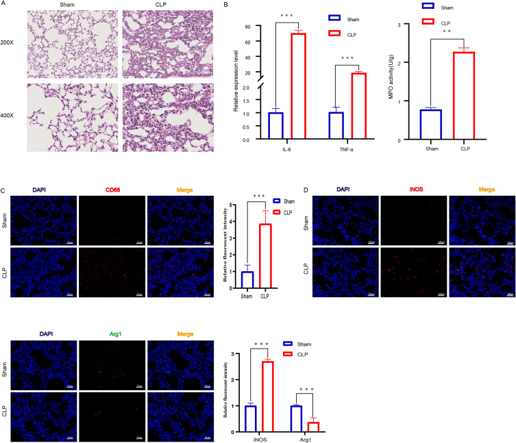
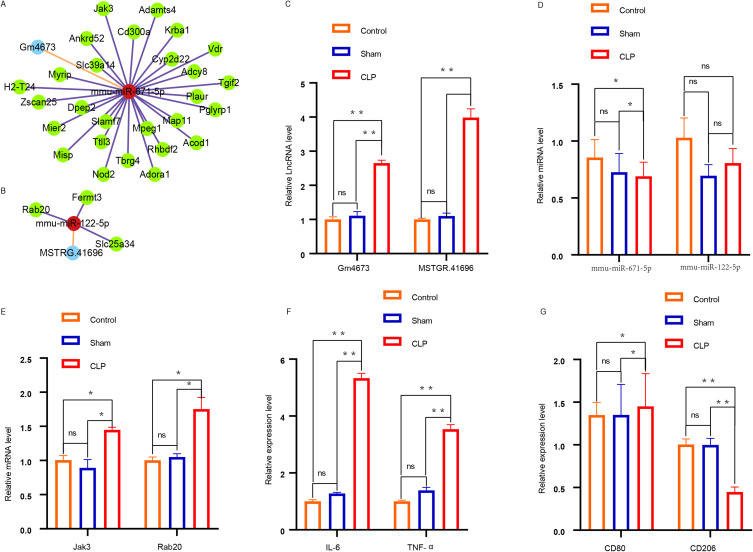
Background: Acute lung injury (ALI) is associated with a high mortality rate; however, the underlying molecular mechanisms are poorly understood. The purpose of this study was to investigate the expression profile and related networks of long noncoding RNAs (lncRNAs), microRNAs (miRNAs), and mRNAs in lung tissue exosomes obtained from sepsis-induced ALI.

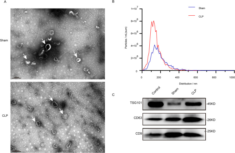
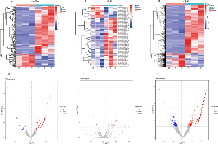
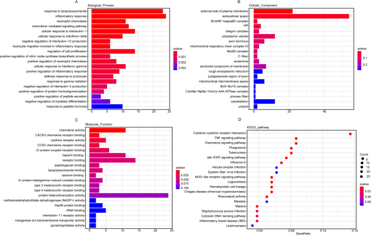
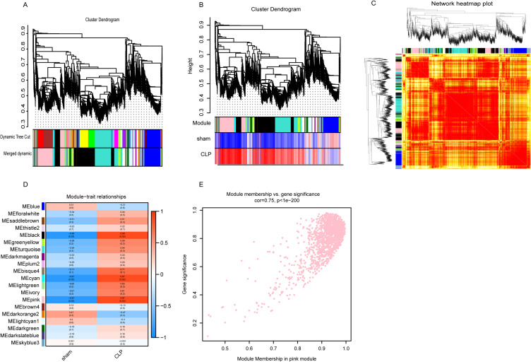
Methods: A mouse model of sepsis was established using the cecal ligation and puncture method. RNA sequencing was performed using lung tissue exosomes obtained from mice in the sham and CLP groups. Hematoxylin-eosin staining, Western blotting, immunofluorescence, quantitative real-time polymerase chain reaction, and nanoparticle tracking analysis were performed to identify relevant phenotypes, and bioinformatic algorithms were used to evaluate competitive endogenous RNA (ceRNA) networks.

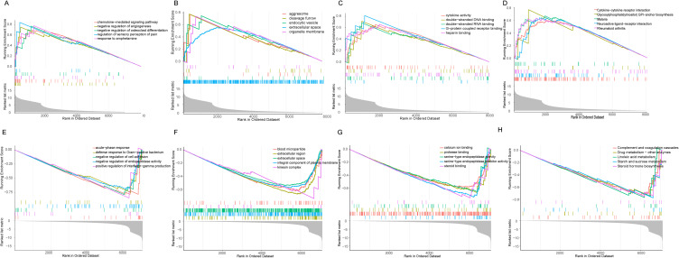
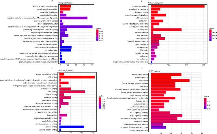
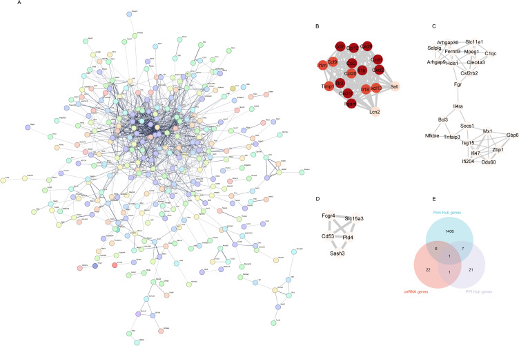
Results: Thirty lncRNA-miRNA-mRNA interactions were identified, including two upregulated lncRNAs, 30 upregulated miRNAs, and two downregulated miRNAs. Based on the expression levels of differentially expressed mRNAs(DEmRNAs), differentially expressed LncRNAs(DELncRNAs), and differentially expressed miRNAs(DEmiRNAs), 30 ceRNA networks were constructed.

Conclusion: Our study revealed, for the first time, the expression profiles of lncRNA, miRNA, and mRNA in exosomes isolated from the lungs of mice with sepsis-induced ALI, and the exosome co-expression network and ceRNA network related to ALI in sepsis.

Keywords: acute lung injury; ceRNA networks; inflammation; lung tissue exosomes; sepsis.


Conflict of interest statement

The authors declare that the research was conducted in the absence of any commercial or financial relationships that could be construed as a potential conflict of interest.

Figures

Figure 1
Figure 1
Macrophages Were Activated in the Lung Tissues of ALI Mice. (A) Histopathological analysis of lung tissues. After CLP modeling for 24 hours, H&E staining was used to evaluate the degree of lung inflammation in mice (n=6 per group), The images taken at 200X magnification correspond to a scale of 100 μm, while the images captured at 400X magnification correspond to a scale of 50 μm. (B) The pro-inflammatory cytokines IL-6 and TNF-α were detected by qPCR (n = 6 per group).The infiltration of inflammatory cells in lung tissue was measured by MPO activity (n=6 per group),**p<0.01, ***p<0.001. (C) After sectioning, the number of macrophages in lung tissue was detected by anti-CD68 antibody staining (n=6 per group). Intensity of immunofluorescence in lung tissues of CD68. (D)Intensity of immunofluorescence in lung tissues of M1 macrophages, (n =6 per group). iNOS (M1, green). Intensity of immunofluorescence in lung tissues of M2 macrophages, (n =6 per group). Arg1 (M2, red).
Figure 2
Figure 2
Isolation and identification of Lung Tissue exosomes. (A) Transmission electron micrographs of lung tissue exo-somes isolated from sham and ALI mice. Scale bar, 200 nm, The white arrows in (A) represent exosome particles. (B) NTA shows the concentration and size distribution of lung tissue exosomes isolated from the sham group and CLP mice (n=6 in each group). (C) Western blot analysis of the lung tissue exosomal markers TSG101, CD63 and CD9.
Figure 3
Figure 3
Differentially expressed lung tissue exosomal lncRNAs, miRNAs and mRNAs in sepsis-induced acute lung Inflammation. (AC) Heatmaps of lncRNA, miRNA and mRNA according to the value of |logFC|, in the Sham and CLP group. (DF) Volcano maps of differentially expressed lung tissue exosomal lncRNAs, miRNAs and mRNAs (DEGs). Blue pixels indicate decreased gene levels in the indicated sample, whereas red pixels indicate increased abundance. S2, S3 and S4 are three biological duplicate samples of Sham group, and C1, C2 and C3 are three biological duplicate samples of CLP group.
Figure 4
Figure 4
Functional enrichment analysis of differentially expressed mRNAs. (AC) The top 20 GO terms in the enrichment analysis of dysregulated mRNAs between Sham and CLP groups. (D) The top 20 KEGG pathways in the enrichment analysis of dysregulated mRNAs between Sham and CLP groups. An enrichment factor is calculated by dividing the number of differentially expressed genes by the total number of genes annotated in this pathway. Number of genes is represented by bubble scale, and P value is represented by bubble color depth.
Figure 5
Figure 5
Functional enrichment analysis of mRNAs. (AD) Between Sham group and CLP group, the first five up-regulated GO biological processes and the first five up-regulated KEGG pathway mRNAs enrichment.(EH) Between Sham group and CLP group, the first five down-regulated GO biological processes and the first five down-regulated KEGG pathway mRNAs enrichment.
Figure 6
Figure 6
Construction and module analysis of weighted gene co-expression network analysis (WGCNA).(A) Based on topological overlap and assigned module colors, clustering dendrogram of dissimilar genes. (B) Cluster analysis based on gene expression profile. (C) Clustering of genes using the average linkage hierarchical clustering method. (D) A module-trait association consists of a single row for each eigengene and a single column for each trait. (E) Scatterplot of gene significance related to module membership in the pink co-expression modules.
Figure 7
Figure 7
Functional enrichment analysis of differentially expressed Hub gene in the pink. (AC) GO classification of differentially expressed genes in biological processes, cellular components and molecular functions. (D) The KEGG pathways in the enrichment analysis of differentially expressed Hub genes. Number of genes is represented by bubble scale, and P value is represented by bubble color depth.
Figure 8
Figure 8
Protein-protein interaction (PPI) network of differentially expressed mRNAs (A), the PPI network consists of 317 nodes and 143 edges. (BD) The top three clusters of PPIs. (E) The intersection gene of the pink module Hub gene, PPI Hub gene and ceRNA gene.
Figure 9
Figure 9
lncRNA- miRNA - mRNA ceRNA network. (A and B) lncRNA–miRNA–mRNA co-expression network. Light blue, red and green are representative of lncRNAs, miRNAs, and mRNAs, respectively. (CE) Detection of LncRNA, miRNA, mRNA expression in ceRNA using qpcr. (F) The pro-inflammatory cytokines IL-6 and TNF-α were detected by qpcr. (G)The expression of CD206 and CD80 was measured using quantitative qpcr. *p<0.05,**p<0.01, ns p>0.05.

Similar articles

References

    1. Sevransky JE, Martin GS, Shanholtz C, et al. Mortality in sepsis versus non-sepsis induced acute lung injury. Crit Care. 2009;13(5):R150. doi:10.1186/cc8048 - DOI - PMC - PubMed
    1. Killien EY, Huijsmans RLN, Ticknor IL, et al. Acute respiratory distress syndrome following pediatric trauma: application of pediatric acute lung injury consensus conference criteria. Crit Care Med. 2020;48(1):e26–e33. doi:10.1097/CCM.0000000000004075 - DOI - PMC - PubMed
    1. Fleischmann C, Scherag A, Adhikari NK, et al. International forum of acute care T: assessment of global incidence and mortality of hospital-treated sepsis. Current estimates and limitations. Am J Respir Crit Care Med. 2016;193(3):259–272. doi:10.1164/rccm.201504-0781OC - DOI - PubMed
    1. Singer M, Deutschman CS, Seymour CW, et al. The third international consensus definitions for sepsis and septic shock (Sepsis-3). JAMA. 2016;315(8):801–810. doi:10.1001/jama.2016.0287 - DOI - PMC - PubMed
    1. Li Y, Cao Y, Xiao J, et al. Inhibitor of apoptosis-stimulating protein of p53 inhibits ferroptosis and alleviates intestinal ischemia/reperfusion-induced acute lung injury. Cell Death Differ. 2020;27(9):2635–2650. doi:10.1038/s41418-020-0528-x - DOI - PMC - PubMed

Grants and funding

This work was supported by the National Natural Science Foundation of China (82160363 and 81871548).