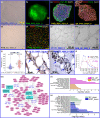
Endothelial cells differentiated from patient dermal fibroblast-derived induced pluripotent stem cells resemble vascular malformations of port-wine birthmark
- PMID: 37672656
- PMCID: PMC10653332
- DOI: 10.1093/bjd/ljad330
Endothelial cells differentiated from patient dermal fibroblast-derived induced pluripotent stem cells resemble vascular malformations of port-wine birthmark
Abstract
Lesional induced pluripotent stem cell-derived endothelial cells can resemble pathological vascular phenotypes of port-wine birthmark (PWB). Our data demonstrate that multiple pathways, including Hippo and Wnt, NFκB, TNF, MAPK and cholesterol metabolism, are dysregulated. These data suggest new therapeutics can be developed to target such dysregulated pathways in the treatment of PWB.
Conflict of interest statement
Conflicts of interest A.G.J. is a member of the Scientific Advisory Board of Gen1E Lifesciences, USA. W.T. is a shareholder of TritaliMed, USA. The other authors declare that they have no conflicts of interest.
Figures

Update of
-
Supporting materials: Endothelial cells differentiated from patient dermal fibroblast-derived induced pluripotent stem cells resemble vascular malformations of Port Wine Birthmark.bioRxiv [Preprint]. 2023 Aug 24:2023.07.02.547408. doi: 10.1101/2023.07.02.547408. bioRxiv. 2023. Update in: Br J Dermatol. 2023 Nov 16;189(6):780-783. doi: 10.1093/bjd/ljad330 PMID: 37662218 Free PMC article. Updated. Preprint.
Similar articles
-
Supporting materials: Endothelial cells differentiated from patient dermal fibroblast-derived induced pluripotent stem cells resemble vascular malformations of Port Wine Birthmark.bioRxiv [Preprint]. 2023 Aug 24:2023.07.02.547408. doi: 10.1101/2023.07.02.547408. bioRxiv. 2023. Update in: Br J Dermatol. 2023 Nov 16;189(6):780-783. doi: 10.1093/bjd/ljad330 PMID: 37662218 Free PMC article. Updated. Preprint.
-
Verrucous carcinoma arising in a port wine stain.Br J Oral Maxillofac Surg. 2016 Sep;54(7):842. doi: 10.1016/j.bjoms.2015.12.021. Epub 2016 Jan 19. Br J Oral Maxillofac Surg. 2016. PMID: 26809361 No abstract available.
-
Delayed ulceration following combination pulse dye laser and topical sirolimus treatment for port wine birthmarks: A case series.Pediatr Dermatol. 2024 Jan-Feb;41(1):108-111. doi: 10.1111/pde.15409. Epub 2023 Aug 12. Pediatr Dermatol. 2024. PMID: 37571864
-
Nevus roseus: a distinct vascular birthmark.Eur J Dermatol. 2005 Jul-Aug;15(4):231-4. Eur J Dermatol. 2005. PMID: 16048748 Review.
-
Vascular anomalies: portwine stains and hemangiomas.J Cutan Pathol. 2010 Apr;37 Suppl 1:88-95. doi: 10.1111/j.1600-0560.2010.01519.x. J Cutan Pathol. 2010. PMID: 20482681 Review. No abstract available.
Cited by
-
Perturbations of Glutathione and Sphingosine Metabolites in Port Wine Birthmark Patient-Derived Induced Pluripotent Stem Cells.Metabolites. 2023 Aug 31;13(9):983. doi: 10.3390/metabo13090983. Metabolites. 2023. PMID: 37755263 Free PMC article.
-
Perturbations of glutathione and sphingosine metabolites in Port Wine Birthmark patient-derived induced pluripotent stem cells.bioRxiv [Preprint]. 2023 Jul 19:2023.07.18.549581. doi: 10.1101/2023.07.18.549581. bioRxiv. 2023. Update in: Metabolites. 2023 Aug 31;13(9):983. doi: 10.3390/metabo13090983 PMID: 37503303 Free PMC article. Updated. Preprint.
References
-
- Lever WF, Schaumburg-Lever G. Histopathology of the Skin, 7th edn. Philadelphia, PA: J.B. Lippincott; , 1990.
-
- Williams J et al. Embryonic stem cell-like population in hypertrophic port-wine stain. JOVA 2021; 2:e006.

