Loading [MathJax]/jax/output/PreviewHTML/jax.js
Skip to main page content
U.S. flag

An official website of the United States government

Access keys NCBI Homepage MyNCBI Homepage Main Content Main Navigation
. 2023 Aug 10;26(9):107586.
doi: 10.1016/j.isci.2023.107586. eCollection 2023 Sep 15.

Genome-wide association study identifies several loci for HEV seropositivity

Affiliations

Genome-wide association study identifies several loci for HEV seropositivity

Maria K Smatti et al. iScience. .

Abstract

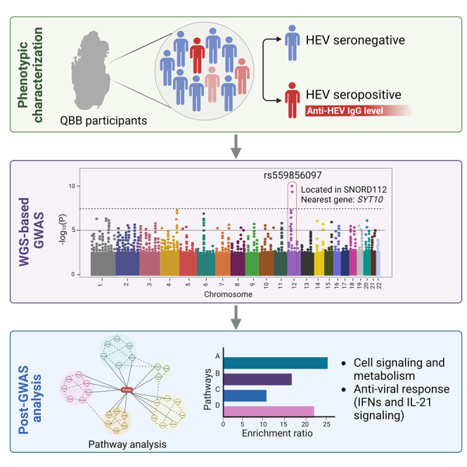
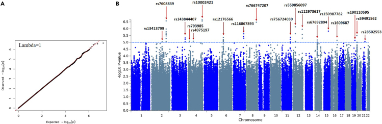
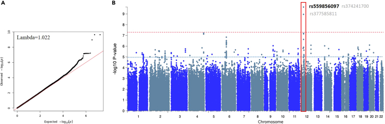
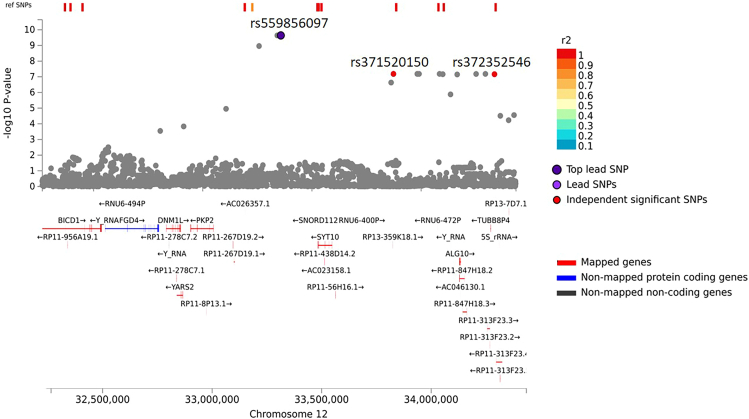
Hepatitis E viral (HEV) infection imposes a heavy global health burden. The variability in the prevalence of serological markers of HEV infection between different ethnic groups proposes a host genetic influence. Here, we report genetic polymorphisms associated with anti-HEV antibody positivity and level using binary- and quantitative-trait genome-wide association studies (GWAS) on a population from Qatar (n = 5829). We identified a region in 12p11.1 (lead SNP: rs559856097, allele: A, p = 2.3 × 10⁻¹⁰) significantly associated with anti-HEV antibodies level. This intergenic variant is located near SNORD112, a small nucleolar RNA (snoRNA). Additional gene-set and pathway enrichment analyses highlighted a strong correlation with anti-viral response-related pathways, including IFNs (alpha/beta) and interleukin-21 (IL-21) signaling. This is the first GWAS on the response to HEV infection. Further replication and functional experimentation are warranted to validate these findings.

Keywords: Association analysis; Immune response; Quantitative genetics; Virology.


Conflict of interest statement

The authors declare no competing interests.

Figures

None
Graphical abstract
Figure 1
Figure 1
Quantile-Quantile (QQ) and Manhattan plots of the GWAS on HEV seropositivity binary trait (A) QQ plot of the association test statistics. The regression line (in red) represents the expected distribution of p values under the null hypothesis. Lambda value is the genomic inflation factor calculated as the ratio of the median of the observed distribution of the test statistic to the expected values. (B) Observed −log10 p values (y axis) are shown for all SNPs on each autosomal chromosome (x axis). The blue dotted line indicates suggestive significant of p value <1× . The top SNPs at each suggestive genomic loci are labeled with rs identifiers.
Figure 2
Figure 2
Quantile-Quantile (QQ) and Manhattan plots of the GWAS on anti-HEV antibodies level quantitative trait (A) QQ plot of the association test statistics. The regression line (in red) represents the expected distribution of p values under the null hypothesis. Lambda value is the genomic inflation factor calculated as the ratio of the median of the observed distribution of the test statistic to the expected values. (B) Observed −log10 p values (y axis) are shown for all SNPs on each autosomal chromosome (x axis). The red dotted line indicates GWAS significant of p value < 1× 10⁸, while the black line indicates suggestive significant at p value < 1× 10⁵. The GWAS significant genomic locus (12p11.1) is bordered with red, and the top SNPs are labeled with the rs identifiers. The top lead SNP is in black, while the two SNPs in the locus (dependent) are in light gray.
Figure 3
Figure 3
Regional association plot for the top signal at rs559856097 Genes were mapped to this locus by positional, eQTLs, and chromatin interaction mapping using FUMA platform (https://fuma.ctglab.nl/). The top lead and the independent significant SNPs are labeled with rs identifier.
Figure 4
Figure 4
Heatmaps of the average gene expression per tissue (GTEx v8 30 general tissue types) for the top genes (highest p value) identified by MAGMA analysis (A) Top 10 MAGMA genes from the binary trait GWAS. (B) Top 10 MAGMA genes from the quantitative trait GWAS. The expression in blood and liver tissues are particularly bordered in black. FUMA platform (https://fuma.ctglab.nl/) was used for gene annotation and representation.
Figure 5
Figure 5
Gene set enrichment analysis of the binary trait HEV GWAS using WebGestalt (A) GO enrichment analysis on the candidate protein coding genes mapped by FUME on the binary GWAS gene set (n = 189 genes mapped from 18 genomic loci). (B) ORA pathway analysis using Reactome database on the same set of genes (n = 189). The top ten enriched pathways are presented in each bar graph. Categories with significant enrichment (FDR <0.05) are colored in dark blue.
Figure 6
Figure 6
Gene set enrichment analysis of the quantitative trait HEV GWAS using WebGestalt (A) GO enrichment analysis on the candidate protein coding genes mapped by FUME on quantitate GWAS gene set (n = 441 candidate genes covering the significant and suggestive GWAS loci). (B) ORA pathway analysis using Reactome database on the same set of genes (n = 441). The top ten enriched pathways are presented in each bar graph. Categories with significant enrichment (FDR <0.05) are colored in dark blue.
Figure 7
Figure 7
Gene set enrichment analysis of immune-related genes from the binary and quantitative trait HEV GWASs using WebGestalt (A–C) A list of 43 immune genes filtered from our candidate genes list was used for: A.GO enrichment analysis. B.ORA pathway analysis. C. ORA in GLAD4U disease database. The top ten enriched pathways are presented in each bar graph. Categories with significant enrichment (FDR <0.05) are colored in dark blue.

Similar articles

References

    1. Wißing M.H., Brüggemann Y., Steinmann E., Todt D. Virus-Host Cell Interplay during Hepatitis E Virus Infection. Trends Microbiol. 2021;29:309–319. doi: 10.1016/j.tim.2020.07.002. - DOI - PMC - PubMed
    1. Kenney S.P. The Current Host Range of Hepatitis E Viruses. Viruses. 2019;11:452. doi: 10.3390/v11050452. - DOI - PMC - PubMed
    1. Wedemeyer H., Pischke S., Manns M.P. Pathogenesis and treatment of hepatitis e virus infection. Gastroenterology. 2012;142:1388–1397.e1. doi: 10.1053/j.gastro.2012.02.014. - DOI - PubMed
    1. Ditah I., Charlton M. Genetic susceptibility to hepatitis E viral infection: An enigmatic virus gives up a secret. Hepatology. 2015;62:1337–1338. doi: 10.1002/hep.28106. - DOI - PubMed
    1. Hepatitis E. 2022. https://www.who.int/en/news-room/fact-sheets/detail/hepatitis-e

LinkOut - more resources