Assessing MTT and sulforhodamine B cell proliferation assays under multiple oxygen environments
- PMID: 37655276
- PMCID: PMC10465423
- DOI: 10.1007/s10616-023-00584-0
Assessing MTT and sulforhodamine B cell proliferation assays under multiple oxygen environments
Abstract
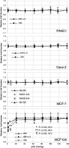
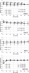
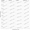
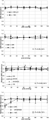
Cell proliferation can be measured directly by counting cells or indirectly using assays that quantitate total protein or metabolic activity. However, for comparing cell proliferation under varying oxygen conditions it is not clear that these assays are appropriate surrogates for cell counting as cell metabolism and protein synthesis may vary under different oxygen environments. We used permeable bottom tissue culture ware to compare proliferation assays as a function of static oxygen concentrations under oxygen partial pressure (pO2) levels ranging from 2 to 139 mmHg. Cell proliferation was measured by cell counting and compared to surrogate methods measuring cell metabolism (3-(4,5-dimethylthiazol-2-yl)-2,5-diphenyltetrazolium bromide, MTT) and total protein (sulforhodamine B) assays under these different environments in Caco-2, MCF-7, MCF-10A and PANC-1 human cell lines. We found that the MTT readings do not correlate with cell number for the Caco-2 and PANC-1 cell lines under different oxygen conditions, whereas the sulforhodamine B protein assays perform well under all conditions. However, within a given oxygen environment, both proliferation assays show a correlation with cell number. Therefore, the MTT assay must be used with caution when comparing cell growth or drug response for cells grown in different oxygen environments.
Supplementary information: The online version contains supplementary material available at 10.1007/s10616-023-00584-0.
Keywords: Cancer; Hypoxia; MTT assay; Oxygen partial pressure; Proliferation; SRB.
© The Author(s), under exclusive licence to Springer Nature B.V. 2023. Springer Nature or its licensor (e.g. a society or other partner) holds exclusive rights to this article under a publishing agreement with the author(s) or other rightsholder(s); author self-archiving of the accepted manuscript version of this article is solely governed by the terms of such publishing agreement and applicable law.
Conflict of interest statement
Competing interestsThe authors have no relevant financial or non-financial interests to disclose.
Figures


Similar articles
-
Mixing and delivery of multiple controlled oxygen environments to a single multiwell culture plate.Am J Physiol Cell Physiol. 2018 Nov 1;315(5):C766-C775. doi: 10.1152/ajpcell.00276.2018. Epub 2018 Sep 5. Am J Physiol Cell Physiol. 2018. PMID: 30183322 Free PMC article.
-
Assessment of Sertoli Cell Proliferation by 3-(4,5-Dimethylthiazol-2-yl)-2,5-Diphenyltetrazolium Bromide and Sulforhodamine B Assays.Curr Protoc Toxicol. 2019 Sep;81(1):e85. doi: 10.1002/cptx.85. Curr Protoc Toxicol. 2019. PMID: 31529795
-
Limitations of the 3-(4,5-dimethylthiazol-2-yl)-2,5-diphenyl-2H-tetrazolium bromide (MTT) assay when compared to three commonly used cell enumeration assays.BMC Res Notes. 2015 Feb 20;8:47. doi: 10.1186/s13104-015-1000-8. BMC Res Notes. 2015. PMID: 25884200 Free PMC article.
-
Schiff Bases and their Metal Complexes as Potential Anticancer Candidates: A Review of Recent Works.Anticancer Agents Med Chem. 2019;19(15):1786-1795. doi: 10.2174/1871520619666190227171716. Anticancer Agents Med Chem. 2019. PMID: 30827264 Review.
-
The MTT assay application to measure the viability of spermatozoa: A variety of the assay protocols.Open Vet J. 2021 Apr-Jun;11(2):251-269. doi: 10.5455/OVJ.2021.v11.i2.9. Epub 2021 May 8. Open Vet J. 2021. PMID: 34307082 Free PMC article. Review.
Cited by
-
Multitarget Pharmacology of Sulfur-Nitrogen Heterocycles: Anticancer and Antioxidant Perspectives.Antioxidants (Basel). 2024 Jul 25;13(8):898. doi: 10.3390/antiox13080898. Antioxidants (Basel). 2024. PMID: 39199144 Free PMC article. Review.
-
New 6-nitro-4-substituted quinazoline derivatives targeting epidermal growth factor receptor: design, synthesis and in vitro anticancer studies.Future Med Chem. 2024;16(19):2025-2041. doi: 10.1080/17568919.2024.2389772. Epub 2024 Sep 4. Future Med Chem. 2024. PMID: 39230501
-
Improving the power of drug toxicity measurements by quantitative nuclei imaging.Cell Death Discov. 2024 Apr 18;10(1):181. doi: 10.1038/s41420-024-01950-3. Cell Death Discov. 2024. PMID: 38637526 Free PMC article.
References
Grants and funding
LinkOut - more resources
Full Text Sources

