. 2023 Nov;63(5):116.
doi: 10.3892/ijo.2023.5564. Epub 2023 Sep 1.

Human cytomegalovirus infection enhances 5‑lipoxygenase and cycloxygenase‑2 expression in colorectal cancer

Affiliations

Human cytomegalovirus infection enhances 5‑lipoxygenase and cycloxygenase‑2 expression in colorectal cancer

Mattia Russel Pantalone et al. Int J Oncol. 2023 Nov.

Abstract

Colorectal cancer (CRC) is one of the most common and fatal types of cancer. Inflammation promotes CRC development, however, the underlying etiological factors are unknown. Human cytomegalovirus (HCMV), a virus that induces inflammation and other cancer hallmarks, has been detected in several types of malignancy, including CRC. The present study investigated whether HCMV infection was associated with expression of the pro‑inflammatory enzymes 5‑lipoxygenase (5‑LO) and cyclooxygenase‑2 (COX‑2) and other molecular, genetic and clinicopathological CRC features. The present study assessed 146 individual paraffin‑embedded CRC tissue microarray (TMA) cores already characterized for TP53 and KRAS mutations, microsatellite instability (MSI) status, Ki‑67 index and EGFR by immunohistochemistry (IHC). The cores were further analyzed by IHC for the expression of two HCMV proteins (Immediate Early, IE and pp65) and the inflammatory markers 5‑LO and COX‑2. The CRC cell lines Caco‑2 and LS‑174T were infected with HCMV strain VR1814, treated with antiviral drug ganciclovir (GCV) and/or anti‑inflammatory drug celecoxib (CCX) and analyzed by reverse transcription‑quantitative PCR and immunofluorescence for 5‑LO, COX‑2, IE and pp65 transcripts and proteins. HCMV IE and pp65 proteins were detected in ~90% of the CRC cases tested; this was correlated with COX‑2, 5‑LO and KI‑67 expression, but not with EGFR immunostaining, TP53 and KRAS mutations or MSI status. In vitro, HCMV infection upregulated 5‑LO and COX‑2 transcript and proteins in both Caco‑2 and LS‑174T cells and enhanced cell proliferation as determined by MTT assay. Treatment with GCV and CCX significantly decreased the transcript levels of COX‑2, 5‑LO, HCMV IE and pp65 in infected cells. HCMV was widely expressed in CRC and may promote inflammation and serve as a potential new target for CRC therapy.

Keywords: celecoxib; colorectal cancer; ganciclovir; human cytomegalovirus; inflammation.


Conflict of interest statement

CSN holds a patent on diagnostics and treatment of a CMV variant strain found in cancer (patent no. US9701943B2). The other authors declare that they have no competing interests.

Figures

Figure 1
Figure 1
Representative micrographs of immunostaining for human Cytomegalovirus IE and late protein pp65 in CRC and normal colorectal mucosa. (A) IE is expressed in the cytoplasm of cancer cells in a punctate pattern and as indicated by the arrows in tumor associated vessels' endothelial cells; (B) magnification. (C) Pp65 shows a diffuse cytoplasmic immunostaining evident in colorectal cancer cells and as indicated by the cirle in tumor-associated interstitial cells and macrophages; (D) magnification of C. (E) IE immunostaining is not found in non-tumorous mucosa. (F) Pp65 immunostaining is not found in non-tumorous mucosa. IE, immediate early; CRC, colorectal cancer.
Figure 2
Figure 2
Representative micrographs of COX-2 and 5-LO immunostaining in colorectal cancer and non-tumorous colorectal mucosa. (A) Cytoplasmic COX-2 is strongly expressed in colorectal cancer cells; (B) magnification of A. (C) Primarily nuclear 5-LOis strongly and diffusely expressed in colorectal cancer cells. (D) magnification of D. (E) COX-2 is not expressed in non-tumorous crypt cells. (F) 5-LO is not expressed in non-tumorous crypt cells (Scale bar, 50 μm. COX-2, cyclooxygenase-2; 5-LO, 5-lipoxygenase.
Figure 3
Figure 3
HCMV IE and pp65 transcript expression in HCMV-infected Caco-2 and LS-174T cells is reduced by GCV and CCX. Relative IE and pp65 expression levels were determined by reverse transcription-quantitative PCR. The bars represent COX-2 relative expression to the housekeeping gene. (A) No IE transcripts were detected at 1 dpi in HCM-infected or mock-infected Caco-2 cells; (B) IE transcripts were detected at 3 dpi in HCMV infected Caco-2 cells (VR) and they were reduced by treamtents with GCV and CCX. (C) IE transcripts were detected at 7 dpi in VR) and they were reduced by treamtent with GCV and CCX. (D) Pp65 transcripts were detected at 7 pi in HCMV-infected Caco-2 cells and reduced by GCV and CCX treatments. Only the 7 dpi timepoint is shown for pp65 since no transcripts were detected at earlier timepoints (E) No IE transcripts were detected at 1 dpi in HCM-infected or mock-infected LS-174T cells; (F) IE transcripts were detected at 3 dpi in HCMV infected LS-174T cells (VR) and they were reduced by treamtents with GCV and CCX; (G) IE transcripts were detected at 7 dpi in HCMV infected LS-174T cells (VR) and they were reduced by treamtents with GCV and CCX; (H) Pp65 transcripts were detected at 7 pi in HCMV-infected LS-174T cells and reduced by GCV and CCX treatments. Only the 7 dpi timepoint is shown for pp65 since no transcripts were detected at earlier timepoints. Data are presented as the mean ± SD. Statistical significance was determined by ANOVA test. *P≤0.05, **P≤0.01, ***P≤0.001, ****P≤0.0001 vs. VR. HCMV, human cytomegalovirus; IE, immediate early; pp65, posphoprotein 65; GCV, ganciclovir; CCX, celecoxib; MI, mock-infected; VR, virus-infected; dpi, days post infection; ns, not significant.
Figure 4
Figure 4
HCMV-infected Caco-2 and LS-174T cells exhibit increased COX-2 expression. Relative COX-2 expression was determined by reverse transcription-quantitative PCR at 1, 3 and 7 dpi. Data are presented as the mean ± SD. Caco-2 infected with HCMV (VR) shows higher COX-2 transcripts than mock-infected cells (MI) at 1 dpi (A), 3 dpi (B) and 7 dpi (C). Treatment with GCV, CCX or a combination of the two drugs significantly reduced COX-2 transcripts at all time points in Caco-2 HCMV-infected cells. LS-174 cells infected with HCMV (VR) shows higher COX-2 transcripts than mock-infected cells (MI) at 1 dpi (D), 3 dpi (E) and 7 dpi (F). Treatment with GCV, CCX or a combination of the two drugs significantly reduced COX-2 transcripts at all time points in LS-174 HCMV-infected cells. Statistical significance was determined by ANOVA test. Statistical significance was determined by ANOVAtest. *P≤0.05, **P≤0.01, ***P≤0.001, ****P≤0.0001 vs. VR. HCMV, human cytomegalovirus; COX-2, cyclooxygenase-2; GCV, ganciclovir; CCX, celecoxib; MI, mock-infected; VR, virus-infected; dpi, days post infection.
Figure 5
Figure 5
HCMV increases 5-LO expression in Caco-2 and LS-174T cells. Relative 5-LO expression was determined by qPCR at 1, 3 and 7 dpi. The bars represent 5-LO relative expression to the housekeeping gene. Data are presented as the mean ± SD. (A) At 1 dpi Caco-2 infected with HCMV (VR) do not show higher 5-LO transcripts than MI); GCV treatment on infected cells did not decrease 5-LO transcripts at 1 dpi, on the contrary CCX and a combination of GCV and CCX reduce 5-LO transcripts in HCMV-infected cells. At 3 dpi (B) and 7 dpi (C) Caco-2 infected with HCMV (VR) shows higher 5-LO transcripts than mock-infected cells (MI). Treatment with GCV, CCX or a combination of the two drugs significantly reduced 5-LO transcripts at 3 dpi (B) and 7 dpi (C) in Caco-2 HCMV-infected cells. LS-174 cells infected with HCMV (VR) shows higher 5-LO transcripts than mock-infected cells (MI) at 1 dpi (D), 3 dpi (E) and 7 dpi (F). Treatment with GCV, CCX or a combination of the two drugs significantly reduced 5-LO transcripts at all time points in LS-174 HCMV-infected cells. Statistical significance was determined by ANOVA test. ***P≤0.001, ****P≤0.0001 vs. VR. HCMV, human cytomegalovirus; 5-LO, 5-lipoxygenase; GCV, ganciclovir; CCX, celecoxib; MI, mock-infected; VR, virus-infected; dpi, days post infection; ns, not significant.
Figure 6
Figure 6
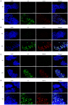
Immunofluorescence analysis of 5-LO and COX-2 protein staining in human cytomegalovirus (HCMV) infected colorectal cancer cells. Caco-2 and LS-174T cells at 7 days post-infection were stained for of IE (green) and 5-LO or COX-2 (red). IE was detectable only in infected cells. 5-LO was upregulated in infected (A) Caco-2 and (B) LS-174T cells. COX-2 was upregulated in infected (C) Caco-2 and (D) LS-174T cells. Scale bar, 100 μm. 5-LO, 5-lipoxygenase; COX-2, cyclooxygenase-2; IE, immediate early; MI, mock-infected; VR, virus-infected.
Figure 7
Figure 7
Increased proliferation in VR. Proliferation of HCMV-infected colorectal cancer cells (VR was analyzed by MTT assay and values were normalized to mock-infected cells (MI). Data are presented as the mean ± SD. *P≤0.05, **P≤0.01, ****P≤0.0001 vs. MI. VR) do not show a higher proliferation rate than MI cells at 1 dpi (A) or at 3 (dpi). HCMV-infected Caco-2 cells VR) show a higher proliferation rate than MI cells at (B) 3 dpi (B) and at 7 dpi (C). HCMV-infected LS-174T cells (VR) do not show a higher proliferation rate than MI cells at 1 dpi (D) or at 3 dpi (E). HCMV-infected LS-174T cells (VR) show a higher proliferation rate than MI cells at 7 dpi (F). HCMV, human cytomegalovirus; dpi, days post-infection; ns, not significant; MI, mock-infected; VR,virus-infected.

Similar articles

Cited by

References

    1. Sung H, Ferlay J, Siegel RL, Laversanne M, Soerjomataram I, Jemal A, Bray F. Global cancer statistics 2020: GLOBOCAN estimates of incidence and mortality worldwide for 36 cancers in 185 countries. CA Cancer J Clin. 2021;71:209–249. doi: 10.3322/caac.21660. - DOI - PubMed
    1. Jeon J, Du M, Schoen RE, Hoffmeister M, Newcomb PA, Berndt SI, Caan B, Campbell PT, Chan AT, Chang-Claude J, et al. Determining risk of colorectal cancer and starting age of screening based on lifestyle, environmental, and genetic factors. Gastroenterology. 2018;154:2152–2164.e2119. doi: 10.1053/j.gastro.2018.02.021. - DOI - PMC - PubMed
    1. Kulaylat MN, Dayton MT. Ulcerative colitis and cancer. J Surg Oncol. 2010;101:706–712. doi: 10.1002/jso.21505. - DOI - PubMed
    1. Jensen AB, Larsen M, Gislum M, Skriver MV, Jepsen P, Nørgaard B, Sørensen HT. Survival after colorectal cancer in patients with ulcerative colitis: A nationwide population-based Danish study. Am J Gastroenterol. 2006;101:1283–1287. doi: 10.1111/j.1572-0241.2006.00520.x. - DOI - PubMed
    1. Dolin TG, Christensen IJ, Johansen AZ, Nielsen HJ, Jakobsen HL, Klein MF, Lund CM, Bojesen SE, Nielsen DL, Jensen BV, Johansen JS. Pre- and perioperative inflammatory biomarkers in older patients resected for localized colorectal cancer: Associations with complications and prognosis. Cancers (Basel) 2022;14:161. doi: 10.3390/cancers14010161. - DOI - PMC - PubMed

Grants and funding

The present study was supported by Swedish Medical Research Council (grant no. 2019-01736), The Cure Cancer Foundation and Italian Association for Cancer Research (grant no. 24501).