. 2023 Nov;167(3):362-375.
doi: 10.1111/jnc.15948. Epub 2023 Aug 31.

Plk2 promotes synaptic destabilization through disruption of N-cadherin adhesion complexes during homeostatic adaptation to hyperexcitation

Affiliations

Plk2 promotes synaptic destabilization through disruption of N-cadherin adhesion complexes during homeostatic adaptation to hyperexcitation

Mai Abdel-Ghani et al. J Neurochem. 2023 Nov.

Abstract

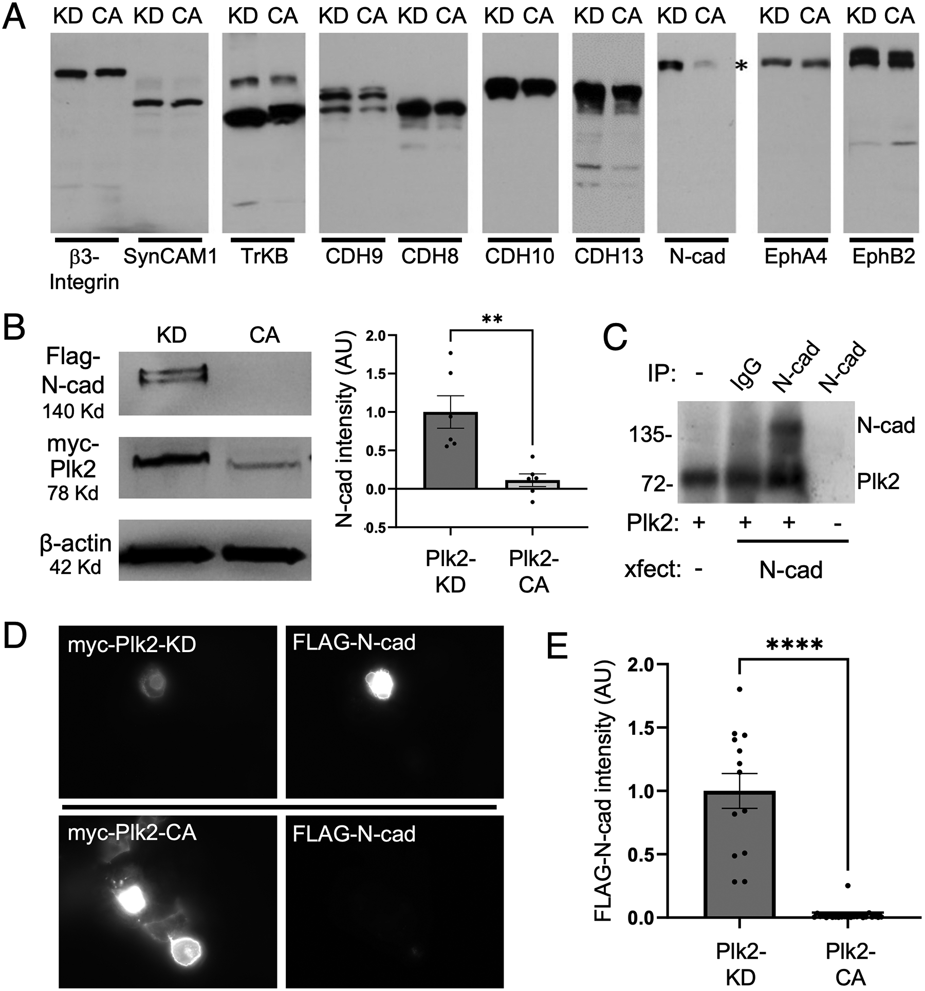
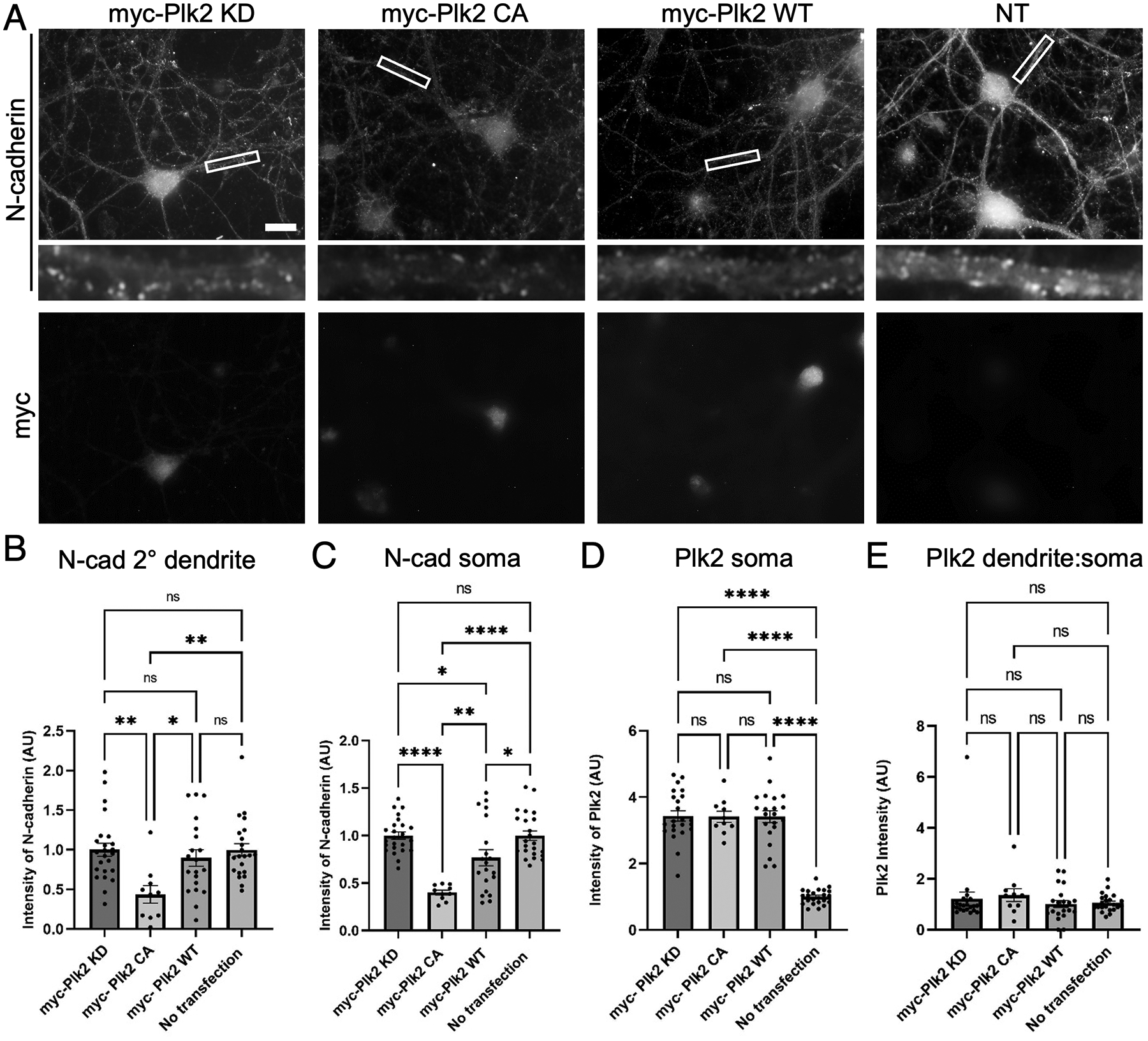
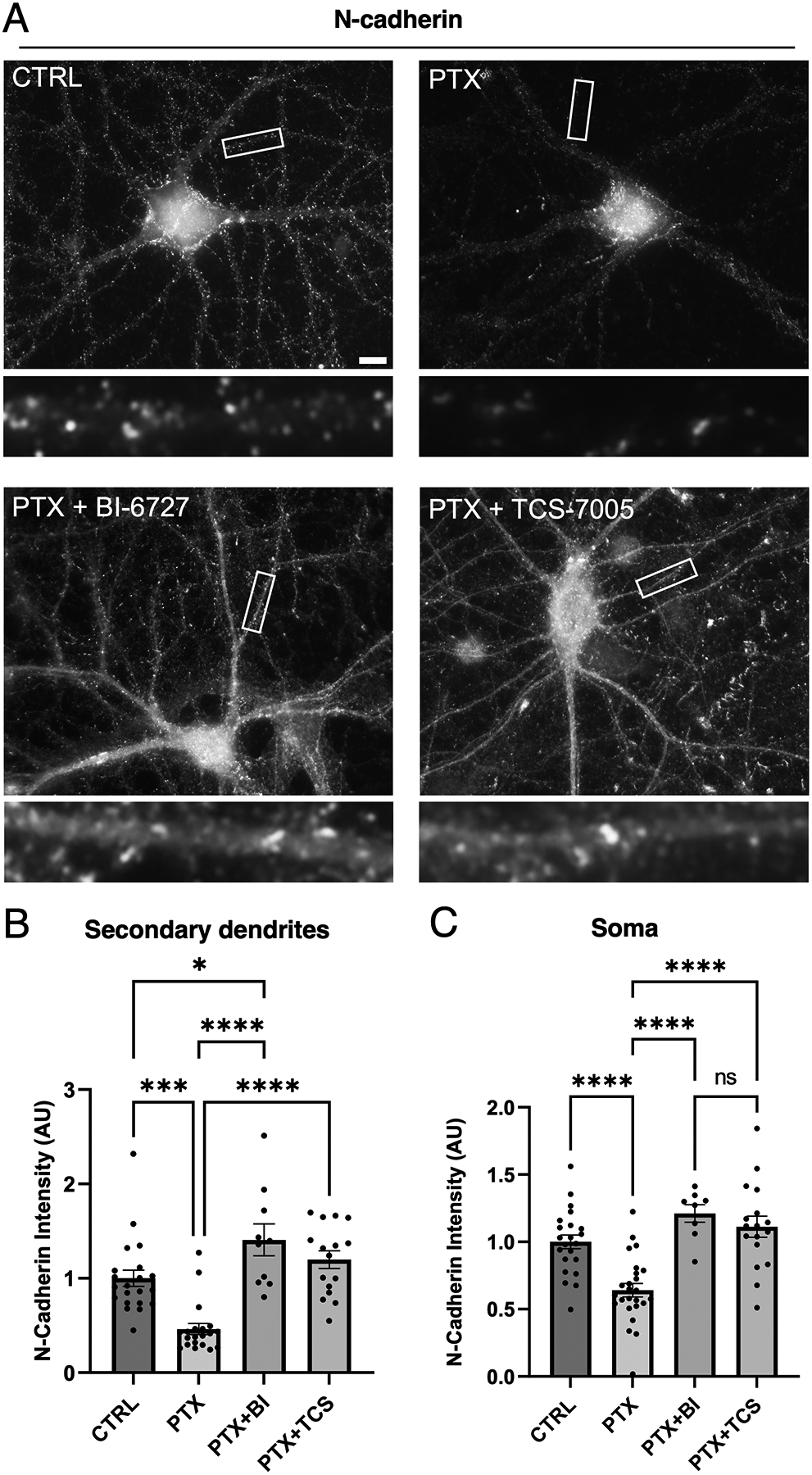
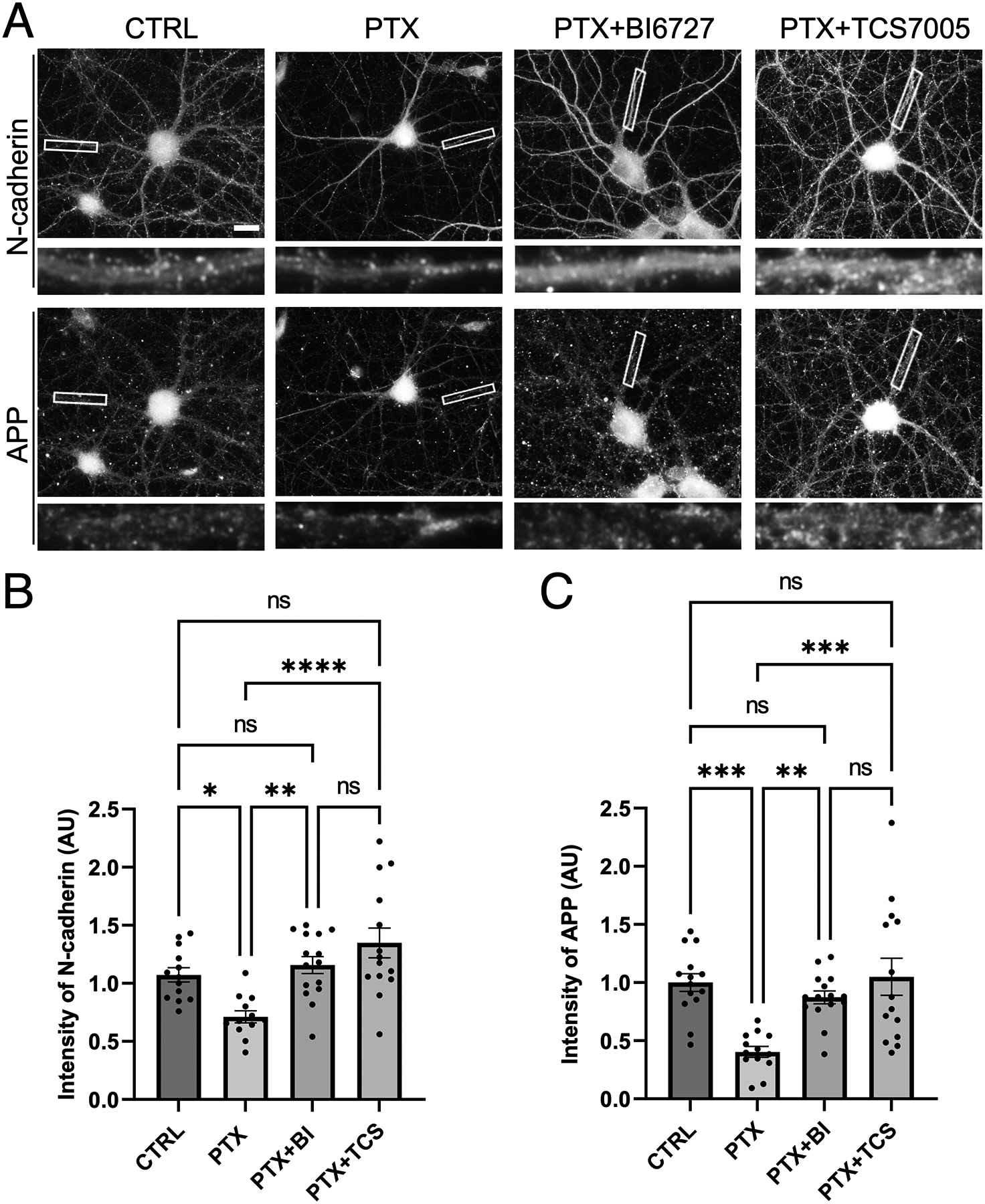
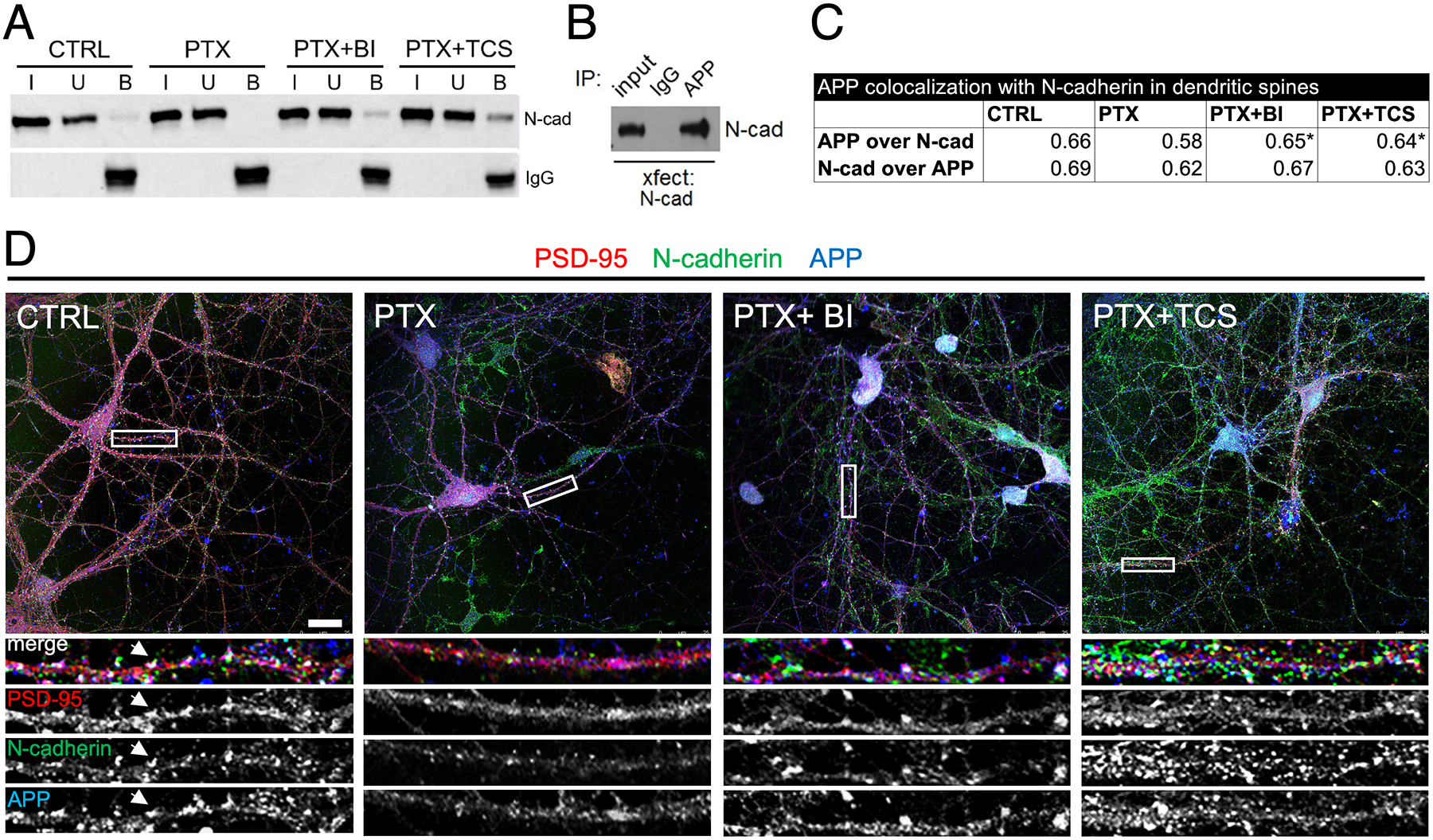
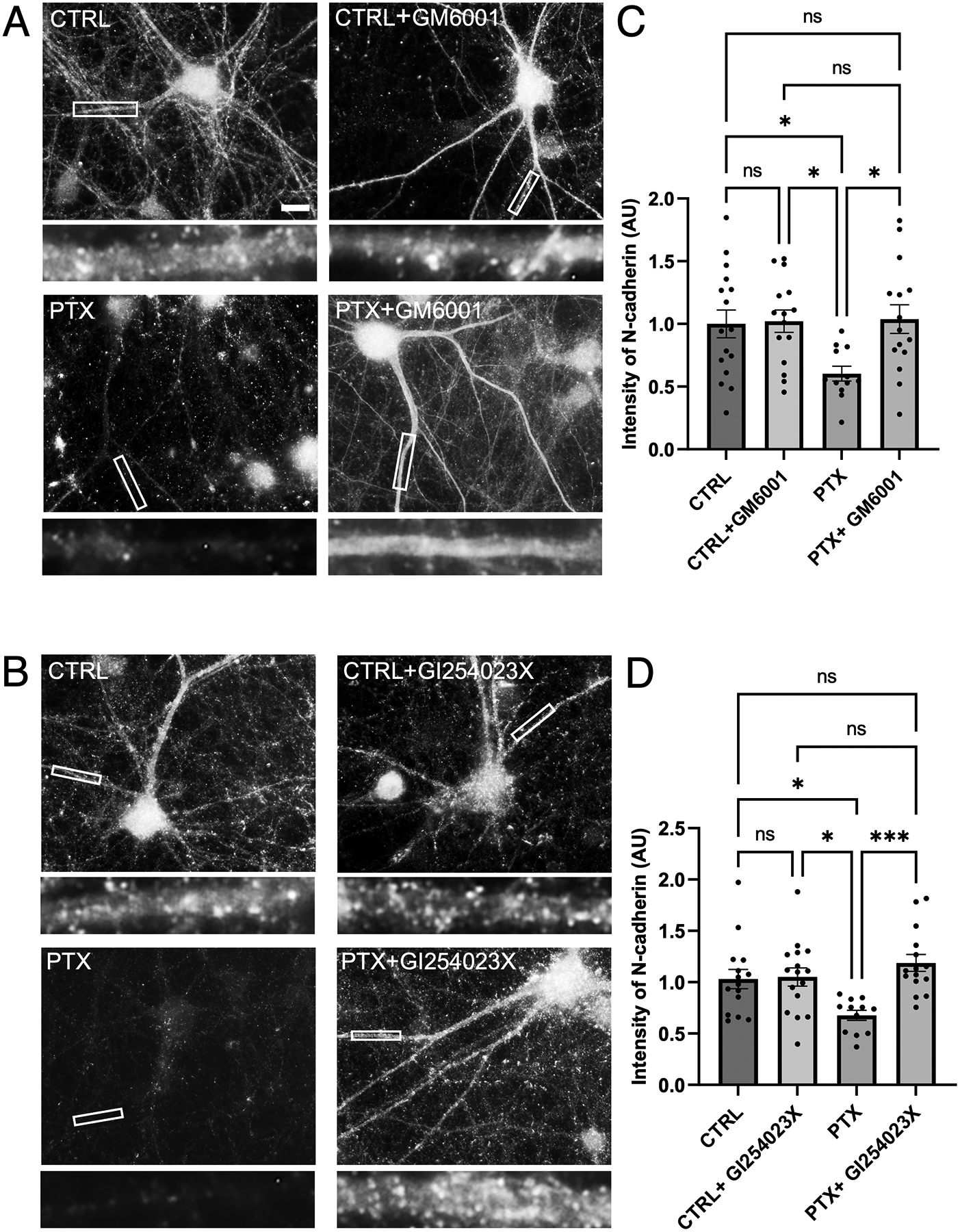
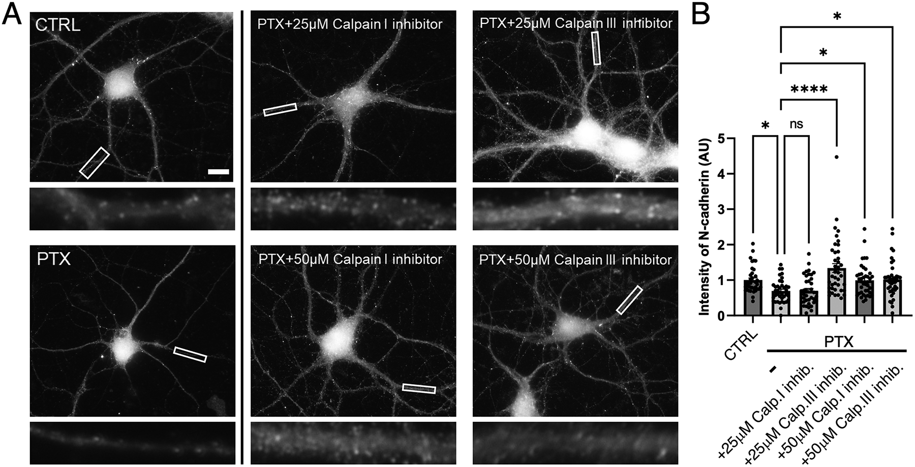
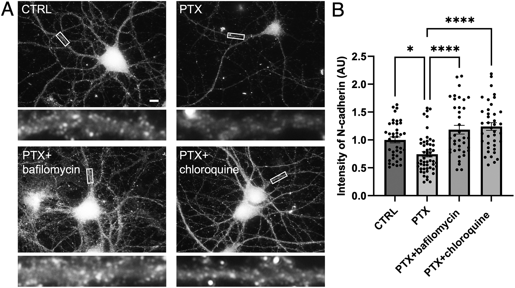
Synaptogenesis in the brain is highly organized and orchestrated by synaptic cellular adhesion molecules (CAMs) such as N-cadherin and amyloid precursor protein (APP) that contribute to the stabilization and structure of synapses. Although N-cadherin plays an integral role in synapse formation and synaptic plasticity, its function in synapse dismantling is not as well understood. Synapse weakening and loss are prominent features of neurodegenerative diseases, and can also be observed during homeostatic compensation to neuronal hyperexcitation. Previously, we have shown that during homeostatic synaptic plasticity, APP is a target for cleavage triggered by phosphorylation by Polo-like kinase 2 (Plk2). Here, we found that Plk2 directly phosphorylates N-cadherin, and during neuronal hyperexcitation Plk2 promotes N-cadherin proteolytic processing, degradation, and disruption of complexes with APP. We further examined the molecular mechanisms underlying N-cadherin degradation. Loss of N-cadherin adhesive function destabilizes excitatory synapses and promotes their structural dismantling as a prerequisite to eventual synapse elimination. This pathway, which may normally help to homeostatically restrain excitability, could also shed light on the dysregulated synapse loss that occurs in cognitive disorders.

Keywords: APP; N-cadherin; Plk2; homeostatic synaptic plasticity; hyperexcitation; synapse loss.


Conflict of interest statement

CONFLICT OF INTEREST STATEMENT

The authors have no conflicts of interest to declare.

Figures

Figure 1.
Figure 1.. (A) Plk2-dependent degradation screen of synaptic cellular adhesion molecules.
Candidate cellular adhesion molecules (CAMs) as indicated above the blots were transfected into COS-7 cells together with either kinase dead (KD) or constitutively active (CA) Plk2. Immunoblotting for each CAM demonstrated that only N-cadherin levels (highlighted by asterisk) were decreased by active Plk2. (B) Immunoblots of FLAG-N-cadherin and myc-tagged Plk2-KD or -CA co-transfected in COS-7 cells. Loading control is β-actin. Quantification is normalized to Plk2-KD; **p=0.0030, t=3.902, DF=10, unpaired two-tailed student’s t-test (n=6 cell culture preparations). (C) Direct phosphorylation of N-cadherin by Plk2. Autoradiogram of in vitro kinase assays using N-cadherin immunopurified from transfected COS-7 cells added to recombinant Plk2. Nonimmune IgG negative control to demonstrate immunoprecipitation specificity. Note Plk2 is autophosphorylated (lower band). (D) Immunostaining of COS-7 co-transfected with FLAG-N-cadherin and either myc-Plk2-KD (upper panels) or -CA (lower panels). (E) Quantification of images in (D) normalized to Plk2-KD; ***p<0.0001, t=9.702, DF=35, unpaired student’s t-test (n=13–24 cells).
Figure 2.
Figure 2.. Loss of endogenous N-cadherin is dependent on Plk2 kinase activity in primary hippocampal neurons.
(A) Hippocampal neurons were transfected with myc-Plk2 constructs as indicated above images and immunostained for endogenous N-cadherin (upper panels) or for myc epitope (lower panels). Higher magnification views of representative dendrites (boxed region) are shown below each neuron for N-cadherin. Scale, 10 μm. (B-C) Quantification of N-cadherin intensity across transfection conditions measured (B) in secondary dendrites (P= 0.0024, F=5.281, DF=3) and (C) in soma (P< 0.0001, F=16.14, DF=3). (D-E) Plk2 intensity in (D) soma (p< 0.0001, F=79.52, DF=3) and (E) proximal dendrites across the various transfection conditions (p=0.6680, F=0.5228, DF=3) One-way ANOVA and Tukey’s multiple comparison test, n=10–24 neurons; *p<0.05, **p<0.01, ***p<0.001, ****p<0.0001, ns=nonsignificant.
Figure 3.
Figure 3.. Hyperactivity-induced loss of endogenous N-cadherin in primary hippocampal neurons requires Plk2 kinase activity.
(A) Neurons were unstimulated (CTRL), treated with PTX alone, or treated with PTX in combination with Plk2 inhibitors BI-6727 or TCS-7005, as indicated, and immunostained for native N-cadherin. Higher magnification views of representative secondary dendrites (boxed regions) are shown below each neuron. Scale, 10 μm. (B-C) Quantification of N-cadherin intensity in (B) secondary dendrites (p<0.0001, F=18.20, DF=3) and (C) soma (p<0.0001, F=16.75, DF=3) (n=10–21 neurons per group, one-way ANOVA and Tukey’s multiple comparison test; *p<0.05, ***p<0.001, ****p<0.0001, ns=nonsignificant).
Figure 4.
Figure 4.. APP and N-cadherin are co-diminished by hyperactivity in a Plk2-dependent manner.
(A) Immunostaining of endogenous APP and N-cadherin in cultured hippocampal neurons left unstimulated (CTRL) or treated with PTX, PTX+BI 6727, or PTX+TCS-7005 (n=12–15 neurons). Higher magnification views of representative secondary dendrites (boxed regions) are shown below each neuron. Scale, 10 μm. (B-C) Quantification of images in (A) for (B) N-cadherin (p<0.0001, F=8.913, DF=3) or (C) APP (p< 0.0001, F=9.047, DF=3); (n=12–15 neurons for N-cadherin, 13–15 neurons for APP; one-way ANOVA and Tukey’s multiple comparison test, *p<0.05, **p<0.01, ***p<0.001, ****p<0.0001, ns=nonsignificant).
Figure 5.
Figure 5.. Association of APP and N-cadherin is disrupted by hyperactivity in a Plk2-dependent manner.
(A) Cultured hippocampal neuron lysates were prepared following treatments as shown at top, immunoprecipitated for APP, and probed for N-cadherin. I, input; U, unbound; B, bound. CTRL, unstimulated; BI, BI-6727; TCS, TCS-7005. (B) COS-7 cells were transfected with N-cadherin, immunoprecipitated with APP antibodies or nonimmune IgG, and probed for N-cadherin. (C) Quantification of mean overlap using Mander’s coefficient between APP and N-cadherin (upper line) (p= 0.0127, F=3.638, DF=3), or between N-cadherin and APP (lower line) (p= 0.0076, F=4.021, DF=3), within PSD-95 puncta across the different treatments. One-way ANOVA and Tukey’s multiple comparison test; *p<0.05 vs. PTX. (D) Triple label immunostaining of PSD-95 (red), N-cadherin (green), and APP (blue) to label dendritic spines under the different treatment conditions as indicated. Arrowheads highlight white puncta indicating triple co-localization at synapses. Higher magnification views of representative secondary dendrites (boxed in full neuron image) are shown below each neuron in merged color as well as in gray scale of separate channels for better visualization of individual distributions. Scale, 10 μm.
Figure 6.
Figure 6.. Metalloproteases and ADAM10 proteolytic cleave N-cadherin during hyperexcitation.
(A-B) Immunostaining of endogenous N-cadherin in cultured hippocampal neurons left unstimulated (CTRL) or treated with PTX, either with or without broad spectrum MMP inhibitor GM6001 or specific ADAM10 inhibitor GI254023X. Higher magnification views of representative secondary dendrites (boxed regions) are shown below each neuron. Scale, 10 μm. (C-D) Quantification of (C) GM6001 (p= 0.0143, F=3.856, DF=3) and (D) GI254023X treatments (p=0.0012, F= 6.059, DF=3) (n=12–16 neurons for GM6001, 12–26 neurons for GI254023X; one-way ANOVA and Tukey’s multiple comparison test; *p<0.05, ***p<0.001, ns=nonsignificant).
Figure 7.
Figure 7.. Calpain proteolytically cleaves N-cadherin during hyperexcitation.
(A) Immunostaining of endogenous N-cadherin in cultured hippocampal neurons left unstimulated (CTRL), treated with PTX by itself, or treated with PTX in combination with calpain inhibitors as indicated. Higher magnification views of representative secondary dendrites (boxed regions) are shown below each neuron. Scale, 10 μm. (B) Quantification of images from (A) (p < 0.0001, F=9.159, DF=5) (n=36–40 neurons; one-way ANOVA and Sidak’s post-hoc test, *p<0.05, ****p<0.0001, ns=nonsignificant).
Figure 8.
Figure 8.. N-cadherin is degraded by the lysosome.
(A) Immunostaining of endogenous N-cadherin in cultured hippocampal neurons left unstimulated (CTRL), treated with PTX alone, or treated with PTX in combination with lysosome inhibitor bafilomycin or chloroquine. Higher magnification views of representative secondary dendrites (boxed regions) are shown below each neuron. Scale, 10 μm. (B) Quantification of images in (A) (p< 0.0001, F=14.87, DF=3) (n=39–52 neurons; one-way ANOVA and Tukey’s multiple comparison test, *p<0.05, ****p<0.0001, ns=nonsignificant).
Figure 9.
Figure 9.. Inhibition of APP and N-cadherin cleavage rescues loss of surface GluA2 during hyperexcitation.
(A-E) Immunostaining of hippocampal neurons for surface GluA2 (sGluA2) (A) left unstimulated (CTRL), or treated with (B) PTX alone, (C) PTX and beta secretase inhibitor (BSI), (D) PTX and ADAM10 inhibitor GI254023X, or (E) PTX and both BSI+GI254023X (combo). Higher magnification views of representative secondary dendrites (boxed regions) are shown below each neuron. Scale, 10 μm. (F) Quantification of images in (A-E) (p< 0.0001, F= 17.29, DF=3) n=14–21; one way ANOVA and Tukey’s multiple comparison test, *p<0.05, **p<0.001, ****p<0.0001, ns=nonsignificant).

Similar articles

References

    1. Aiga M, Levinson JN, & Bamji SX (2011). N-cadherin and neuroligins cooperate to regulate synapse formation in hippocampal cultures. The Journal of Biological Chemistry, 286(1), 851–858. 10.1074/jbc.M110.176305 - DOI - PMC - PubMed
    1. Amini M, Ma C-L, Farazifard R, Zhu G, Zhang Y, Vanderluit J, Zoltewicz JS, Hage F, Savitt JM, Lagace DC, Slack RS, Beique J-C, Baudry M, Greer PA, Bergeron R, & Park DS (2013). Cellular/Molecular Conditional Disruption of Calpain in the CNS Alters Dendrite Morphology, Impairs LTP, and Promotes Neuronal Survival following Injury. 10.1523/JNEUROSCI.4247-12.2013 - DOI - PMC - PubMed
    1. Ando K, Uemura K, Kuzuya A, Maesako M, Asada-Utsugi M, Kubota M, Aoyagi N, Yoshioka K, Okawa K, Inoue H, Kawamata J, Shimohama S, Arai T, Takahashi R, & Kinoshita A (2011). N-cadherin regulates p38 MAPK signaling via association with JNK-associated leucine zipper protein: implications for neurodegeneration in Alzheimer disease. The Journal of Biological Chemistry, 286(9), 7619–7628. 10.1074/jbc.M110.158477 - DOI - PMC - PubMed
    1. Arikkath J, & Reichardt LF (2008). Cadherins and catenins at synapses: roles in synaptogenesis and synaptic plasticity. Trends in Neurosciences, 31(9), 487. 10.1016/J.TINS.2008.07.001 - DOI - PMC - PubMed
    1. Asada-Utsugi M, Uemura K, Kubota M, Noda Y, Tashiro Y, Uemura TM, Yamakado H, Urushitani M, Takahashi R, Hattori S, Miyakawa T, Ageta-Ishihara N, Kobayashi K, Kinoshita M, & Kinoshita A (2021). Mice with cleavage-resistant N-cadherin exhibit synapse anomaly in the hippocampus and outperformance in spatial learning tasks. Molecular Brain, 14(1), 1–16. 10.1186/S13041-021-00738-1/FIGURES/8 - DOI - PMC - PubMed

Publication types