. 2023 Sep;9(35):eadh8939.
doi: 10.1126/sciadv.adh8939. Epub 2023 Aug 30.

Endothelial RIPK1 protects artery bypass graft against arteriosclerosis by regulating SMC growth

Affiliations

Endothelial RIPK1 protects artery bypass graft against arteriosclerosis by regulating SMC growth

Yao Lu et al. Sci Adv. 2023 Sep.

Abstract

RIPK1 is crucial in the inflammatory response. The process of vascular graft remodeling is also involved in endothelial inflammation, which can influence the behavior of smooth muscle cells. However, the role of endothelial RIPK1 in arterial bypass grafts remains unknown. Here, we established an arterial isograft mouse model in wild-type and endothelial RIPK1 conditional knockout mice. Progressive vascular remodeling and neointima formation occurred in the graft artery, showing SMC accumulation together with endothelial inflammatory adhesion molecule and cytokine expression. Endothelial RIPK1 knockout exacerbated graft stenosis by increasing secretion of N-Shh. Mechanistically, RIPK1 directly phosphorylated EEF1AKMT3 at Ser26, inhibiting its methyltransferase activity and global protein synthesis, which further attenuated N-Shh translation and secretion. Consistently, treatment with the Hedgehog pathway inhibitor GDC0449 markedly alleviated RIPK1 knockout-induced graft stenosis. Our results demonstrated that endothelial RIPK1 played a protective role in arterial bypass graft vascular remodeling, highlighting that targeting Hedgehog pathway may be an attractive strategy for graft failure in the future.


Figures
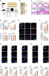
Fig. 1.
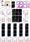
Fig. 1.. Atlas of vascular remodeling changes in arterial bypass isograft.
(A) Schematic procedure for isograft transplantation experiments. Aortic segments from C57BL/6J mice were transplanted into another C57BL/6J mice, and grafts were collected at 2/4/8 weeks after surgery. (B) The macroscopic pictures of graft vessels were collected at 4 weeks. (C) Hematoxylin and eosin (H&E) staining of 2/4/8-week isograft tissues. (D) Quantification of neointimal and luminal areas. n = 6 in each group. Scale bars, 100 μm. (E) Grafts from WT C57BL/6J mice were harvested at 2/4/8 weeks after surgery, and immunofluorescence staining of indicated antibodies was performed. (F) Quantification data. n = 6 in each group. Scale bars, 100 μm. (G to P) Representative immunofluorescence images of WT isograft showing staining of VCAM1 (G), ICAM1 (I), CD45 (K), MCP1 (H), and pP65 (O), with DAPI labeling of the nuclei. The quantification data are shown below, demonstrated as all positive cell counting in intima and/or medium layers of each section. Data are means and SEM; *P < 0.05, **P < 0.01, ***P < 0.001.
Fig. 2.
Fig. 2.. RIPK1 KO in vascular endothelium exacerbates graft stenosis.
(A) Schematic procedure for transplanting WT or RIPK1 cKO aortic segments to WT C57BL/6J mice for 4/8 weeks. (B) RIPK1 and SMC22 were examined by immunofluorescence, with DAPI labeling of the nuclei. (C) H&E staining of 4/8-week WT or RIPK1 cKO isograft tissues. (D) Quantification of neointimal and luminal areas. n = 6 in each group. Scale bars, 100 μm. (E) WT or RIPK cKO grafts were harvested at 4/8 weeks after surgery, and immunofluorescence staining of indicated antibodies was performed. (F) Quantification data. n = 6 in each group. Scale bars, 100 μm. (G to P) Representative immunofluorescence images of WT or RIPK1 cKO isograft showing staining of VCAM1 (G), ICAM1 (I), CD45 (K), MCP1 (H), and pP65 (O), with DAPI labeling of the nuclei. The quantification data are shown below, demonstrated as all positive cell counting in intima and/or medium layers of each section. Data are means and SEM; *P < 0.05, **P < 0.01, ***P < 0.001.
Fig. 3.
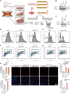
Fig. 3.. RIPK1 knockdown in ECs promotes SMC proliferation.
(A) Schematic model to show the strategy to investigate the effect of ECs on SMC aberrant proliferation. (B) HUVECs were transfected with siRIPK1 or control siRNA, and the expression of RIPK1 was checked by Western blot. (C) The primary ECs were isolated from the mice. The expression of RIPK1 was checked by Western blot. (D and F) HUVECs were treated with indicated siRNAs and cocultured with CFDA-SE pretreated SMCs for 3 days, followed by digestion and flow cytometry analysis. (E and G) The primary ECs were cocultured with CFDA-SE pretreated SMCs for 3 days, followed by digestion and flow cytometry analysis. (H) Quantification of percentage of proliferating cells. n = 3 in each group. (I) HUVECs treated with indicated siRNAs or primary ECs cocultured with SMCs for 3 days, followed by immunofluorescence staining of Ki67 and DAPI in SMC. (J) Quantification of percentage of Ki67+ cells. n = 6 in each group. Scale bars, 50 μm. Data are means and SEM; *P < 0.05, **P < 0.01, ***P < 0.001.
Fig. 4.
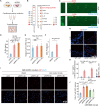
Fig. 4.. Endothelial RIPK1 KO promotes SMC proliferation by regulating N-Shh secretion.
(A) Schematic procedure for cytokine array. The HUVECs were transfected with indicated siRNAs, and conditioned medium was collected and loaded to cytokine array chips. (B) Dot plot displaying average scaled expression levels of indicated proteins. Dot size reflected the relative expression level of respective proteins. (C) Representative images of cytokine array chips. Amplified area selected in blue and red frames represented N-Shh and positive control proteins, respectively. (D) Conditioned medium was collected from HUVECs transfected with indicated siRNAs, followed by ELISA to measure N-Shh level. n = 3 per group. (E) Quantitative reverse transcription polymerase chain reaction (qRT-PCR) analysis of relative mRNA level of N-Shh from HUVECs treated by siRIPK1 or control siRNA. (F) SMCs were treated with N-Shh or DMSO for 3 days, followed by CCK8 analysis to examine the proliferation. (G) Gli1 expression of WT or RIPK1 cKO isograft was demonstrated through immunofluorescence. (H) Quantification data, demonstrated as all positive cell counting in intima and/or medium layers of each section. n = 6 in each group. Scale bars, 50 μm. (I) HUVECs were transfected with indicated siRNAs and cocultured with SMCs, 100 nM GDC0449, or DMSO used to treat SMCs for 3 days. Immunofluorescence showed staining of Ki67 with DAPI labeling of the nuclei. (J) Quantification of percentage of Ki67+ cells. n = 9 in each group. Scale bars, 50 μm. Data are means and SEM; *P < 0.05, **P < 0.01, ***P < 0.001.
Fig. 5.
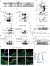
Fig. 5.. RIPK1 phosphorylates EEF1AKMT3 at S26.
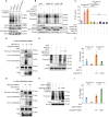
(A) RIPK1-interacting proteins predicted by BioPlex Interactome. (B) 293T cells were transfected with indicated Flag-tagged plasmids, followed by co-IP and Western blot with indicated antibodies. (C and D) 293T cells were transfected with indicated plasmids, followed by co-IP and Western blot. (E) HUVECs were lysed, followed by co-IP with indicated antibodies and Western blot. (F) In vitro phosphorylation kinase assays with purified Flag-EEF1AKMT3, recombinant RIPK1, and ATP-γ-S. The phosphorylation of EEF1AKMT3 was detected with thiophosphate ester antibody. (G) The in vitro phosphorylation kinase assay products were analyzed by LC-MS/MS to find the EEF1AKMT3 phosphorylation site. The mass spectrum of a tryptic fragment that matched the peptide 19-EVGLFADpSYSEK-30 containing phosphorylated S26 is shown. This phosphopeptide was detected with a high peptide confidence (false discovery rate of less than 1%) in the product adding ATP. Amino acid sequences of EEF1AKMT3 from different species were aligned. Conserved serines at position 26 (red arrow) are highlighted. (H) In vitro kinase assay was performed with purified Flag-EEF1AKMT3 WT or S26A mutant and ATP-γ-S. The phosphorylation of EEF1AKMT3 was detected with thiophosphate ester antibody. (I) In vitro kinase assay was performed with purified Flag-EEF1AKMT3 WT or S26A mutant and ATP. The phosphorylation of EEF1AKMT3 was detected with pEEF1AKMT3 S26 antibody. (J) RIPK1 was deleted with single guide RNA (sgRNA) in HUVECs then treated with TNF-α, followed by Western blot with indicated antibodies. (K) WT or RIPK1 cKO isografts were stained with CD31, pEEF1AKMT3 S26 antibodies, and DAPI. (L) Quantification data, demonstrated as all positive cell counting in intima and/or medium layers of each section. n = 6 in each group. Scale bars, 100 μm. Data are means and SEM; *P < 0.05, **P < 0.01, ***P < 0.001.
Fig. 6.
Fig. 6.. RIPK1 regulates Shh secretion in ECs through EEF1AKMT3 S26 phosphorylation–EEF1A K165 methylation–ribosomal protein synthesis pathway.
(A and B) SUnSET assays were performed in HUVECs transfected with indicated siRNAs. Whole-cell extracts were isolated and probed with the indicated antibodies. (C) The level of N-Shh was measured by ELISA in conditioned medium of HUVECs treated with indicated siRNAs. (D) In vitro methylation assay was performed with purified proteins and SAM, followed by detecting EEF1A methylation with pan-methylation antibodies. (E) SUnSET assays were performed in HUVECs transfected with indicated siRNAs and plasmids, followed by detection with anti-puromycin antibody. (F) The level of N-Shh was measured by ELISA in conditioned medium of HUVECs transfected with indicated siRNAs and plasmids. n = 3 in each group. (G) In vitro methylation assay was performed with purified EEF1AKMT3 WT or S26A mutant, followed by detecting EEF1A methylation with pan-methylation antibodies. (H) SUnSET assays were performed in HUVECs transfected with indicated siRNAs and plasmids, followed by detection with anti-puromycin antibody. (I) The level of N-Shh was measured by ELISA in conditioned medium of HUVECs transfected with indicated siRNAs and plasmids. n = 3 in each group. Data are means and SEM; *P < 0.05, **P < 0.01, ***P < 0.001.
Fig. 7.
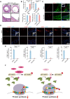
Fig. 7.. RIPK1-EEF1AKMT3 affects graft vascular remodeling through the Shh pathway.
(A) RIPK1 cKO artery was transplanted for 4 weeks in the treatment with DMSO or GDC0449, and RIPK1 cKO grafts were examined by H&E staining. (B) Quantification of neointimal and luminal areas. n = 6 in each group. Scale bars, 100 μm and 25 μm as indicated. (C) Representative immunofluorescence images of RIPK1 cKO grafts stained with CD31 and SM22 antibodies. Scale bars, 100 μm. (D) RIPK1 cKO grafts treated with GDC0449 or DMSO were stained with VCAM1, ICAM1, MCP1, and CD45, with DAPI labeling of the nuclei. (E) Quantification data, demonstrated as all positive cell counting in intima and/or medium layers of each section. Scale bars, 100 μm. Data are means and SEM; *P < 0.05, **P < 0.01, ***P < 0.001. (F) Proposed model for the protective role of RIPK1 in arterial bypass graft vascular remodeling by regulating EEF1AKMT3 phosphorylation in ECs and Hedgehog pathway in SMC.

Similar articles

Cited by

References

    1. D. Thuijs, A. Kappetein, P. Serruys, F. Mohr, M. Morice, M. Mack, D. Holmes, N. Curzen, P. Davierwala, T. Noack, M. Milojevic, K. Dawkins, B. da Costa, P. Jüni, S. Head, Percutaneous coronary intervention versus coronary artery bypass grafting in patients with three-vessel or left main coronary artery disease: 10-year follow-up of the multicentre randomised controlled SYNTAX trial. Lancet 394, 1325–1334 (2019). - PubMed
    1. M. Gaudino, U. Benedetto, S. Fremes, G. Biondi-Zoccai, A. Sedrakyan, J. Puskas, G. Angelini, B. Buxton, G. Frati, D. Hare, P. Hayward, G. Nasso, N. Moat, M. Peric, K. Yoo, G. Speziale, L. Girardi, D. Taggart, Radial-artery or saphenous-vein grafts in coronary-artery bypass surgery. N. Engl. J. Med. 378, 2069–2077 (2018). - PubMed
    1. M. Gaudino, U. Benedetto, S. Fremes, K. Ballman, G. Biondi-Zoccai, A. Sedrakyan, G. Nasso, J. Raman, B. Buxton, P. Hayward, N. Moat, P. Collins, C. Webb, M. Peric, I. Petrovic, K. Yoo, I. Hameed, A. Di Franco, M. Moscarelli, G. Speziale, J. Puskas, L. Girardi, D. Hare, D. Taggart, Association of radial artery graft vs saphenous vein graft with long-term cardiovascular outcomes among patients undergoing coronary artery bypass grafting: A systematic review and meta-analysis. JAMA 324, 179–187 (2020). - PMC - PubMed
    1. R. Mitchell, P. Libby, Vascular remodeling in transplant vasculopathy. Circ. Res. 100, 967–978 (2007). - PubMed
    1. J. Pober, W. Sessa, Evolving functions of endothelial cells in inflammation. Nat. Rev. Immunol. 7, 803–815 (2007). - PubMed