. 2023 Aug 12:31:136-150.
doi: 10.1016/j.bioactmat.2023.08.009. eCollection 2024 Jan.

Modeling human brain rhabdoid tumor by inactivating tumor suppressor genes in induced pluripotent stem cells

Affiliations

Modeling human brain rhabdoid tumor by inactivating tumor suppressor genes in induced pluripotent stem cells

Timothy Hua et al. Bioact Mater. .

Abstract

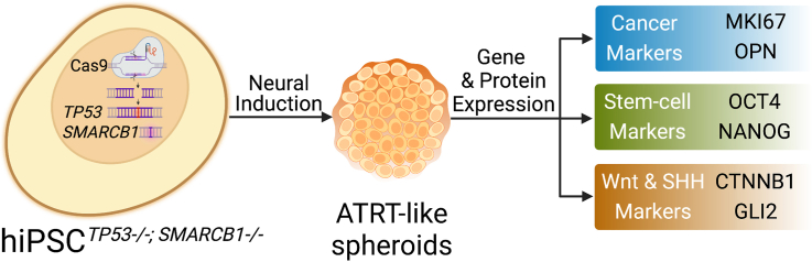
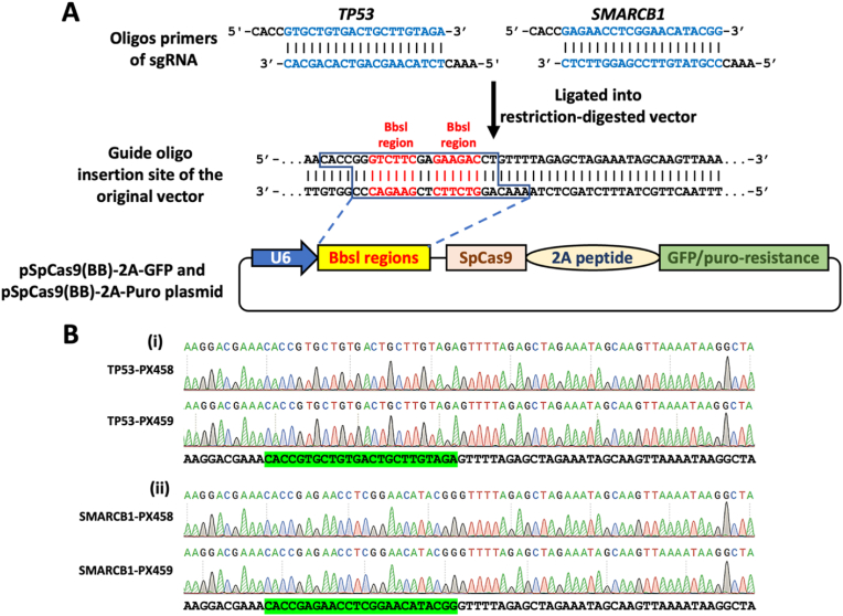
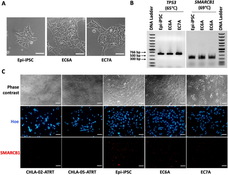
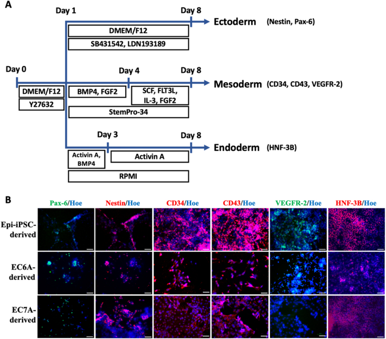
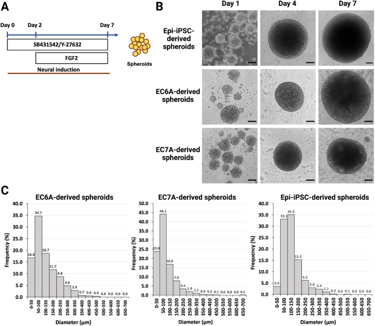
Atypical teratoid/rhabdoid tumor (ATRT) is a rare childhood malignancy that originates in the central nervous system. Over ninety-five percent of ATRT patients have biallelic inactivation of the tumor suppressor gene SMARCB1. ATRT has no standard treatment, and a major limiting factor in therapeutic development is the lack of reliable ATRT models. We employed CRISPR/Cas9 gene-editing technology to knock out SMARCB1 and TP53 genes in human episomal induced pluripotent stem cells (Epi-iPSCs), followed by brief neural induction, to generate an ATRT-like model. The dual knockout Epi-iPSCs retained their stemness with the capacity to differentiate into three germ layers. High expression of OCT4 and NANOG in neurally induced knockout spheroids was comparable to that in two ATRT cell lines. Beta-catenin protein expression was higher in SMARCB1-deficient cells and spheroids than in normal Epi-iPSC-derived spheroids. Nucleophosmin, Osteopontin, and Ki-67 proteins were also expressed by the SMARCB1-deficient spheroids. In summary, the tumor model resembled embryonal features of ATRT and expressed ATRT biomarkers at mRNA and protein levels. Ribociclib, PTC-209, and the combination of clofilium tosylate and pazopanib decreased the viability of the ATRT-like cells. This disease modeling scheme may enable the establishment of individualized tumor models with patient-specific mutations and facilitate high-throughput drug testing.

Keywords: Atypical teratoid/rhabdoid tumor; Brain tumor modeling; CRISPR/Cas9 gene editing; Human induced pluripotent stem cells; SMARCB1; Tumor suppressor genes.


Conflict of interest statement

None.

Figures

Image 1
Graphical abstract
Fig. 1
Fig. 1
Insertion of guide RNA oligos into PX458 and PX459 vectors. (A) Oligos for single guide RNA (sgRNA) targeting TP53 and SMARCB1 genes were inserted separately into the BbsI regions of two different vectors, PX458 (GFP) and PX459 (Puromycin resistance), to construct a total of 4 distinct plasmids. (B) These plasmids were checked for the presence of the sgRNA-coding sequences that targets (i)TP53 and (ii)SMARCB1. The green highlighted sequences are the inserts for sgRNA.
Fig. 2
Fig. 2
Assessment of morphology, on-target plasmid DNA insertion, and nuclear SMARBC1 expression in knockout clones. (A) Phase contrast images of normal Epi-iPSC, EC6A, and EC7A show no major morphological differences. Scale bar: 25 μm. (B) Target DNA amplification yielded PCR products of 604 bp (TP53) and 341 bp (SMARCB1), implying no on-target plasmid DNA insertion. (C) SMARCB1 staining in two negative control cell lines, CHLA-02-ATRT and CHLA-05-ATRT; one positive control, Epi-iPSC; and two SMARCB1 knockout clones, EC6A and EC7A. Scale bar: 50 μm.
Fig. 3
Fig. 3
Sequencing around the TP53 target site for the clones (A)TP53 target site was sequenced for (i) normal Epi-iPSC and (ii)TP53-knockout EC6 and EC7 clones to confirm the addition of an extra adenosine nucleotide to exon 4 in knockout cells. (B) TP53 protein contains a DNA binding region (highlighted in yellow) and Zn2+ binding amino acids (underlined red). EC6 and EC7 clones with the additional adenosine nucleotide in the TP53 gene have a premature stop codon in place of Tyr163 (underlined blue).
Fig. 4
Fig. 4
Sequencing around the SMARCB1 target site for the clones. (A)SMARCB1 target site was sequenced for (i) normal Epi-iPSC and (ii) dual-knockout EC6A and EC7A clones to confirm the deletion of 9 nucleotides from exon 2 in knockout clones. (B) The deleted nucleotides affected three contiguous codons for the amino acids Met38-Phe39-Arg40 (strikethrough and red-colored). (C) The cartoon representation of the first 115 amino acids of SMARCB1. They are highly conserved across metazoans and possibly bind DNA.
Fig. 5
Fig. 5
Differentiation capacity of EC6A and EC7A. (A) Schematic illustration of germ layer differentiation protocols. 10 μM of ROCK inhibitor was added to the human iPSCs, EC6A, and EC7A in the first 24 h. The endoderm differentiation was performed in RPMI medium with Activin A and BMP4. On day 3, the media was changed to fresh media with only Activin A till day 8. For the mesoderm differentiation, the cells were cultured in StemPro™-34 with factors BMP4 and FGF2 on day 1. These factors were replaced on day 4 with SCF, FLT3L, IL-3, and FGF2 till day 8. For the ectoderm differentiation, the cells were plated in DMEM/F12 with SB431542 and LDN193189 from day 1 to day 8. (B) Immunostaining reveals the expression of germ layer-specific markers by both clones. CD34, CD43, and VEGFR-2 are markers for mesoderm. HNF–3B is an endoderm marker. The markers for ectoderm are Pax-6 and Nestin. Separate ICC images for each marker can be found in Supplementary Fig. S5. BMP4: bone morphogenetic protein 4; FGF2: fibroblast growth factor 2; SCF: stem cell factor; FLT3L: FMS-like tyrosine kinase 3 ligand; IL-3: interleukin 3; CD34: hematopoietic progenitor cell antigen CD34; CD43: leukosialin; VEGFR-2: vascular endothelial growth factor receptor 2; Pax-6: paired box protein Pax-6; HNF–3B: hepatocyte nuclear factor 3-beta; Hoe: Hoechst 33342. Scale bar: 50 μm.
Fig. 6
Fig. 6
Neural induction of Epi-iPSC, EC6A, and EC7A. (A) Schematic illustration of the neural induction procedure. SB431542 is the inhibitor of the transforming growth factor (TGF)-beta, and Y-27632 is the inhibitor of rho-associated protein kinase signaling pathway. (B) Phase contrast images of spheroids undergoing neural induction at days 1, 4, and 7. (C) The size distribution of EC6A-derived spheroids (n = 1084), EC7A-derived spheroids (n = 1233), and Epi-iPSC-derived spheroids (n = 876). Scale bar: 50 μm.
Fig. 7
Fig. 7
Live-cell images of Epi-iPSC-, EC6A-, and EC7A-derived spheroid cells with intracellular calcium transients. On day 7, spheroids were dissociated and cultured on Matrigel™ for 24 h. The transients were measured every 30 s using a fluorescent microscope with 20x magnification. Noticeable signal changes between two frames are circled. Scale bar: 50 μm.
Fig. 8
Fig. 8
Characterization of the ATRT-like model with RT-PCR. Normal Epi-iPSC and both the knockout clones (EC6A and EC7A) were subjected to neural induction. The spheroids were harvested post-induction and characterized for (A) SHH and β-catenin pathways, (B) embryonic, (C) neuronal, and (D) potential ATRT biomarkers. *: p-value <0.05 when compared with the control, Epi-iPSC-SP; **: p-value <0.01 when compared with the control, Epi-iPSC-SP. Epi-iPSC-SP: normal Epi-iPSC-derived spheroids; EC6A-SP: EC6A-derived ATRT-like spheroids; EC7A-SP: EC7A-derived ATRT-like spheroids; GLI1: glioma-associated oncogene family zinc finger 1; GLI2: glioma-associated oncogene family zinc finger 2; GSK3B: glycogen synthase kinase 3 beta; CTNNB1: catenin beta-1; LEF1: lymphoid enhancer-binding factor 1; OCT4: octamer-binding protein 4; NANOG: Nanog homeobox; ENO2: enolase 2; NEPH3: kirre-like nephrin family adhesion molecule 2; IDH1: isocitrate dehydrogenase NADP(+) 1; OPN: osteopontin; RB1: retinoblastoma transcriptional corepressor 1; MKI67: marker of proliferation Ki-67; STAT3: signal transducer and activator of transcription 3.
Fig. 9
Fig. 9
Immunocytochemistry images for ATRT biomarker detection. ICC was performed for ATRT cell lines CHLA-02-ATRT and CHLA-05-ATRT, normal Epi-iPSC-derived spheroid cells, and EC6A- and EC7A-derived spheroid cells. Separate ICC images for each marker can be found in Supplementary Fig. S6. NPM: nucleophosmin; GLI1: zinc finger protein GLI1; GLI2: zinc finger protein GLI2; Ki-67: proliferation marker protein Ki-67; Pax-6: paired box protein Pax-6; Oct-4: octamer-binding transcription factor 4; NANOG: homeobox protein NANOG; Hoe: Hoechst 33342. Scale bar: 50 μm.
Fig. 10
Fig. 10
Determining the IC50valuesfor drug treatments of the ATRT-like model (A) The drug treatment scheme for EC6A- and EC7A-derived ATRT-like spheroids. The spheroids were collected, dissociated, and replated into wells of a 96-well plate to test the drug effect 48 h after the treatment. (B) Reduction of MTT inside live cells to elicit a color change. (C) IC50 curves for different drug treatments of EC6A- and EC7A-derived spheroids: (i) ribociclib, (ii) PTC-209, and (iii) the combination of clofilium tosylate and pazopanib. (D) The reported IC50 values and hillslope values. SB431542: SMAD protein inhibitor; Y-27632: rho-associated protein kinase inhibitor; FGF2: fibroblast growth factor 2; MTT: 3-(4,5-dimethylthiazol-2-yl)-2,5-diphenyl tetrazolium bromide; PBS: phosphate-buffered saline; SDS: sodium dodecyl sulfate; IC50: half-maximal inhibitory concentration; RB: ribociclib; PTC: PTC-209; CT + PZ: the combination of clofilium tosylate and pazopanib.

Similar articles

References

    1. Ramli R.A., et al. Identification of the cellular origin and" stemness" phenotype of Malignant Rhabdoid Tumors (MRT) may represent a new therapeutic approach in paediatric oncology. Cancer Res. 2017;77 4875-4875.
    1. Terada Y., et al. Human pluripotent stem cell-derived tumor model uncovers the embryonic stem cell signature as a key driver in atypical teratoid/rhabdoid tumor. Cell Rep. 2019;26:2608–2621. e2606. - PubMed
    1. Kim K.H., Roberts C.W. Mechanisms by which SMARCB1 loss drives rhabdoid tumor growth. Cancer Genet. 2014;207:365–372. - PMC - PubMed
    1. Kohashi K., Oda Y. Oncogenic roles of SMARCB1/INI1 and its deficient tumors. Cancer Sci. 2017;108:547–552. - PMC - PubMed
    1. Rorke L.B., Packer R.J., Biegel J.A. Central nervous system atypical teratoid/rhabdoid tumors of infancy and childhood: definition of an entity. J. Neurosurg. 1996;85:56–65. - PubMed