Review
. 2023 Aug 19;21(1):282.
doi: 10.1186/s12951-023-01974-4.

Next-generation nanomaterials: advancing ocular anti-inflammatory drug therapy

Affiliations
Review

Next-generation nanomaterials: advancing ocular anti-inflammatory drug therapy

Jing Wei et al. J Nanobiotechnology. .

Abstract

Ophthalmic inflammatory diseases, including conjunctivitis, keratitis, uveitis, scleritis, and related conditions, pose considerable challenges to effective management and treatment. This review article investigates the potential of advanced nanomaterials in revolutionizing ocular anti-inflammatory drug interventions. By conducting an exhaustive analysis of recent advancements and assessing the potential benefits and limitations, this review aims to identify promising avenues for future research and clinical applications. The review commences with a detailed exploration of various nanomaterial categories, such as liposomes, dendrimers, nanoparticles (NPs), and hydrogels, emphasizing their unique properties and capabilities for accurate drug delivery. Subsequently, we explore the etiology and pathophysiology of ophthalmic inflammatory disorders, highlighting the urgent necessity for innovative therapeutic strategies and examining recent preclinical and clinical investigations employing nanomaterial-based drug delivery systems. We discuss the advantages of these cutting-edge systems, such as biocompatibility, bioavailability, controlled release, and targeted delivery, alongside potential challenges, which encompass immunogenicity, toxicity, and regulatory hurdles. Furthermore, we emphasize the significance of interdisciplinary collaborations among material scientists, pharmacologists, and clinicians in expediting the translation of these breakthroughs from laboratory environments to clinical practice. In summary, this review accentuates the remarkable potential of advanced nanomaterials in redefining ocular anti-inflammatory drug therapy. We fervently support continued research and development in this rapidly evolving field to overcome existing barriers and improve patient outcomes for ophthalmic inflammatory disorders.

Keywords: Bioavailability; Biocompatibility; Controlled release; Nanomaterials; Ophthalmic inflammatory diseases; Targeted delivery; Toxicity.


Conflict of interest statement

The authors declare that they have no competing interests.

Figures
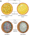
Fig. 1
Fig. 1
A Schematic overview of the structural organization of first-generation SLNs and second-generation lipid NPs-NLCs. B The structural design of nanocapsules and nanospheres. This is reprinted from Ref. [342] with permission from MDPI
Fig. 2
Fig. 2
The diagram elucidates the multifaceted applications of AuNP in the domain of ophthalmology. This is reproduced from Ref. [23] with the authorization of John Wiley and Sons
Fig. 3
Fig. 3
The image illustrates the varying capacities of different nano-formulations to traverse distinct barriers and reach diverse tissues within the eye, as dictated by their individual properties. This is referenced from Ref. [343], reproduced with permission from Royal Society of Chemistry Advances
Fig. 4
Fig. 4
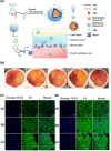
The effectiveness of PBA-CS-VE-VRC in treating fungal keratitis is illustrated, highlighting its role in minimizing ocular irritation while enhancing corneal permeability and extending immediate retention time for the administration of topical ocular medications. A The structure of PBA-CS-VE-VRC nanocelles and their role in treating corneal diseases is diagrammed. B The HET-CAM assay, an in vitro surrogate for ocular stimulation, is employed to assess the irritation potential of various preparations, namely: Sanitary saline, Sol-VRC, CS-VE-VRC, PBA-CS-VE-VRC, and 0.1 M NaOH solution on the chick embryo chorioallantoic membrane. C Fluorescent preparations of Sol-C6 (a), CS-VE-C6 (b), or PBA-CS-VE-C6 (c) were prepared and their respective uptake rates by the HCE-T cell line (human immortalized corneal epithelial cells) were observed using confocal fluorescence microscopy at 2 h and 4 h D intervals. Scale bar equals 20 μm. This figure has been reprinted from Ref. [208] with permission from Elsevier
Fig. 5
Fig. 5
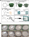
The electrospinning process for the creation of polymer-free and free-standing acyclovir/cyclodextrin nanofibers, notable for their exceptional histocompatibility and facilitation of drug release in the treatment of viral keratitis. A Chemical structures of (a) HP-βCD, (b) PVP, and (c) acyclovir are presented, (d-e) alongside schematic diagrams demonstrating their interrelationships. B Experimental data confirms the solubility of acyclovir/HP-βCD nanofibers and acyclovir/PVP nanofibers in an artificial saliva environment. Adapted from Ref. [218] with permission from Elsevier
Fig. 6
Fig. 6
The schematic diagram demonstrates the pathway of a drug through the conjunctiva, sclera, and onward to the choroid and retinal tissue following the injection of 50 μl of 0.2% Rapamycin MNF into the conjunctival sac of rabbits. Reproduced from Ref. [247] with permission from Springer Nature
Fig. 7
Fig. 7
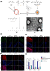
The use of Tacrolimus-loaded mPEG-b-PLGA micelles in the treatment of corneal immune rejection subsequent to allogeneic penetrating corneal transplantation in rats A. (a) presents a schematic diagram of the composition and ocular application of Tacrolimus-loaded mPEG-b-PLGA micelles, while (b) shows the detection of mPEG-b-PLGA by a 1H NMR spectrometer. (c) provides a scanning electron microscope (SEM) image of Tacrolimus-loaded mPEG-b-PLGA micelles with a scale bar denoting 50 nm. The immunofluorescence assay was used to observe the inhibitory effect of Tacrolimus-loaded mPEG-b-PLGA micelles (administered either via subconjunctival injection, or solution drops) on the phosphorylation of NFAT B, CD4 C and CD8 D, key factors in immune rejection, across various layers of corneal tissue in comparison to blank controls and standard 0.05% tacrolimus eye drops. E showcases a statistical graph of the percentage of fluorescence intensity. Values are represented as mean ± SD, with statistical significance denoted as *p < 0.05, **p < 0.01, ***p < 0.001. This figure is reproduced from Ref. [249] with permission from Elsevier
Fig. 8
Fig. 8
The synthesis of PLGA-GA2-CUR and its resultant therapeutic impact on a beagle uveitis model. A The schematic diagram illustrates the composition of PLGA-GA2-CUR. B A dynamic size distribution of light scattering describes the model particle of PLGA-GA2-CUR. C SEM provides microstructure images of PLGA-GA2-CUR. D The anti-inflammatory effect of topical PLGA-GA2-CUR is demonstrated in a canine model of acute endophthalmitis. Following an intraocular injection of lens protein at t = 0 h, the semiquantitative preclinical ocular toxicology scoring (SPOTS) was employed, incorporating scores for aqueous flare (a), pupillary light reflex (b), and conjunctival swelling (c). Local administration of PLA-GA2-CUR showed statistical significance when compared to topical prednisolone acetate (PA) and untreated controls, as assessed by two-way ANOVA. Statistical significance is denoted as *p < 0.05, **p < 0.01, ***p < 0.001, and ****p < 0.0001. This figure is reproduced from Ref. [186] with permission from the American Association for the Advancement of Science
Fig. 9
Fig. 9
A schematic illustration of the diverse aspects of nanomaterial application in ocular biomedicine. This figure is reproduced from Ref [344]. with permission from John Wiley and Sons
Fig. 10
Fig. 10
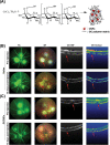
The inhibitory effect of GCCNPs on neovascularization in a laser-induced choroidal neovascularization mouse model. A A schematic diagram depicting the synthesis, structure, and morphology of GCCNP is presented. The protective role of GCCNP on laser-induced lesions is highlighted in a mouse model of choroidal neovascularization. The control group received a vitreal injection of saline B, while the experimental group was administered a vitreal injection of GCCNPs C. Observations of the repair of damaged fundus vessels by the drug were conducted both prior to and 14 days post-injection using fundus fluorescein angiography, plain fundus photography, and OCT. Laser-induced lesion sites are indicated by red arrows. The representations include fluorescein angiography (FA) and bright field (BF) images. This figure is reproduced from Ref. [305] with permission from the American Chemical Society
Fig. 11
Fig. 11
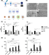
The therapeutic impact of surface-modified melphalan nanoparticles (NPs) on retinoblastoma (RB). A A schematic depiction of the preparation and concentration of melphalan is provided. B Structural schematic diagrams and scanning electron microscope (SEM) images of four distinct NP preparations are depicted: Surface-unmodified PLGA NPs loaded with melphalan (a), surface-modified PLGA NPs loaded with melphalan by TET1 (b), surface-modified PLGA NPs loaded with melphalan by PEG (c), and surface-modified PLGA NPs loaded with melphalan by MPG (d), scale bar = 200 nm. C In an in vitro cellular assay, four different NP formulations in 1 mg/mL and 10 mg/mL melphalan configurations were used to treat Y79 cells for 24 h to observe their cytotoxic effects. MPG NPs were identified as the most effective treatment group. IC50 values for TET1, PEG, and unmodified NPs were higher than those for free melphalan, while MPG NPs demonstrated statistically similar efficacy to free melphalan. IC50 values are displayed as mean ± SD; statistical significance is indicated by *P ≤ 0.05, **P ≤ 0.01, ***P ≤ 0.001, ****P ≤ 0.0001. D The influence of surface modification of NPs on Y79 cell binding (black) and internalization (gray) was assessed using flow cytometry. Surface-modified NPs showed increased cell binding and internalization compared to unmodified NPs at 1.5 h (a) and 24 h (b). Data is represented as mean ± SD; statistical significance is indicated by *P ≤ 0.05, **P ≤ 0.01, ***P ≤ 0.001, ****P ≤ 0.0001. This figure is reproduced from Ref. [345] with permission from the Association for Research in Vision and Ophthalmology Inc
Fig. 12
Fig. 12
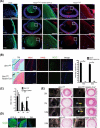
The pivotal roles of Pax6, Bmi1, and Ki67 in ocular development and lens-induced regeneration. A This panel illustrates the role of Pax6 in eye and lens development. In progeny derived from crossing the ROSAmTmG membrane-bound GFP reporter strain with (P0-3.9-GFPcre) mice, robust GFP expression was noted in lens epithelial cells (LECs) under fluorescence microscopy, indicating that Pax6 LECs from embryonic or adult lenses contribute to the regeneration of mouse lens fiber cells. B The part shows that deletion of Bmi-1 results in diminished Pax6 + and Sox2 + expression in LECs. The vertical axis represents the percentage of Pax6 + and SOX2 + cells. Data are presented as mean ± SD; statistical significance is denoted by *P < 0.001. The part C reveals that the absence of Bmi1 leads to a decrease in LEC proliferation. The proportion of BrdU + LECs was calculated for each eye at 2 m, 7 m, and 12 m. Statistical significance was determined via a two-tailed Student’s t-test; *P < 0.05. Part D displays Nestin (green) staining images of wild-type mice at E13.5, E18.5, and 2 months of age, as observed through fluorescence microscopy. Part E depicts the histological examination (HE staining) of eyeballs from 2 m, 7 m, and 12-month-old Bmi1fl/fl control mice and Nestin-cre; Bmi1fl/fl mice, to observe the development of cataracts. After using Nestn-creER to delete Bmi-1 in 6-week-old mice and following 10 months of tamoxifen treatment, the HE morphology of mouse eyes showed no cataract phenotype. All scale bars equal 100 μm. Reproduced from Ref. [346] with permission from Springer Nature
Fig. 13
Fig. 13
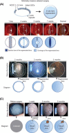
A novel minimally invasive surgical technique for promoting lens regeneration. A A minimally invasive ocular surgery conducted on a rabbit eye, employing a capsulorhexis size of 1–1.5 mm. The procedure targets a 1.2 mm2 region surrounding the lens, with photographic evidence of lens regeneration observed via a slit-lamp microscope from day 1 to 4 weeks post-surgery. B A similar minimally invasive ocular operation was executed in a macaque model. Slit lamp microscopy demonstrates the regenerated lens tissue expanding from the periphery towards the lens center between 2–5 months post-surgery. Direct illumination reveals a translucent visual axis. C The minimally invasive procedure was also performed on infants with congenital cataracts. Lens regeneration was observed from week 1 to 5 months post-surgery, with almost all eyes (95.8%) attaining visual axis transparency. The surgical incision remained peripheral, and the anterior capsule wound scar tissue kept away from the visual axis was less than 1.5 mm in diameter. This novel surgical approach significantly mitigated visual axis opacity compared to the current standard surgical approach. Reproduced from Ref. [346] with permission from Springer Nature
Fig. 14
Fig. 14
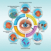
Ocular pathologies induced by the improper utilization of nanomaterials. The image showcases irritation and toxic responses elicited by nanomaterials interacting with the eye surface (cornea, conjunctiva), intraocular structures (e.g., iris, ciliary body, choroid, and lens), or various regions of the retina, macula, and optic nerve. Reproduced from Ref. [347] with permission from John Wiley and Sons

Similar articles

Cited by

References

    1. Huerva V, Ascaso FJ, Grzybowski A. Ocular inflammation. Mediators Inflamm. 2015;2015:398076. - PMC - PubMed
    1. Shan J, Li L, Du L, Yang P. Association of TBX21 gene polymorphisms and acute anterior uveitis risk in a Chinese population: a case-control study. Exp Eye Res. 2023;229:109417. - PubMed
    1. Xu TT, Reynolds MM, Hodge DO, Smith WM. Epidemiology and clinical characteristics of episcleritis and scleritis in Olmsted County. Minnesota Am J Ophthalmol. 2020;217:317–324. - PMC - PubMed
    1. Voss E, Raab P, Trebst C, Stangel M. Clinical approach to optic neuritis: pitfalls, red flags and differential diagnosis. Ther Adv Neurol Disord. 2011;4(2):123–134. - PMC - PubMed
    1. Gaballa SA, Kompella UB, Elgarhy O, Alqahtani AM, Pierscionek B, Alany RG, et al. Corticosteroids in ophthalmology: drug delivery innovations, pharmacology, clinical applications, and future perspectives. Drug Deliv Transl Res. 2021;11(3):866–893. - PubMed