. 2023 Aug 17;83(16):2959-2975.e7.
doi: 10.1016/j.molcel.2023.07.023.

Molecular mechanism for activation of the 26S proteasome by ZFAND5

Affiliations

Molecular mechanism for activation of the 26S proteasome by ZFAND5

Donghoon Lee et al. Mol Cell. .

Abstract

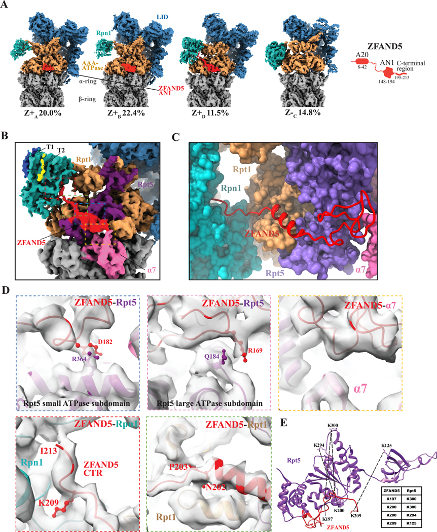
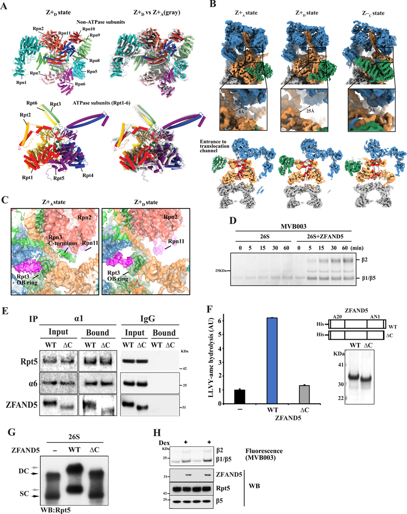
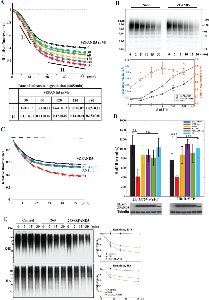
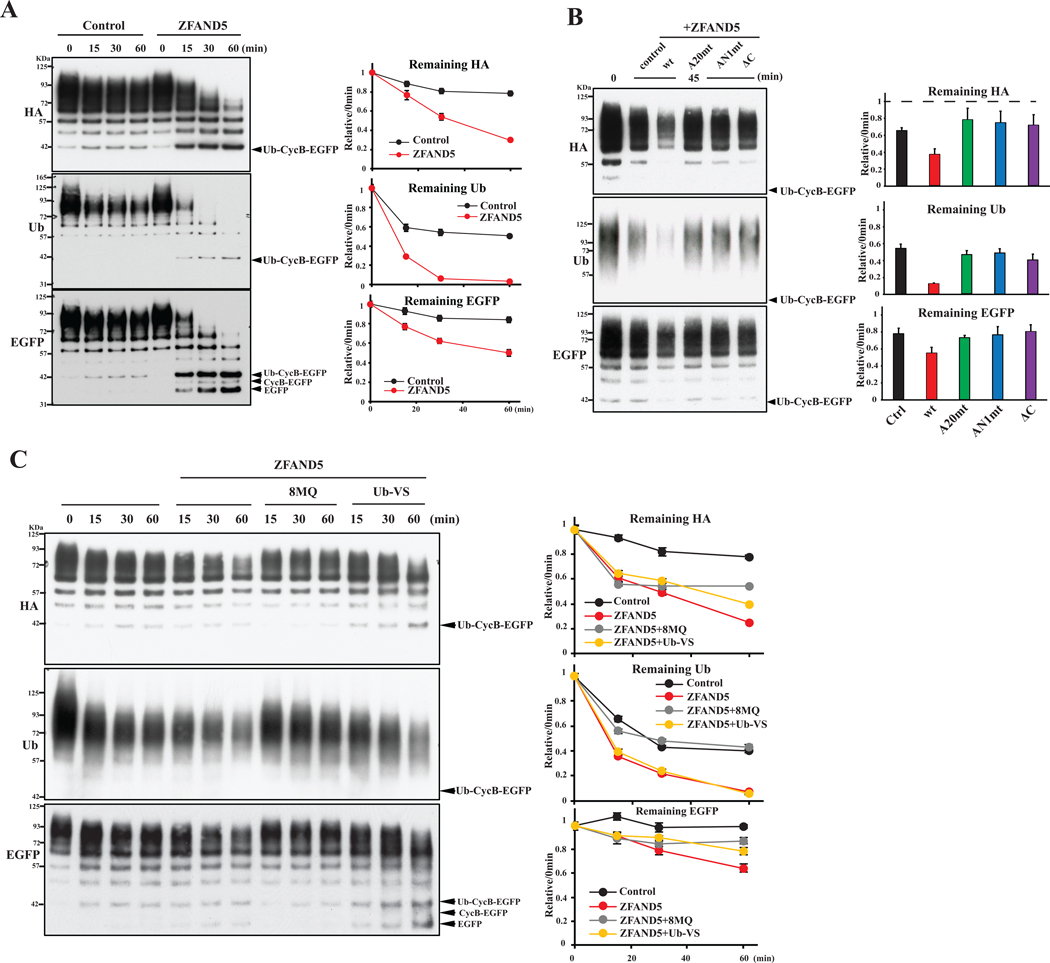
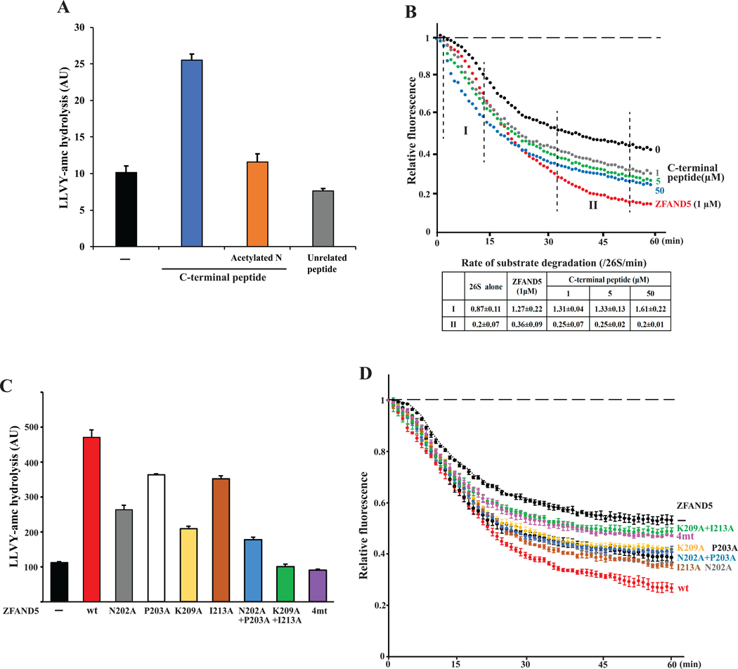
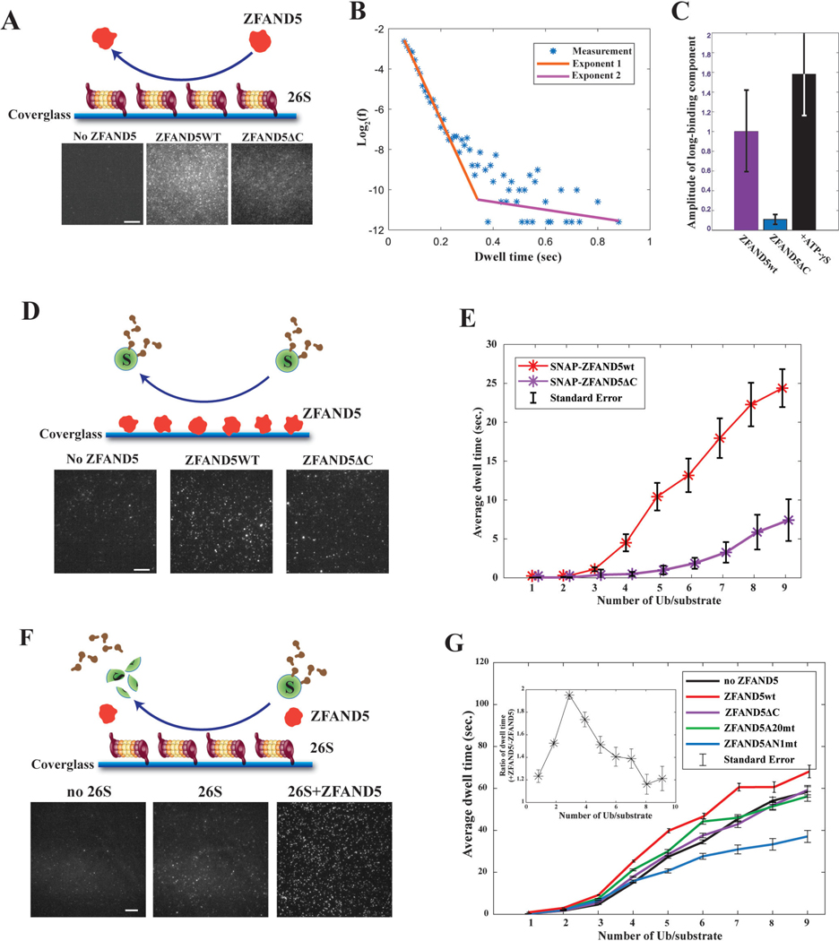
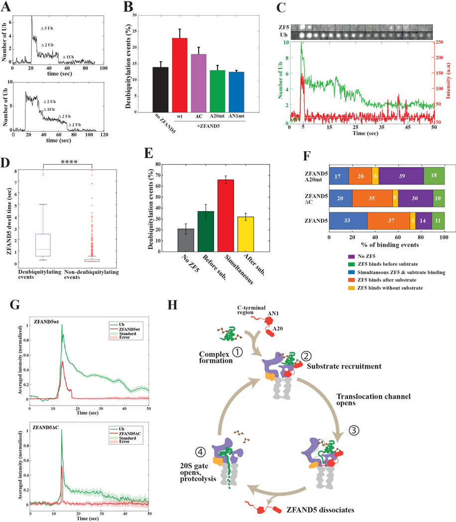
Various hormones, kinases, and stressors (fasting, heat shock) stimulate 26S proteasome activity. To understand how its capacity to degrade ubiquitylated proteins can increase, we studied mouse ZFAND5, which promotes protein degradation during muscle atrophy. Cryo-electron microscopy showed that ZFAND5 induces large conformational changes in the 19S regulatory particle. ZFAND5's AN1 Zn-finger domain interacts with the Rpt5 ATPase and its C terminus with Rpt1 ATPase and Rpn1, a ubiquitin-binding subunit. Upon proteasome binding, ZFAND5 widens the entrance of the substrate translocation channel, yet it associates only transiently with the proteasome. Dissociation of ZFAND5 then stimulates opening of the 20S proteasome gate. Using single-molecule microscopy, we showed that ZFAND5 binds ubiquitylated substrates, prolongs their association with proteasomes, and increases the likelihood that bound substrates undergo degradation, even though ZFAND5 dissociates before substrate deubiquitylation. These changes in proteasome conformation and reaction cycle can explain the accelerated degradation and suggest how other proteasome activators may stimulate proteolysis.

Keywords: ZFAND5; muscle atrophy; proteasome activation; proteasomes; protein degradation; ubiquitin.


Conflict of interest statement

Declaration of interests The authors claim no competing interests.

Figures

Figure 1.
Figure 1.. Cryo-EM structures of the human 26S proteasome complexed with ZFAND5/ZNF216.
(A) Selected cryo-EM density maps of human 26S proteasomes in the presence of ZFAND5 (red). The relative abundances of each 3D class (% total). Z+ refers to each class containing a ZFAND5 density and Z‒ to classes lacking ZFAND5. As shown below, the domain architecture of ZFAND5 is shown on the right. (B) A local view of ZFAND5 in the cryo-EM density map of the Z+D state. Close-up views of ZFAND5’s interfaces with various proteasome subunits are shown in D, with regions of same color indicated in the top panel. (C) A local view of the Z+D model highlighting the molecular path that ZFAND5 follows on by Rpt5, Rpt1 and Rpn1 and the positions of its C-terminal residues in Z+D. (D) Close-up views of ZFAND5’s interfaces with various proteasome subunits are shown in the lower panels with the same colors as in B. (E) Chemically-crosslinked residue pairs in a proteasome-ZFAND5 sample were identified by mass spectrometry and are represented as dotted lines. See also Figures S1–S4 and Tables S1 and S2.
Figure 2.
Figure 2.. ZFAND5 induces a 19S conformation with an open entry to the substrate translocation channel in the Z+D state.
(A) Comparison of the structures of the ATPase and the non-ATPase parts in Z+D (left panel) and Z+A states. Superimposed on Z+D conformation (right panel) are shown in gray the locations of the corresponding subunits in Z+A, which are very similar in Z+B and Z+C. (B) Comparison of the entrance to the substrate translocation channel in three different 19S states. The diameter of the unobstructed part of the entrance is indicated by an arrow. Lower: vertical cross sections of cryo-EM maps. Substrate translocation channel’s interior residues are colored red, surface delineated by dashed lines. (C) Close-up views showing relocation of Rpn3’s C-terminal domain leading to unimpeded entry to the ATPase channel in Z+D states. (D) ZFAND5 increases access to active sites in the CP. An activity-based probe MVB003 for the active sites in the CP was incubated with 26S proteasome in the presence or absence of ZFAND5. Samples were analyzed by SDS electrophoresis and fluorescence imaging. (E) ZFAND5ΔC mutant lacking its 19 C-terminal residues binds to the 26S proteasome. Co-immunoprecipitation with immobilized anti-α1 antibody or control IgG. The bound samples were analyzed by Western blotting (WB). (F) The peptidase activity of 26S proteasome in the presence of ZFAND5 or its ΔC mutant. Right: Coomassie staining of purified ZFAND5. Error bars represent SD of three replicates. (G) Change in migration of proteasomes by ZFAND5 requires its C-terminus. 26S proteasomes were incubated with ZFAND5 or its ΔC mutant and were analyzed by native electrophoresis and by WB. (H) Induction of ZFAND5 by dexamethasone leads to proteasome gate opening shown by reactivity with MVB003. C2C12 myotubes were treated with dexamethasone (50mM) for 1 day and with MVB003 for 1h before harvesting. The labeled 20S subunits were resolved in SDS-PAGE, and proteasome contents were compared by WB. See also Figures S5–S9 and Tables S1 and S2.
Figure 3.
Figure 3.. ZFAND5 stimulates the degradation of Ub conjugates by the 26S proteasome.
(A) Intensities of a fluorescent substrate, ubiquitylated cycB-cpGFP, upon degradation reactions with purified 26S proteasomes and ZFAND5 at various concentrations. CycB-cpGFP’s degradation rates calculated from the slopes at the initial (“I”) or late (“II”) stage of the reaction are listed in the table. Shown are the mean rates ± the standard deviations (SD) of five replicates. The degradation rates for the indicated time frames (I and II) are shown ± SD from three independent experiments. (B) Effect of ZFAND5 on the degradation of substrates containing increasing numbers of Ub molecules. 32P-labeled ubiquitylated cycB-cpGFP was incubated with 26S proteasomes and ZFAND5. Samples were analyzed by autoradiography and the degradation rate of each ubiquitylated species was plotted on the right. Error bars represent uncertainty in quantification. (C) ZFAND5’s C-terminal 19 residues, A20 domain and AN1 domain are essential for the stimulation of degradation. Reactions were performed as in A but with ZFAND5 mutants. Error bars represent the SD of five replicates. (D) Effects of ZFAND5 and its mutants on the mean half-lives of, Ub-R-YFP, a substrate of the N-end rule pathway, and Ub-(G76V)-YFP, a substrate of the UFD pathway in cells. Degradation of these substrates was monitored using time-lapse microscopy in a cycloheximide chase experiment in HEK293 cells cotransfected with ZFAND5 WT or mutants. Cells expressing 5~15uM reporters were selected. 15~50 cells were analyzed in each category. Error bars represent the standard errors and the statistical significance is marked by “*”. The expression levels of FLAG-ZFAND5WT and mutants were examined by WB. (E) ZFAND5 stimulates the degradation of naturally ubiquitylated proteins, which were isolated from HEK293 cells expressing polyHis-HA-tagged Ub, followed by incubation with purified 26S proteasome with or without ZFAND5. The samples were analyzed for K48 Ub linkage and HA epitope by WB. Error bars represent the SD of three replicates. See also Figure S10.
Figure 4.
Figure 4.. ZFAND5 stimulates deubiquitylation and degradation of unfolding-resistant substrate by the 26S proteasome.
(A) Ubiquitylated cycB-EGFP was incubated with 26S proteasome in the presence of WT and mutant ZFAND5 in (B). Levels of cycB, EGFP and ubiquitylation were determined by WB. Error bars represent the SD of three replicates. (C) Stimulation of degradation and deubiquitylation requires Rpn11. Degradation assays were performed as in (A), but in the presence of the cysteine DUB inhibitor Ub-VS or the Rpn11 inhibitor 8MQ. Error bars represent uncertainty in quantification. See also Figure S11.
Figure 5.
Figure 5.. A 19-residue peptide from ZFAND5’s C-terminus by itself activates proteasomal degradation of short peptides and Ub conjugates.
(A) The chymotryptic peptidase activity of purified 26S proteasome was determined in the presence of indicated peptides. Error bars represent the SD of three replicates. (B) The CTR peptide stimulates degradation of ubiquitylated substrates. Increasing concentrations of the CTR peptide were incubated with ubiquitylated cycB-cpGFP and 26S, and degradation was measured by fluorescence intensity. The degradation rates during the indicated time frame are presented ± SD from three replicates. (C) Mutagenesis of the proteasome-interacting residues of ZFAND5 and the effects on proteasome activation. The peptidase activity was determined as in A, but with indicated ZFAND5 variants. (D) As in B, but in the presence of indicated ZFAND5 variants. Results are presented as the mean ± SD of three replicates. See also Figure S12.
Figure 6.
Figure 6.. ZFAND5 exhibits two binding modes on the 26S proteasome and enhances substrate-proteasome interaction.
(A) ZFAND5’s C-terminal region is essential for the stimulation of substrate association with the proteasome. Schematic and typical images of the single-molecule assay testing the interaction of fluorescent ZFAND5 with surface-immobilized 26S particles. Scale bars = 5μm (B) Distribution of ZFAND5’s dwell times on the proteasome. The measurements were fitted with a double exponential function and the two exponents are plotted as straight lines on a semi-log scale. (C) Amplitude of the second (i.e. long-binding mode) exponential component of ZFAND5’s dwell time distribution. Measurement and data processing were performed as in B in the presence of ZFAND5, or ZFAND5ΔC, or upon addition of ATP-γS. Error bars represent fitting uncertainty. (D) Schematic and sample images of the single-molecule assay to study the interactions of securin conjugated with Dy550-Ub and immobilized ZFAND5 WT or ΔC. The average dwell-time of a securin molecule on ZFAND5 is shown in (E). (F) Schematic and sample images of the single-molecule fluorescence analysis of the kinetics of substrate processing by immobilized 26S proteasomes. (G) The average dwell-time of a securin molecule conjugated with Dy550-Ub on the 26S proteasome in the presence of 500μM ZFAND5 or its mutants was plotted vs. the number of Ub molecules per substrate molecule. Inset: the ratio of the dwell-time values in the presence and absence of ZFAND5. See also Figure S13.
Figure 7.
Figure 7.. ZFAND5 ferries Ub conjugates to the proteasome and promotes their deubiquitylation and proteolysis.
(A) Examples of single-molecule traces exhibiting processive deubiquitylation. Shown are the number of Ub molecules lost with each deubiquitylation step. (B) Effects of ZFAND5 and its mutants on the fraction of all substrate-proteasome encounters leading to processive deubiquitylation. Substrates containing at least four Ubs were analyzed. Error bars represent the SD of three replicates. (C) An example montage and the time trajectory from a single-molecule measurement of ZFAND5 and ubiquitylated securin interacting with immobilized 26S proteasomes and undergoing dissociation or deubiquitylation. ZFAND5 was labeled with a JF646 dye via a SNAP tag; Ub was labeled with Dy550. (D) Comparison of ZFAND5 dwell times on proteasomes when leading to processive deubiquitylation or not. “*” indicates the statistical significance. (E) The fraction of substrate binding events leading to processive deubiquitylation when ZFAND5 binds with, before or after the substrate. See methods for the definition of each category. Error bars represent the SD of three replicates. (F) ZFAND5, but not its A20 mutant or C-terminal deletion, increases the frequency of simultaneous binding with substrate to the proteasome. The fraction of times that the substrate binds together with, before, or after ZFAND5 or its mutants. (G) The averaged single-molecule kinetics of ZFAND5 and ubiquitylated securin on proteasome, suggesting that ZFAND5 dissociates from proteasomes before securin undergoes deubiquitylation. Single-molecule events recorded in the presence of ZFAND5 (N=440) or ZFAND5ΔC (N=97) were aligned by the moment of securin binding to proteasome, and the average fluorescence intensities among these events were plotted for the Ub and ZFAND5 channels. (H) ZFAND5 stimulates Ub conjugate degradation through a multistep reaction cycle 1) Formation of ZFAND5-substrate complex. 2) Its association with the proteasome RP. 3) RP assumes an open-channel conformation (Z+D) that favors deubiquitylation and degradation. 4) ZFAND5 dissociation leads to CP-gate opening, alignment with ATPase channel and substrate translocation into CP. See also Figure S14.

Update of

Similar articles

References

    1. VerPlank JJS, Lokireddy S, Zhao J, and Goldberg AL (2019). 26S Proteasomes are rapidly activated by diverse hormones and physiological states that raise cAMP and cause Rpn6 phosphorylation. Proc Natl Acad Sci U S A 116, 4228–4237. 10.1073/pnas.1809254116. - DOI - PMC - PubMed
    1. Lee D, and Goldberg AL (2022). 26S proteasomes become stably activated upon heat shock when ubiquitination and protein degradation increase. 10.1073/pnas. - DOI - PMC - PubMed
    1. Lee D, Takayama S, and Goldberg AL (2018). ZFAND5/ZNF216 is an activator of the 26S proteasome that stimulates overall protein degradation. Proc Natl Acad Sci U S A 115, E9550–E9559. 10.1073/pnas.1809934115. - DOI - PMC - PubMed
    1. Hishiya A, Iemura S, Natsume T, Takayama S, Ikeda K, and Watanabe K. (2006). A novel ubiquitin-binding protein ZNF216 functioning in muscle atrophy. EMBO J 25, 554–564. 10.1038/sj.emboj.7600945. - DOI - PMC - PubMed
    1. de la Pena AH, Goodall EA, Gates SN, Lander GC, and Martin A. (2018). Substrate-engaged 26S proteasome structures reveal mechanisms for ATP-hydrolysis-driven translocation. Science (1979) 362. 10.1126/science.aav0725. - DOI - PMC - PubMed

Publication types

Substances