. 2023 Aug 22;120(34):e2302901120.
doi: 10.1073/pnas.2302901120. Epub 2023 Aug 17.

Reconstitution of phytochrome A-mediated light modulation of the ABA signaling pathways in yeast

Affiliations

Reconstitution of phytochrome A-mediated light modulation of the ABA signaling pathways in yeast

Hong Li et al. Proc Natl Acad Sci U S A. .

Abstract

Abscisic acid (ABA), a classical plant hormone, plays an essential role in plant adaptation to environmental stresses. The ABA signaling mechanisms have been extensively investigated, and it was shown that the PYR1 (PYRABACTIN RESISTANCE1)/PYL (PYR1-LIKE)/RCAR (REGULATORY COMPONENT OF ABA RECEPTOR) ABA receptors, the PP2C coreceptors, and the SnRK2 protein kinases constitute the core ABA signaling module responsible for ABA perception and initiation of downstream responses. We recently showed that ABA signaling is modulated by light signals, but the underlying molecular mechanisms remain largely obscure. In this study, we established a system in yeast cells that was not only successful in reconstituting a complete ABA signaling pathway, from hormone perception to ABA-responsive gene expression, but also suitable for functionally characterizing the regulatory roles of additional factors of ABA signaling. Using this system, we analyzed the roles of several light signaling components, including the red and far-red light photoreceptors phytochrome A (phyA) and phyB, and the photomorphogenic central repressor COP1, in the regulation of ABA signaling. Our results showed that both phyA and phyB negatively regulated ABA signaling, whereas COP1 positively regulated ABA signaling in yeast cells. Further analyses showed that photoactivated phyA interacted with the ABA coreceptors ABI1 and ABI2 to decrease their interactions with the ABA receptor PYR1. Together, data from our reconstituted yeast ABA signaling system provide evidence that photoactivated photoreceptors attenuate ABA signaling by directly interacting with the key components of the core ABA signaling module, thus conferring enhanced ABA tolerance to light-grown plants.

Keywords: ABA; light; phyA; reconstitution; yeast.


Conflict of interest statement

The authors declare no competing interest.

Figures

Fig. 1.
Fig. 1.
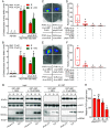
Reconstitution of an ABA signaling pathway in yeast cells. (A) Immunoblots showing the expression of ABF4, OST1, ABI1, and PYR1 in yeast. Red arrow indicates the phosphorylated form of ABF4. Anti-H3 was used as a sample loading control. (B and C) In vivo plate assays (B) and liquid culture assays (C) showing expression of the LacZ reporter gene with or without 20 μM ABA. In (B), selected transformants were streaked on appropriate synthetic dropout medium containing X-gal as substrate. In (C), β-galactosidase activities were measured using ONPG as substrate. Error bars represent SD of four independent yeast clones. Different letters represent significant differences by one-way ANOVA with Duncan’s post hoc test (P < 0.05). (D) Effects of ABA concentrations on the activation of the RD29B promoter in yeast reconstitution system. Yeast cultures were supplied with the indicated concentrations of ABA and incubated at 30 °C, then β-galactosidase activities were measured using ONPG as the substrate. Error bars represent SD of four independent yeast clones. Different letters represent significant differences by one-way ANOVA with Duncan’s post hoc test (P < 0.05).
Fig. 2.
Fig. 2.
phyA negatively regulates ABA signaling in yeast cells. (AC) phyA (A) and phyB (B) inhibit ABA signaling, but COP1 (C) promotes ABA signaling in the yeast reconstitution system. (D) phyA-Pfr inhibited the PYR1–ABI1–OST1–ABF4 pathway more strongly than phyA-Pr in yeast cells. (E) phyA-Pfr inhibited the ABA signaling pathways initiated by PYR1, PYL4, and PYL9 in yeast cells. (F) phyA-Pfr inhibited the PYR1–ABI2–OST1–ABF4 pathway more strongly than phyA-Pr in yeast cells. In (A, B, and DF), yeast cells incubated in the dark in appropriate synthetic dropout medium containing 20 μM ABA (with or without 10 μM PCB) were irradiated with 5 min of R light alone, or 5 min of R light immediately followed by 5 min of FR light, and then incubated in darkness for additional 2 h. In (C), yeast cells were incubated in appropriate synthetic dropout medium containing 20 μM ABA. The β-galactosidase activities were measured by liquid culture assays using ONPG as substrate. Error bars indicate SD of four independent yeast clones. Different letters represent significant differences by one-way ANOVA with Duncan’s post hoc test (P < 0.05).
Fig. 3.
Fig. 3.
phyA physically interacts with ABI1 and ABI2. (A and B) Y2H assays showing that the Pfr form of phyA preferentially interacts with ABI1 and ABI2. Transformants coexpressing phyA-LexA and AD-FHY1 were used as positive controls. Yeast cells incubated in the dark in appropriate synthetic dropout medium (containing 10 μM PCB) were irradiated with 5 min of R light alone, or 5 min of R light immediately followed by 5 min of FR light, and then incubated in darkness for 2 h. The β-galactosidase activities were measured by liquid culture assays using ONPG as the substrate. Error bars indicate SD of four independent yeast clones. Different letters represent significant differences by one-way ANOVA with Duncan’s post hoc test (P < 0.05). (C and D) LCI assays showing that cLuc-ABI1/2 interacted with phyA-nLuc in Nicotiana benthamiana leaves. GUS-nLuc was used as a negative control. A representative picture is shown in (C), and relative luciferase activities are shown in (D). Error bars represent SD of three independent biological replicates. Different letters represent significant differences by one-way ANOVA with Duncan’s post hoc test (P < 0.05). (E) Co-IP assays showing that the Pfr form of phyA preferentially associated with ABI1 and ABI2 in vivo. ABI1/2-GFP and phyA-FLAG fusion proteins were expressed in Arabidopsis protoplasts, then the protoplasts were treated with R light (R) or remained in the dark (D) for 30 min. Total proteins were then extracted and incubated with anti-GFP beads (AlpaLife, Shenzhen, China). Total and precipitated proteins were analyzed by immunoblotting using antibodies against FLAG, GFP, and HSP, respectively. Numbers below the immunoblots indicate the relative band intensities of coprecipitated phyA-FLAG normalized to those of precipitated ABI1/2-GFP, respectively. (F) Schematic diagram of bait proteins (PHYA-N, PHYA-C, PHYA-C1, and PHYA-C2 fused with LexA DNA-binding domains). NTE, N-terminal extension; H, hinge; PRD, PAS-related domain; HKRD, histidine kinase-related domain. (G) Y2H assays showing that ABI1 and ABI2 interacted with the N-terminal, C-terminal, and HKRD domains of PHYA in yeast cells.
Fig. 4.
Fig. 4.
phyA inhibited interactions between PYR1 and ABI1/2. (A and B) Y3H assays showing that phyA-Pfr inhibited PYR1 interactions with ABI1 (A) and ABI2 (B) more strongly than phyA-Pr. Yeast cells incubated in the dark in appropriate synthetic dropout medium containing 10 μM ABA (with 10 μM PCB) were irradiated with 5 min of R light alone, or 5 min of R light immediately followed by 5 min of FR light, and then incubated in darkness for additional 2 h. The β-galactosidase activities were measured by liquid culture assays using ONPG as the substrate. Error bars indicate SD of four independent yeast clones. Different letters represent significant differences by one-way ANOVA with Duncan’s post hoc test (P < 0.05). (CF) LCI assays showing that phyA inhibited PYR1 interactions with ABI1 (C and D) and ABI2 (E and F) in Nicotiana benthamiana leaves. Representative pictures are shown in (C) and (E), and relative luciferase activities are shown in (D) and (F). Error bars represent SD of six independent biological replicates. Different letters represent significant differences by one-way ANOVA with Duncan’s post hoc test (P < 0.05). (G and H) Semi-in vivo pull-down assays showing that phyA inhibited PYR1 interaction with ABI1. Total proteins were extracted from 4-d-old Col and phyA-211 mutant seedlings grown in the dark, then treated with R light (R) or remained in the dark (D) for 30 min. Then, equivalent amounts of recombinant GST-ABI1 (with or without 10 μM ABA) were added into the protein extracts as indicated for pull-down assays. The pulled-down proteins were analyzed by immunoblotting with antibodies against PYR1, GST, phyA, and RPN6, respectively. Representative pictures are shown in (G) and the relative levels of coprecipitated PYR1 are shown in (H). Error bars indicate SD from three independent assays. Different letters represent significant differences by one-way ANOVA with Duncan’s post hoc test (P < 0.05).
Fig. 5.
Fig. 5.
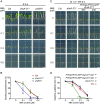
phyA and phyB negatively regulate ABA signaling in plants. (A) Seedling establishment of phyB-9 mutant was sensitive to ABA in R light. Col, phyA-211, and phyB-9 seedlings were grown vertically on MS medium containing the indicated concentrations of ABA in R light for 5 d. (Bar, 5 mm.) (B) Measurements of seedling establishment rates in (A). Error bars represent SD of four independent pools of seedlings. *P < 0.05, ***P < 0.001 (Student’s t test). (C) Seedling establishment of phyA-211 mutant was sensitive to ABA in FR light. Col, phyA-211 mutant, and two phyA-211 complementation lines were grown vertically on the MS medium containing the indicated concentrations of ABA in white light for 1 d, then transferred to FR light, and incubated for additional 6 d. (Bar, 5 mm.) (D) Measurements of seedling establishment rates in (C). Error bars represent SD of four independent pools of seedlings. **P < 0.01, ***P < 0.001 (Student’s t test).
Fig. 6.
Fig. 6.
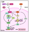
A working model depicting that photoactivated phyA negatively regulates ABA signaling. In the dark, the COP1-PIF module promotes ABA signaling by transcriptionally and posttranslationally mediating ABA-induced ABI5 accumulation (27, 42). In the light, photoactivated phyA attenuates ABA signaling by inhibiting the COP1-PIF module, and by interacting with the ABA coreceptors (such as ABI1 and ABI2) to decrease their interactions with the ABA receptors (such as PYR1). Therefore, photoactivated photoreceptors confer enhanced ABA tolerance to light-grown plants.

Similar articles

Cited by

References

    1. Chen X., et al. , Protein kinases in plant responses to drought, salt, and cold stress. J. Integr. Plant Biol. 63, 53–78 (2021). - PubMed
    1. Hubbard K. E., Nishimura N., Hitomi K., Getzoff E. D., Schroeder J. I., Early abscisic acid signal transduction mechanisms: Newly discovered components and newly emerging questions. Genes Dev. 24, 1695–1708 (2010). - PMC - PubMed
    1. Chen K., et al. , Abscisic acid dynamics, signaling, and functions in plants. J. Integr. Plant Biol. 62, 25–54 (2020). - PubMed
    1. Gong Z., et al. , Plant abiotic stress response and nutrient use efficiency. Sci. China Life Sci. 63, 635–674 (2020). - PubMed
    1. Zhang H., Zhu J., Gong Z., Zhu J. K., Abiotic stress responses in plants. Nat. Rev. Genet. 23, 104–119 (2022). - PubMed

Publication types