. 2023 Aug 31;97(8):e0075623.
doi: 10.1128/jvi.00756-23. Epub 2023 Aug 14.

The atlastin ER morphogenic proteins promote formation of a membrane penetration site during non-enveloped virus entry

Affiliations

The atlastin ER morphogenic proteins promote formation of a membrane penetration site during non-enveloped virus entry

Madison Pletan et al. J Virol. .

Abstract

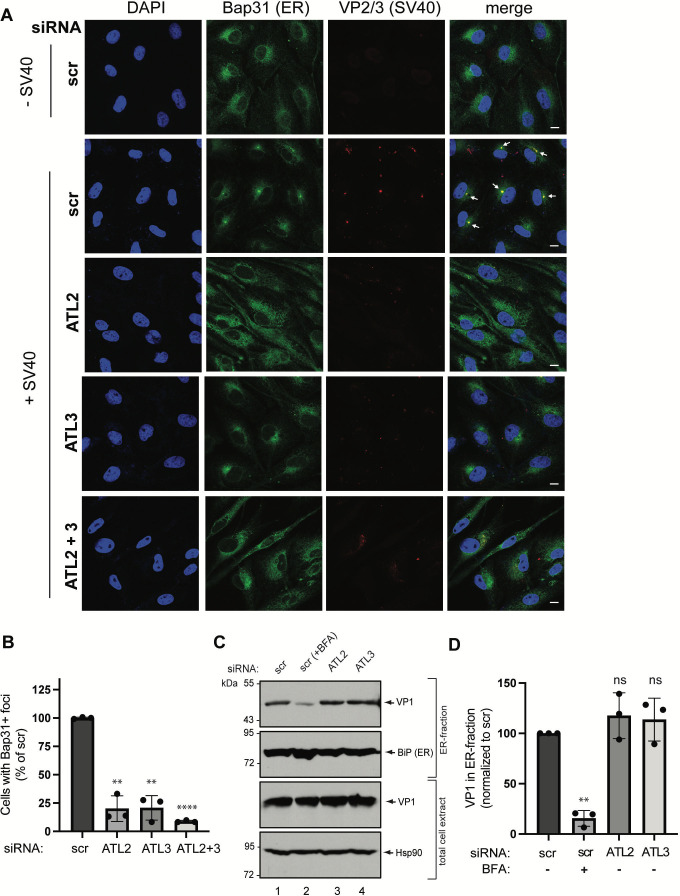
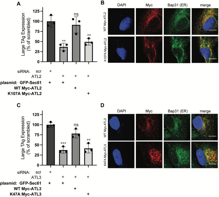
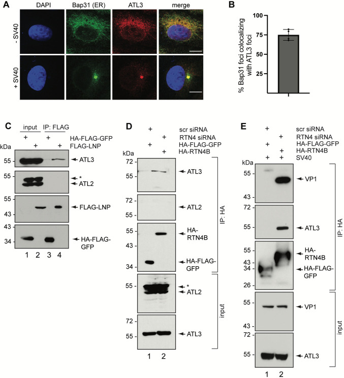
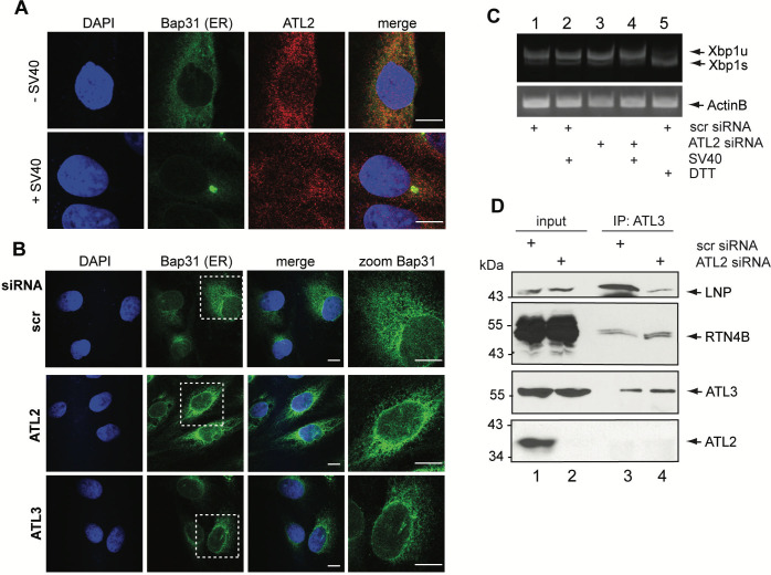
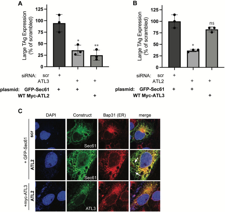
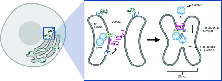
During entry, non-enveloped viruses penetrate a host membrane to cause infection, although how this is accomplished remains enigmatic. Polyomaviruses (PyVs) are non-enveloped DNA viruses that penetrate the endoplasmic reticulum (ER) membrane to reach the cytosol en route to the nucleus for infection. To penetrate the ER membrane, the prototype PyV simian virus 40 (SV40) induces formation of ER-escape sites, called foci, composed of repeating units of multi-tubular ER junctions where the virus is thought to exit. How SV40 triggers formation of the ER-foci harboring these multi-tubular ER junctions is unclear. Here, we show that the ER morphogenic atlastin 2 (ATL2) and ATL3 membrane proteins play critical roles in SV40 infection. Mechanistically, ATL3 mobilizes to the ER-foci where it deploys its GTPase-dependent membrane fusion activity to promote formation of multi-tubular ER junctions within the ER-foci. ATL3 also engages an SV40-containing membrane penetration complex. By contrast, ATL2 does not reorganize to the ER-foci. Instead, it supports the reticular ER morphology critical for the integrity of the ATL3-dependent membrane complex. Our findings illuminate how two host factors play distinct roles in the formation of an essential membrane penetration site for a non-enveloped virus. IMPORTANCE Membrane penetration by non-enveloped viruses, a critical infection step, remains enigmatic. The non-enveloped PyV simian virus 40 (SV40) penetrates the endoplasmic reticulum (ER) membrane to reach the cytosol en route for infection. During ER-to-cytosol membrane penetration, SV40 triggers formation of ER-associated structures (called ER-foci) that function as the membrane penetration sites. Here, we discover a role of the ATL ER membrane proteins-known to shape the ER morphology-during SV40-induced ER-foci formation. These findings illuminate how a non-enveloped virus hijacks host components to construct a membrane penetration structure.

Keywords: endoplasmic reticulum; entry; non-enveloped virus; simian virus 40.


Conflict of interest statement

The authors declare no conflict of interest.

Figures

Fig 1
Fig 1
ATL2 and ATL3 are important for SV40 infection. (A) CV-1 cells were transfected with scrambled control siRNA (scr) or siRNA against ATL2, ATL3, or both. After 48 h of transfection, cell extracts were assessed by SDS-PAGE and immunoblotting with the indicated antibodies. * indicates unidentified protein. (B) CV-1 cells transfected with the indicated siRNA were infected with SV40 (multiplicity of infection [MOI] ~0.3), fixed, and stained for large T antigen (TAg). TAg expression was scored by immunofluorescence microscopy. Data were normalized to the mean of the scrambled controls. (C and D) As in (B), except cells were transfected with the indicated constructs after the initial siRNA transfection. Cells were fixed and stained for TAg and Myc, and at least 100 GFP/Myc dual-expressing cells were counted in each condition for each biological replicate. Graphs represent mean ± standard deviation (SD) from three independent experiments. The significance was determined via Student’s two-tailed t test. *, P ≤ 0.05; **, P ≤ 0.01; ***, P ≤ 0.001.
Fig 2
Fig 2
ATL2 and ATL3 are critical for SV40-induced ER-foci formation. (A) CV-1 cells transfected with the indicated siRNA were infected (or left uninfected) with SV40 (MOI ~10), fixed, stained with the indicated antibodies, counterstained with 4´,6-diamidino-2-phenylindole (DAPI), and subjected to confocal immunofluorescence microscopy. Scale bars, 10 μm. (B) Quantification of (A). Cells were scored as positive if at least one Bap31 focus was present in the cell. Data were normalized to the scrambled control. (C) CV-1 cells were transfected with the indicated siRNA. After transfection, either dimethylsulfoxide (DMSO) control or brefeldin A (BFA) was added to the cells and infected with SV40 (MOI ~5). Cells were then harvested and subjected to a fractionation assay (see Materials and Methods) to isolate an ER-containing fraction. This fraction and the total cell extract were subjected to SDS-PAGE and immunoblotting with the indicated antibodies. (D) Quantification of relative VP1 band intensities in the ER-fractions of (C). Bands were quantified with FIJI software, normalized relative to the total levels of VP1, and expressed as a percentage of the band intensity of the scrambled control. Graphs represent mean ± SD from three independent experiments. The significance was determined via Student’s two-tailed t test. **, P ≤ 0.01; ****, P ≤ 0.0001.
Fig 3
Fig 3
The GTPase-dependent membrane fusion activity of ATL2 and ATL3 is crucial for SV40 infection. (A and C) CV-1 cells were transfected with the indicated siRNA, then transfected with the indicated constructs. Cells were then infected with SV40 (MOI ~0.3), fixed, stained for TAg and Myc, then assessed for infection via confocal microscopy as in Fig. 1. At least 100 GFP/Myc dual-expressing cells were counted in each condition for each biological replicate. Data were normalized to the mean of the scrambled controls. Graphs represent mean ± SD from three independent experiments. The significance was determined via Student’s two-tailed t test. **, P ≤ 0.01; ***, P ≤ 0.001. (B and D) CV-1 cells were transfected with the indicated constructs, fixed, stained for Myc and Bap31, and counterstained with DAPI. Scale bars, 10 μm.
Fig 4
Fig 4
ATL3 mobilizes to the SV40-induced ER-foci to participate in an ER membrane penetration complex. (A) CV-1 cells were infected (or left uninfected) with SV40 (MOI ~50), fixed, stained with the indicated antibodies, and then counterstained with DAPI. Scale bars, 10 μm. (B) Quantification of (A): a Bap31 + focus was scored as positive if it colocalized with an ATL3 + focus. Graph represents mean ± SD from four independent experiments. (C) COS-7 cells were transfected with the indicated constructs and the cell extracts were subjected to immunoprecipitation with FLAG M2 antibody-conjugated beads. Immunoprecipitated material was subjected to SDS-PAGE and immunoblotting with the indicated antibodies. Input represents 2.5% of the total sample used for immunoprecipitation. * indicates unidentified protein. (D) COS-7 cells were transfected with the indicated siRNAs and then transfected with the indicated constructs. Cell extracts were subjected to immunoprecipitation with HA antibody-conjugated beads. Immunoprecipitated material was subjected to SDS-PAGE and immunoblotting with the indicated antibodies. Input represents 2.5% of the total sample used for immunoprecipitation. * indicates unidentified protein. (E) As in (D), except cells were infected with SV40 (MOI ~10) after transfection of the indicated plasmids. Cell extracts were cross-linked using dithiobis-succinimidyl-propionate, then subjected to immunoprecipitation with HA antibody-conjugated beads. Input represents 1% of the total sample used for immunoprecipitation.
Fig 5
Fig 5
ATL2 knockdown alters ER morphology and selectively impairs ATL3-LNP interaction. (A) CV-1 cells were infected (or left uninfected) with SV40 (MOI ~50), fixed, stained with the indicated antibodies, and then counterstained with DAPI. (B) CV-1 cells were transfected with the indicated siRNA, fixed, stained with Bap31 antibody, counterstained with DAPI, and then examined using confocal immunofluorescence. Scale bars, 10 μm. (C) CV-1 cells were transfected with the indicated siRNAs, mock infected or infected (MOI ~50), then treated with dithiothreitol (DTT) or DMSO control. RNA was extracted and reverse transcribed; the resulting cDNA was subjected to PCR amplification and gel electrophoresis. Xbp1u is unspliced Xpb1, while Xbp1s is spliced Xbp1. (D) COS-7 cells were transfected with the indicated siRNAs and the cell extracts were subjected to immunoprecipitation with antibody against endogenous ATL3. Immunoprecipitated material was subjected to SDS-PAGE and immunoblotting with the indicated antibodies. Input represents 2.5% of the total sample used for immunoprecipitation.
Fig 6
Fig 6
Overexpression of ATL3 compensates for ATL2 knockdown, restoring both ER morphology and SV40 infection. (A and B) CV-1 cells were transfected with the indicated siRNA and then transfected with the indicated constructs. Cells were then infected with SV40 (MOI ~0.3), fixed, stained for TAg and Myc, and then assessed for infection via confocal microscopy as in Fig. 1. At least 100 GFP/Myc dual-expressing cells were counted in each condition for each biological replicate. Data were normalized to the mean of the scrambled controls. Graphs represent mean ± SD from three independent experiments. The significance was determined via Student’s two-tailed t test. *, P ≤ 0.05; **, P ≤ 0.01. (C) CV-1 cells were transfected with the indicated siRNAs and then transfected with the indicated constructs. Cells were fixed, stained for Myc and Bap31, and counterstained with DAPI. Scale bars, 10 μm.
Fig 7
Fig 7
Model depicting the roles of ATL2 and ATL3 during SV40-induced ER-foci formation. During entry, SV40 is targeted from the cell surface to the ER. Here, ER-resident host factors impart conformational changes to the virus, generating a hydrophobic SV40 particle that initiates the ER-to-cytosol membrane penetration process. In this manuscript, we found that this viral entry pathway is dependent on ATL2, which serves to establish and maintain the reticular structure of the ER. During membrane penetration itself, SV40 engages the ER morphogenic proteins RTN and ATL3, which recruit LNP, and the virus reorganizes to the ER-foci with this morphogenic protein complex. Importantly, the interaction between LNP and ATL3 is dependent on ATL2. Thus, both ATL2 and ATL3 promote formation of a multi-tubular ER junction site where the virus penetrates to reach the cytosol. Figure created in Biorender.com.

Similar articles

References

    1. Weissenhorn W, Dessen A, Calder LJ, Harrison SC, Skehel JJ, Wiley DC. 1999. Structural basis for membrane fusion by enveloped viruses. Mol Membr Biol 16:3–9. doi:10.1080/096876899294706 - DOI - PubMed
    1. Kumar CS, Dey D, Ghosh S, Banerjee M. 2018. Breach: host membrane penetration and entry by nonenveloped viruses. Trends Microbiol 26:525–537. doi:10.1016/j.tim.2017.09.010 - DOI - PubMed
    1. Moyer CL, Nemerow GR. 2011. Viral weapons of membrane destruction: variable modes of membrane penetration by non-enveloped viruses. Curr Opin Virol 1:44–49. doi:10.1016/j.coviro.2011.05.002 - DOI - PMC - PubMed
    1. Tsai B. 2007. Penetration of nonenveloped viruses into the cytoplasm. Annu Rev Cell Dev Biol 23:23–43. doi:10.1146/annurev.cellbio.23.090506.123454 - DOI - PubMed
    1. DeCaprio JA, Garcea RL. 2013. A cornucopia of human polyomaviruses. Nat Rev Microbiol 11:264–276. doi:10.1038/nrmicro2992 - DOI - PMC - PubMed

Publication types

MeSH terms