[Preprint]. 2023 Aug 7:2023.08.04.551915.
doi: 10.1101/2023.08.04.551915.

Curved crease origami and topological singularities at a cellular scale enable hyper-extensibility of Lacrymaria olor

Affiliations

Curved crease origami and topological singularities at a cellular scale enable hyper-extensibility of Lacrymaria olor

Eliott Flaum et al. bioRxiv. .

Update in

Abstract

Eukaryotic cells undergo dramatic morphological changes during cell division, phagocytosis and motility. Fundamental limits of cellular morphodynamics such as how fast or how much cellular shapes can change without harm to a living cell remain poorly understood. Here we describe hyper-extensibility in the single-celled protist Lacrymaria olor, a 40 μm cell which is capable of reversible and repeatable extensions (neck-like protrusions) up to 1500 μm in 30 seconds. We discover that a unique and intricate organization of cortical cytoskeleton and membrane enables these hyper-extensions that can be described as the first cellular scale curved crease origami. Furthermore, we show how these topological singularities including d-cones and twisted domain walls provide a geometrical control mechanism for the deployment of membrane and microtubule sheets as they repeatably spool thousands of time from the cell body. We lastly build physical origami models to understand how these topological singularities provide a mechanism for the cell to control the hyper-extensile deployable structure. This new geometrical motif where a cell employs curved crease origami to perform a physiological function has wide ranging implications in understanding cellular morphodynamics and direct applications in deployable micro-robotics.

Keywords: Active filaments; Biophysics; Cell-behavior; Non-linear dynamics; Protists; curved crease origami.


Figures
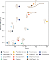
Figure 1:
Figure 1:. Hyper extensible single cells with large strain and strain rates.
Extension rate (μm/s) versus strain (∆L/L) of protrusions in both unicellular and multicellular cell types. All cell types, and the name of their extensible protrusions when applicable from bottom to top: Chlamydomonas (cilia)[90], Tetrahymena (cilia)[90], Euglena[91], Neuron (axon)[35], Fibroblast (lamellipodium)[92], Sea Urchin Embryonic Cell (filopodium)[93], Dictyostelium (pseudopod)[94], Maize Gamete (pollen tube)[95], Vorticella (stalk)[37], Cultured Lacrymaria olor[15], Spirostomum[96], Wildtype Lacrymaria olor[15, 38, 39], Nematocyte (nematocyst)[33], Microsporidium (polar tube)[32].
Figure 2:
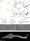
Figure 2:. Lacrymaria olor undergoes extreme extensions at ultra fast speeds.
(A) An overlay of snapshots from one neck extension-contraction cycle over a duration of 27.6 seconds shows an active cell which can reach many unique points around its cell body. Scale bar is 150 μm.(B) Starting from an active, contracted state, the cell can reach an extension of 1210 μm in under 7 seconds then retract just as quickly. (C) Time-points from multiple extension events during an active hunting period show similar extension rates. (D) Extension [+] and contraction [−] rates calculated from these extension events show an extension rate of up to 1420 μm/s. Individual extension/retraction events are noisy while the overall maximum extension rates are slightly faster than retraction rates. (E) Strain calculated for these deformations during extension [+] and contraction [−] show large strain magnitudes [15+] for a single cell. (F) Extension Rate vs. Strain for these same extension events. The blue shading and dotted lines were drawn in to highlight the reversibility of these dynamics. Data in A-F analyzed from publicly available wild type cell videos accessible at [16, 17, 18, 19, 20, 21]). (G) L. olor transitions between a dormant, contracted state and two active behavioral states by extending and contracting a neck-like protrusion. (H) The distinct cell morphologies between active contracted and active elongated states are apparent in live cells under DIC imaging. Scale bar is 40 μm. (I) Confocal fluorescence z-stack projection of α-tubulin stained fixed cells characterizes the helically-arranged cortical cytoskeleton.
Figure 3:
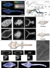
Figure 3:. A helically-arranged cortical cytoskeleton contains layers of microtubule filaments amid membrane folds.
(A) Confocal fluorescence z-stack projections of α-tubulin in fixed contracted and elongated cells. Inset shows measurements taken on these 2d projections of z-stack projections (radius and helix angle).(B) The microtubule structure varies in radius along the length of the cell in both elongated and contracted cells (n=6). (C) The microtubule structure varies in helix angle along the length of the cell in both elongated and contracted cells (N=6). (D) Microtubule filaments occur in doublets which are additionally layered in z. Scale bars are 10μm. (E) These layers are present both in the body and neck region in contracted cells, and in the body region of elongated cells, however they are not observed in the neck region of elongated cells. Scale bars are 10μm. (F) Schematic showing the planes in panels 5–7, which are three rotated images of the α-tubulin in three different planes along the length of the cell. Panels 5–7 confirm multiple layers of α-tubulin in the cortical cytoskeleton. Below are zoom-ins of the images which highlight the layers. (G) Scanning Electron image of the body of an elongated L. olor cell. (H) Confocal fluorescence z-stack projection of centrin stained fixed cells. Scale bars are 30 μm.
Figure 4:
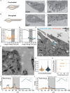
Figure 4:. The membrane is folded into pleats which contain contain microtubule ribbons.
(A-B) TEM images of slices in fixed contracted and elongated cells highlight the presence of membrane folds in the body-to-neck region of a contracted cell which are absent in the same region of an elongated cell. (C) The pleat depth in a contracted cell increases in the body-to-neck region (transition zone). The transition zone is highlighted in grey, and the region of the transition zone within the cell is shown using an outline of the cell in the top of the plot. (D) The pleat depth remains the minimal depth of the ciliary pit depths in the same zone in an elongated cell. The transition zone is highlighted in grey, and the region of the transition zone within the cell is shown using an outline of the cell in the top of the plot. (E) Representative TEM image of a membrane pleat show that these folds contain ciliary pits. (F) There are many microtubule ribbon in each of these pleats which lie along the membrane. (G) The median width of a ribbon is consistent in both contracted and elongated cells. (H) The total number of ribbons per pleat increases with pleat depth in contracted cells. (I-J) Microtubule bending and twisting creates an energy barrier at transition zone.(I) Schematic shows how the bending energy was calculated from the points in each filament. The bending energy has a global maximum in the transition zone in both a contracted and an elongated cell, with a higher energy maxima in the contracted cell. (J) Schematic shows how the twisting energy was calculated from the points in each filament. The twisting energy has a global maximum at the beginning of the transition zone an elongated cell, with a lower energy maxima in the contracted cell.
Figure 5:
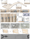
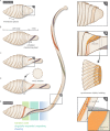
Figure 5:. Membrane-microtubule spooling: curved crease origami in a living cell.
(A) Schematic of the membrane and microtubule of contracted and elongated cells which highlights the sequential nature of the membrane pleat opening during extension. (1) Zoom-in of the membrane and cortical cytoskeleton folds in the body of an elongated cell. (2) Zoom-in of the membrane and cortical cytoskeleton folds in the transition zone of an elongated cell. (3) Zoom-in of the membrane and cortical cytoskeleton folds in the neck of an elongated cell. (B) Folded origami structures which are pulled on to elongate. This elongation process demonstrates that the membrane prevents the microtubules from extending like a normal spring through d-cone singularity propagation. The result is sequential opening of the curved crease pleats. Scale bars are 25 mm. (C) Sequential unspooling was observed for multiple fold angles (15°,30°,45°,60°,75°). (D) The Poisson Ratio for extension at all angle is less than zero. (E) Top-down and Bottom-up views of one origami cylinder show that there is a chirality-induced asymmetry in the pleat structure. (F) Updated energy plot calculated using the geometry of the microtubule filaments in 3D. This updated plot now includes bending energy (circle), twisting energy from the surface of the cell (triangle), and twisting energy from the twisting of the microtubule ribbons (square). (G) Folded cylinders composed of mylar, with bamboo filaments representing the microtubules, and how the ribbons change orientation across folded, transition, and unfolded states. (H) The two d-cone singularities travel, one (B-B’) along a mountain fold and another (C-C’) along a valley fold as force is generated. As the membrane unfolds one of the microtubule ribbons (ribbon 2) twists to accommodate the re-orientation of the membrane. The d-cones and this twist singularity travel as the membrane transitions from folded to unfolded. (I) Image of a cell being held with two micropipettes under suction pressure. The neck was manually elongated while one pipette was kept steady and the second was pulled back. Scale bar is 40μm.
Figure 6:
Figure 6:. During extension, the microtubule filaments and the membrane pass through the transition zone for coordinated, reversible unspooling.
This schematic shows a cell transitioning from a contracted to an elongated state. (A) In the first cell, the membrane is completely folded into pleats. Inset (1) from this schematic depicts microtubules stored in the pleats. (B) In the second cell, the neck is partially elongated, and one pleat is unfolded (shown in purple). In this cell, the transition zone between the cell body and the neck becomes apparent. (C) As the neck continues to elongate, more pleats unfold while in the transition zone. The specific location where the pleats transition from folded to unfolded occurs at point singularities, which is enlarged in inset (2). The synchronization of twist singularity during extension is shown below inset (2). (D) A cross section of the transition zone is shown to illustrate the 3-dimensional shape of the pleats. (E) In the final, most-elongated cell, we highlight the 3 regimes which are required for hyper-extension: the transition zone where the membrane is pleated, the zone where singularities propagate and the pleats sequentially unfold, and the last zone where the neck undergoes limited shear, resulting in thin hyper-extensions. In the neck of this last cell, inset (3) shows that the microtubule ribbons and the basal bodies are now most visible as the pleats are unfolded.

Similar articles

References

    1. Remedios C. G. D. et al. Actin binding proteins: Regulation of cytoskeletal microfilaments. Physiological Reviews 83, 433–473 (2003). - PubMed
    1. Tzur A., Kafri R., LeBleu V. S., Lahav G. & Kirschner M. W. Cell growth and size homeostasis in proliferating animal cells. Science 325, 167–171 (2009). - PMC - PubMed
    1. Nyga A. et al. Dynamics of cell rounding during detachment. iScience 26, 106696 (2023). - PMC - PubMed
    1. Yap B. & Kamm R. D. Cytoskeletal remodeling and cellular activation during deformation of neutrophils into narrow channels. Journal of Applied Physiology 99, 2323–2330 (2005). - PubMed
    1. Weiner O. D., Marganski W. A., Wu L. F., Altschuler S. J. & Kirschner M. W. An actin-based wave generator organizes cell motility. PLoS Biology 5, e221 (2007). - PMC - PubMed

Publication types