Mechanisms and regulation of defensins in host defense
- PMID: 37574471
- PMCID: PMC10423725
- DOI: 10.1038/s41392-023-01553-x
Mechanisms and regulation of defensins in host defense
Abstract
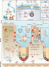
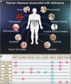
As a family of cationic host defense peptides, defensins are mainly synthesized by Paneth cells, neutrophils, and epithelial cells, contributing to host defense. Their biological functions in innate immunity, as well as their structure and activity relationships, along with their mechanisms of action and therapeutic potential, have been of great interest in recent years. To highlight the key research into the role of defensins in human and animal health, we first describe their research history, structural features, evolution, and antimicrobial mechanisms. Next, we cover the role of defensins in immune homeostasis, chemotaxis, mucosal barrier function, gut microbiota regulation, intestinal development and regulation of cell death. Further, we discuss their clinical relevance and therapeutic potential in various diseases, including infectious disease, inflammatory bowel disease, diabetes and obesity, chronic inflammatory lung disease, periodontitis and cancer. Finally, we summarize the current knowledge regarding the nutrient-dependent regulation of defensins, including fatty acids, amino acids, microelements, plant extracts, and probiotics, while considering the clinical application of such regulation. Together, the review summarizes the various biological functions, mechanism of actions and potential clinical significance of defensins, along with the challenges in developing defensins-based therapy, thus providing crucial insights into their biology and potential clinical utility.
© 2023. West China Hospital, Sichuan University.
Conflict of interest statement
The authors declare no competing interests.
Figures


Similar articles
-
Defensins and Paneth cells in inflammatory bowel disease.Inflamm Bowel Dis. 2007 Oct;13(10):1284-92. doi: 10.1002/ibd.20197. Inflamm Bowel Dis. 2007. PMID: 17567878 Review.
-
Paneth cells, defensins, and the commensal microbiota: a hypothesis on intimate interplay at the intestinal mucosa.Semin Immunol. 2007 Apr;19(2):70-83. doi: 10.1016/j.smim.2007.04.002. Epub 2007 May 7. Semin Immunol. 2007. PMID: 17485224 Review.
-
Defensin-immunology in inflammatory bowel disease.Gastroenterol Clin Biol. 2009 Jun;33 Suppl 3:S137-44. doi: 10.1016/S0399-8320(09)73149-5. Gastroenterol Clin Biol. 2009. PMID: 20117337
-
Paneth cell defensins: key effector molecules of innate immunity.Biochem Soc Trans. 2006 Apr;34(Pt 2):263-6. doi: 10.1042/BST20060263. Biochem Soc Trans. 2006. PMID: 16545089 Review.
-
Inflammatory bowel disease: an impaired barrier disease.Langenbecks Arch Surg. 2013 Jan;398(1):1-12. doi: 10.1007/s00423-012-1030-9. Epub 2012 Nov 18. Langenbecks Arch Surg. 2013. PMID: 23160753 Review.
Cited by
-
Lung antimicrobial proteins and peptides: from host defense to therapeutic strategies.Physiol Rev. 2024 Oct 1;104(4):1643-1677. doi: 10.1152/physrev.00039.2023. Epub 2024 Jul 25. Physiol Rev. 2024. PMID: 39052018 Review.
-
Review: A Contemporary, Multifaced Insight into Psoriasis Pathogenesis.J Pers Med. 2024 May 16;14(5):535. doi: 10.3390/jpm14050535. J Pers Med. 2024. PMID: 38793117 Free PMC article. Review.
-
Porcine β-Defensin 114: Creating a Dichotomous Response to Inflammation.Int J Mol Sci. 2024 Jan 13;25(2):1016. doi: 10.3390/ijms25021016. Int J Mol Sci. 2024. PMID: 38256090 Free PMC article.
-
Antifungal Plant Defensins as an Alternative Tool to Combat Candidiasis.Plants (Basel). 2024 May 29;13(11):1499. doi: 10.3390/plants13111499. Plants (Basel). 2024. PMID: 38891308 Free PMC article. Review.
-
Investigation into the Effectiveness of an Herbal Combination (Angocin®Anti-Infekt N) in the Therapy of Acute Bronchitis: A Retrospective Real-World Cohort Study.Antibiotics (Basel). 2024 Oct 17;13(10):982. doi: 10.3390/antibiotics13100982. Antibiotics (Basel). 2024. PMID: 39452248 Free PMC article.
References
-
- Hilchie AL, Wuerth K, Hancock RE. Immune modulation by multifaceted cationic host defense (antimicrobial) peptides. Nat. Chem. Biol. 2013;9:761–768. - PubMed
-
- Wang J, et al. Antimicrobial peptides: promising alternatives in the post feeding antibiotic era. Med. Res. Rev. 2019;39:831–859. - PubMed
-
- Zaiou M, Nizet V, Gallo RL. Antimicrobial and protease inhibitory functions of the human cathelicidin (hCAP18/LL-37) prosequence. J. Invest. Dermatol. 2003;120:810–816. - PubMed
Publication types
MeSH terms
Substances
LinkOut - more resources
Full Text Sources
Research Materials

