Review
. 2023 Jul 28;24(15):12095.
doi: 10.3390/ijms241512095.

Gating of β-Barrel Protein Pores, Porins, and Channels: An Old Problem with New Facets

Affiliations
Review

Gating of β-Barrel Protein Pores, Porins, and Channels: An Old Problem with New Facets

Lauren A Mayse et al. Int J Mol Sci. .

Abstract

β barrels are ubiquitous proteins in the outer membranes of mitochondria, chloroplasts, and Gram-negative bacteria. These transmembrane proteins (TMPs) execute a wide variety of tasks. For example, they can serve as transporters, receptors, membrane-bound enzymes, as well as adhesion, structural, and signaling elements. In addition, multimeric β barrels are common structural scaffolds among many pore-forming toxins. Significant progress has been made in understanding the functional, structural, biochemical, and biophysical features of these robust and versatile proteins. One frequently encountered fundamental trait of all β barrels is their voltage-dependent gating. This process consists of reversible or permanent conformational transitions between a large-conductance, highly permeable open state and a low-conductance, solute-restrictive closed state. Several intrinsic molecular mechanisms and environmental factors modulate this universal property of β barrels. This review article outlines the typical signatures of voltage-dependent gating. Moreover, we discuss recent developments leading to a better qualitative understanding of the closure dynamics of these TMPs.

Keywords: conformational transitions; electrophysiology; membrane proteins; protein folding; single-molecule dynamics.


Conflict of interest statement

The authors declare no competing interest.

Figures
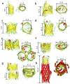
Figure 1
Figure 1
β-barrel proteins of Gram-negative bacteria. (a) OmpA (PDB:1QJP; [15]). (b) OmpT (PDB:6EHD; [39]). (c) OprD (OccD1) (PDB:3SY7; [40]). (d) OmpG(PDB:2F1C; [31]). (e) OmpF(PDB:2ZFG; [41]). (f) FhuA(PDB:1BY3; [16,17]). (g) PapC(PDB:3FIP; [18]). (h) TolC(PDB:7NG9; [42]).
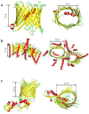
Figure 2
Figure 2
Mitochondrial β-barrel proteins. (a) VDAC-1 (porin) from H. sapiens (PDB:6TIQ; [43]). (b) TOM complex from H. sapiens (PDB:7VD2; [44]). (c) FhaC from E. coli (PDB:4QKY; [45,46,47]).
Figure 3
Figure 3
β-barrel pore-forming toxins. (a) α-hemolysin of S. aureus (PDB:4ANZ; [48,49,50]). (b) Anthrax toxin with lethal factor side and top view (PDB:6PSN; [51]). (c) γ–hemolysin from S. aureus (PDB:3B07; [9]). (d) Aerolysin prepore side and top view from A. hydrophila (PDB: 5JZH/5JZW; [12]). (e) MspA of M. smegmatis (PDB:1UUN; [52]).
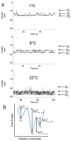
Figure 8
Figure 8
Temperature dependence of conductance substates of OpdK. (a) Single-channel electrical traces collected with the native OpdK at various temperatures. (b) A free energy landscape model illustrating the kinetic transitions among the O1, O2, and O3 open substates. This model shows the activation free energies characterizing various kinetic transitions (ΔGO1→O2, ΔGO2→O1, ΔGO2→O3, and ΔGO3→O2). This figure was adapted from Cheneke and coworkers (2015) [221].
Figure 4
Figure 4
Loop 6 is crucial for the gating dynamics of OmpG. (a) This is a cartoon representation of loop L6 of OmpG being anchored into the lipid bilayer via dodecylation at Cys226. (b) Representative single-channel electrical recordings using the wild-type OmpG (left panel) and an OmpG mutant with the loop L6 immobilized onto the lipid bilayer, as shown in (a) (right panel). This figure was adapted from Zhuang and Tamm (2014) [128].
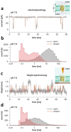
Figure 5
Figure 5
Gating evaluations of OmpG using single-molecule electrophysiology and high-speed AFM height spectroscopy (HS-AFM-HS). (a) A representative single-channel electrical trace of OmpG acquired at a transmembrane potential of +40 mV and pH 7.6. The schematic on the right side provides a scheme of the single-channel electrical recording experimental formulation. OmpG (yellow) is functionally reconstituted into a lipid bilayer (green). Potassium and chloride ions are indicated as red and blue spheres, respectively. The red arrow shows the direction of the ionic flow of cations at a positive applied potential. (b) A semilogarithmic dwell time histogram of the open and closed states, as determined by single-molecule electrophysiology. (c) A representative 60 ms long HS-AFM-HS recording that probes an OmpG protein functionally reconstituted into a lipid bilayer, which was suspended on mica at pH 7.6. The schematic on the right side is the HS-AFM-HS experimental setup. An AFM tip monitors conformational fluctuations of loop L6. (d) A semilogarithmic dwell time histogram of the open and closed states, as determined by HS-AFM-HS. Here, the low state indicates the open state, where the tip navigates within the pore lumen. The high state corresponds to the closed state, precluding the partitioning of the tip into the pore lumen. This figure was adapted from Sanganna Gari and coworkers (2021) [129].
Figure 6
Figure 6
A proposed model for voltage sensing of VDAC1. VDAC1 (in blue) remains in a 4 nS-conductance open state at a zero transmembrane potential (upper, left). Yet, at an amplified applied transmembrane potential greater than 30 mV, regardless of its polarity, an electric force is exerted on the N-terminal helix that acts as a voltage sensor (in red; center). L10 (in green) is the contact residue of the N-terminal helix with the V143 residue on the barrel wall. The reversible dissociation of the rigid N-terminal helix from the pore wall results in a more flexible structure, which is likely to switch the channel into a semi-collapsed, elliptical conformation that leads to a 2 nS conductance closed state (upper, right). The lower panel indicates the correlated values in the open and closed state unitary conductance and ionic selectivity. This figure was adapted from Zachariae and coworkers (2013) [136].
Figure 7
Figure 7
Direct experimental evidence for the implication of a key charged residue in the voltage-dependent gating of VDAC1. (a) Single-channel electrical recordings of mVDAC1 reveal the intense gating activity of the wild-type channel (left traces) but the drastically declined gating activity of the charge-reversal K12E mutant (right traces). Horizontal dashed lines show the zero current. (b) These panels indicate quantitative assessments of the gating activity of different VDAC1 proteins using a multichannel system. The vertical axis indicates the overall multichannel current normalized to the value corresponding to open-state multichannel conductance. The left panel compares the wild-type (WT) protein and the charge-reversal K12E mutant. The right panel compares the WT protein as well as the K12A and K12S mutants. This figure was adapted from Ngo and coworkers (2022) [161].

Similar articles

Cited by

References

    1. Pocanschi C.L., Kleinschmidt J.H. The Thermodynamic Stability of Membrane Proteins in Micelles and Lipid Bilayers Investigated with the Ferrichrom Receptor FhuA. J. Membr. Biol. 2022;255:485–502. doi: 10.1007/s00232-022-00238-w. - DOI - PMC - PubMed
    1. Horne J.E., Radford S.E. A growing toolbox of techniques for studying β-barrel outer membrane protein folding and biogenesis. Biochem. Soc. Trans. 2016;44:802–809. doi: 10.1042/BST20160020. - DOI - PMC - PubMed
    1. Chaturvedi D., Mahalakshmi R. Transmembrane β-barrels: Evolution, folding and energetics. Biochim. Biophys. Acta Biomembr. 2017;1859:2467–2482. doi: 10.1016/j.bbamem.2017.09.020. - DOI - PMC - PubMed
    1. Slusky J.S. Outer membrane protein design. Curr. Opin. Struct. Biol. 2017;45:45–52. doi: 10.1016/j.sbi.2016.11.003. - DOI - PMC - PubMed
    1. Thoma J., Sapra K.T., Müller D.J. Single-Molecule Force Spectroscopy of Transmembrane β-Barrel Proteins. Annu. Rev. Anal. Chem. 2018;11:375–395. doi: 10.1146/annurev-anchem-061417-010055. - DOI - PubMed

MeSH terms

LinkOut - more resources