. 2023 Aug 11;13(1):13079.
doi: 10.1038/s41598-023-38663-z.

Outside-in signaling through the major histocompatibility complex class-I cytoplasmic tail modulates glutamate receptor expression in neurons

Affiliations

Outside-in signaling through the major histocompatibility complex class-I cytoplasmic tail modulates glutamate receptor expression in neurons

Brett A Eyford et al. Sci Rep. .

Abstract

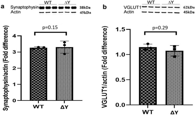
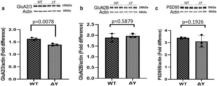
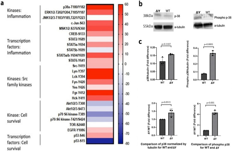
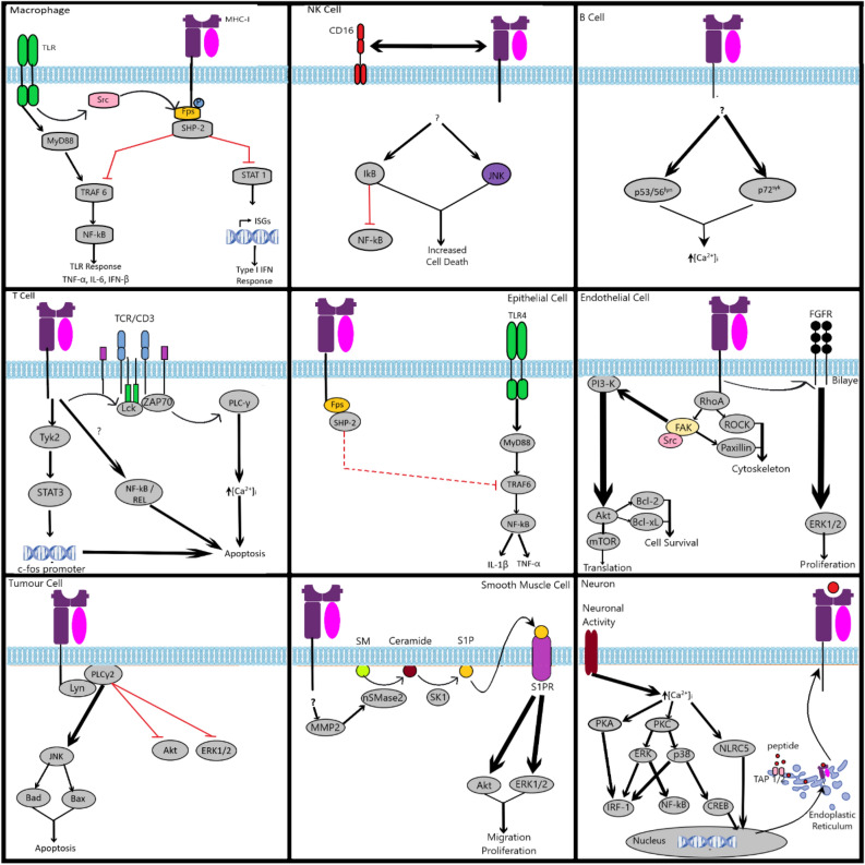
The interplay between AMPA-type glutamate receptors (AMPARs) and major histocompatibility complex class I (MHC-I) proteins in regulating synaptic signaling is a crucial aspect of central nervous system (CNS) function. In this study, we investigate the significance of the cytoplasmic tail of MHC-I in synaptic signaling within the CNS and its impact on the modulation of synaptic glutamate receptor expression. Specifically, we focus on the Y321 to F substitution (Y321F) within the conserved cytoplasmic tyrosine YXXΦ motif, known for its dual role in endocytosis and cellular signaling of MHC-I. Our findings reveal that the Y321F substitution influences the expression of AMPAR subunits GluA2/3 and leads to alterations in the phosphorylation of key kinases, including Fyn, Lyn, p38, ERK1/2, JNK1/2/3, and p70 S6 kinase. These data illuminate the crucial role of MHC-I in AMPAR function and present a novel mechanism by which MHC-I integrates extracellular cues to modulate synaptic plasticity in neurons, which ultimately underpins learning and memory.


Conflict of interest statement

The authors declare no competing interests.

Figures

Figure 1
Figure 1
The ΔY mutation has no effect on the expression of pre-synaptic proteins in the hippocampus. Representative Western blots and analysis of integrated fluorescent intensities of pre-synaptic proteins, synaptophysin and VGlut1, from WT and ΔY mice, n = 3 independent experiments. Each lane for the Western blots represents a different mouse. The data was pooled from each group in the graphs, and statistical analysis was performed between each group. All proteins were normalized to actin, and values represent means ± SEM. (a) Synaptophysin has an apparent molecular weight of 38 kDa, and beta-actin has an apparent molecular weight of 45 kDa. There is no statistical difference between protein expression in VGlut1 expression in ΔY compared to WT mice (p = 0.29). (b) Based on the standard of molecular weight (MW) markers proteins, VGLUT1 has apparent molecular weight of 62 kDa. There is no statistical difference between protein expression in synaptophysin expression in ΔY compared to WT mice (p = 0.15) using an unpaired t test. The full-gels and their analysis are provided in the Supplementary figures, including the migration of each protein relative the MW ladder.
Figure 2
Figure 2
The ΔY mutation reduces the expression of AMPA receptors. Representative Western blots and analysis of integrated fluorescent intensities of post-synaptic proteins GluA2/3, GluN2B, and PSD95 in ΔY and WT mice, n = 3. (a) There was an observed significant decrease in GluA2/3 in ΔY mice compared to WT mice (p = 0.0078) using an unpaired t test. (b) No change in GluN2B in ΔY mice compared to WT mice (p = 0.5879) using an unpaired t test. (c) No changes in PSD95 expression in ΔY mice compared to WT mice (p = 0.1926) using an unpaired t test. All proteins were normalized to beta-actin and values represent means ± SEM. Each lane for the western blots represents a different mouse. The data was pooled from each group in the graphs, and statistical analysis was performed between each group. Based on the standard of molecular weight markers, GluA2/3 has an apparent molecular weight of 100 kDa, GluN2b has an apparent molecular weight of 190 kDa, PSD95 has an apparent molecular weight of 95 kDa, and actin has an apparent molecular weight of 45 kDa. The full-gels and their analysis is provided in the Supplementary figures, including the migration of each protein relative to the MW ladder.
Figure 3
Figure 3
The ΔY mutation alters phosphorylation of various kinases and transcription factors. (a) Relative changes in protein kinase phosphorylation levels of target proteins in brain tissue lysates from ΔY and WT mice represented in a heat map as a ratio of the ΔY/WT phosphokinase levels. Increased phosphorylation was observed in various kinases and transcription factors involved in inflammation and members of the Src family kinases with roles in neuronal and synaptic plasticity, while decreased phosphorylation was observed in kinases involved in cell survival. Kinase phosphoarray assays were performed in two animals and analyzed in duplicate in each kinase assay before establishing the means that are shown in the heatmap. (b) Validation of the protein phosphoarray by western blot analysis for p38a T180/Y182. Based on the standard molecular weight markers, the control alpha-tubulin has an apparent molecular weight of 55 kDa, and p38 has an apparent molecular weight of 38 kDa. (c) Expression levels of phosphorylated and non-phosphorylated p38 in brain tissue lysates from ΔY and WT mice, represented as mean ± SEM. All comparisons were analyzed using a paired t test where p < 0.05 was consider significant. The antibodies used were Anti-p38 alpha/MAPK14 antibody, Anti-p38 (phospho T180 + Y182) antibody and Anti-alpha Tubulin antibody). Secondary antibodies: Goat Anti-Mouse IgG H&L (HRP) preabsorbed (ab97040) and Invitrogen. Goat anti-Rabbit IgG (H + L) Secondary Antibody, HRP (31460) The full-gels and their analysis is provided in the Supplementary figures, including the migration of each protein relative the MW ladder.
Figure 4
Figure 4
Summary of reverse MHC-I signalling pathways in various cell types. Summary of the main reverse (inside-out) MHC-I signalling pathways in immune and non-immune cells. A more detailed description can be found in a review by Valenzuela and Reed as well as Muntjewerff et al..

Similar articles

References

    1. Corriveau RA, Huh GS, Shatz CJ. Regulation of class I MHC gene expression in the developing and mature CNS by neural activity. Neuron. 1998;21(3):505–520. - PubMed
    1. Huh GS, Boulanger LM, Du H, Riquelme PA, Brotz TM, Shatz CJ. Functional requirement for class I MHC in CNS development and plasticity. Science. 2000;290(5499):2155–2159. - PMC - PubMed
    1. Goddard CA, Butts DA, Shatz CJ. Regulation of CNS synapses by neuronal MHC class I. Proc. Natl. Acad. Sci. U.S.A. 2007;104(16):6828–6833. - PMC - PubMed
    1. Needleman LA, Liu XB, El-Sabeawy F, Jones EG, McAllister AK. MHC class I molecules are present both pre- and postsynaptically in the visual cortex during postnatal development and in adulthood. Proc. Natl. Acad. Sci. U.S.A. 2010;107(39):16999–17004. - PMC - PubMed
    1. Hu Y, Pei W, Hu Y, Li P, Sun C, Du J, et al. MiR34a regulates neuronal MHC class I molecules and promotes primary hippocampal neuron dendritic growth and branching. Front. Cell. Neurosci. 2020;14:573208. - PMC - PubMed

Publication types

MeSH terms