Review
. 2023 Aug 31;51(4):1505-1520.
doi: 10.1042/BST20221238.

New insights into the mechanochemical coupling mechanism of kinesin-microtubule complexes from their high-resolution structures

Affiliations
Review

New insights into the mechanochemical coupling mechanism of kinesin-microtubule complexes from their high-resolution structures

Matthieu P M H Benoit et al. Biochem Soc Trans. .

Abstract

Kinesin motor proteins couple mechanical movements in their motor domain to the binding and hydrolysis of ATP in their nucleotide-binding pocket. Forces produced through this 'mechanochemical' coupling are typically used to mobilize kinesin-mediated transport of cargos along microtubules or microtubule cytoskeleton remodeling. This review discusses the recent high-resolution structures (<4 Å) of kinesins bound to microtubules or tubulin complexes that have resolved outstanding questions about the basis of mechanochemical coupling, and how family-specific modifications of the motor domain can enable its use for motility and/or microtubule depolymerization.

Keywords: cryo-electron microscopy; crystallography; kinesin; microtubule; molecular motors; tubulin.


Conflict of interest statement

The authors declare that there are no competing interests associated with the manuscript.

Figures
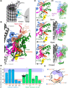
Figure 1.
Figure 1.. Kinesin motor domain conformations.
(a) Illustration of a typical kinesin with microtubule plus-end directed motility walking on a microtubule. These kinesins in general contain two polypeptide chains with the motor domain (also called head, blue) located at the N-terminal end. Next in the sequence after the motor domain is the neck-linker (red), followed by an α-helical coiled coil segment, the neck coil (NC, bright green). Following there are additional coiled coil segments (CC1, green) and regions that vary between kinesin families in the number and length of coiled coil segments and other sequence motifs (dashed lines). At the C-terminal end the tail domain (TD) also varies between kinesins. The neck-coil (NC) and other coiled coil dimerization regions place the two motor domains in close proximity and connected through their neck-linkers. (b) Cartoon representation of the atomic model of a microtubule bound kinesin motor domain (KIF14, PDBid: 6WWO) with distinct functional and secondary structure regions highlighted in different colors. Loop-8 (L8), loop-12 (L12) and helix-4 (H4) in cornflower-blue; Switch-1 in deep-pink; Switch-2 in magenta; nucleotide in orange-red; helix-2 (H2), helix-3 (H3); helix-6 (H6) and the P-loop in forest-green; helix-0 (H0) in yellow; loop-2 (L2) in medium-purple; central β-sheet in light-salmon; neck-linker in crimson-red; α-tubulin in light-gray; β-tubulin in gray. (c) KIF14 motor domain nucleotide-binding pocket close-up views in the semi-closed, open, and closed conformations (PDBid: 4OZQ, 6WWM and 6WWO). Arrows indicate the distance between H4 and H0. Regions colored as in (a). (d) KIF14 motor domain subdomains. These are areas within the motor domain that move relative to each other (green and magenta arrows) as the motor transition between the semi-closed, open, and closed conformations. Each subdomain is enclosed by a semitransparent surface. Plus subdomain (Plus SD) in cornflower blue; Switch subdomain (Switch SD) in magenta and minus subdomain (Minus SD) in forest green. (e) Nucleotide pocket openness (NPO) is expressed as the average distance between α-carbon atoms of six pairs of highly conserved residues across the nucleotide-binding pocket (black lines shown in paned (c)). Residues used are listed in Supplementary Table S1. Each symbol in the plot corresponds to an NPO value calculated from the coordinates of an atomic model and each bar represents the mean of the NPO values in the column. Each column corresponds to a particular kinesin motor domain nucleotide state ADP, Apo, AMPPNP or ADP-AlFx (labeled ADP, APO, ANP and AAF respectively) and the motor domain is microtubule (MT), or curved tubulin (CT) bound. Blue bars correspond to the KIF14 crystal structure and KIF14 microtubule bound cryo-EM structures in the single head bound states. Green bars correspond to the kinesin motor domain crystal structures deposited in the protein data bank except mutant versions and drug bound structures. The NPO values of the first kinesin motor domain high-resolution structures in an open conformation and in the closed catalytic competent conformation are shown respectively with a red diamond and a red triangle symbol (PDBid: 4LNU and 3HQD). The semitransparent blue and red horizontal bars represent the range of values encompassing structures we identified in the open and closed states [16,32,33] (18.3 to 17.2 Å and 15.1 to 14.2 Å, respectively). Values in between these two ranges encompass the semi-closed conformation. The residue pair distances and the NPO values for the structures in this plot and all the ones deposited in the protein databank are listed in Supplementary Table S1. (f) Basic mechanochemical cycle for a single motile kinesin motor domain when the neck-linker is free to dock or undock unencumbered by the partner motor domain or by other kinesin-family specific structural elements. Model structure figures were prepared with Blender (panel a) and UCSF-ChimeraX [70].
Figure 2.
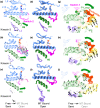
Figure 2.. Microtubule-binding-induced conformational changes in the motor domain of three distinct kinesins.
(ac) View of the microtubule binding region of three ADP-kinesin X-ray crystal structures, kinesin-3 MmKIF14 (a), kinesin-1 HsKIF5B (b), and kinesin-8 CaKIP3 (c). Arrows indicate the direction of structural changes to corresponding microtubule bound open structures (semi-closed to open conformational change). The structures are shown as semitransparent ribbons using the same color scheme as in Figure 1b. Black arrows correspond to the displacement vectors of Cα atoms from the X-ray crystal motor domain structure to the microtubule bound motor domain structure after aligning the plus subdomain (blue colored area) of both structures. For clarity arrows corresponding to the switch-2 region are not shown. (d–f) Views of the kinesin microtubule interface in the microtubule-bound open conformation of the same three kinesins shown in ac. (gi) Same structures and vectors shown in ac but viewed from the bottom of the minus subdomain showing part of the microtubule interface and the nucleotide pocket. The PDBid corresponding to the semi-closed and microtubule bound open structures shown or compared in each row are, respectively: 4OZQ and 6WWM (row 1 panels a, d, g), 1BG2 and 8OJQ (row 2 panels b, e, h), 7LFF and 7TQZ (row 3 panels c, f, i).
Figure 3.
Figure 3.. Relationship between neck-linker conformations and nucleotide pocket openness in a motile kinesin-3.
(a and b) Model of the two-head-bound structure of the kinesin-3 MmKIF14 (PDBid: 6WWL), obtained by cryo-EM (resolution of the kinesin part of the map: 3.3 Å). Both heads are AMPPNP bound. Key parts of KIF14 leading and trailing heads and of the microtubule are labeled with the same color scheme as in Figure 1b. Microtubule polarity is indicated with + and − signs. The views in (a) and (b) are rotated 180° to each other to emphasize respectively the distinct positions of the neck-linkers and the distinct conformations of the nucleotide-binding pockets. (c) Comparison of the neck linker position in four microtubule-bound KIF14 constructs (K) bound to AMPPNP and differing by the length of their C-termini. The number of the last residue of each construct as well as the number of neck-linker residues are indicated. K735: no neck-linker, K743 has the first eight of 15 neck-linker residues, K748 has the full neck-linker. K755 has the full neck-linker and the first coiled-coil residues and is a two-heads-bound state on the microtubule. Semi*: The K735-ANP structure (microtubule bound KIF14 with no neck-linker in the presence of AMPPNP, PDBid: 6WWV) was previously called open* but due to its slightly more closed nucleotide-binding pocket than other structures in the open conformation [16], it falls within the semi conformation group according to the NPO boundaries defined in Figure 1. (d) Quantification of the nucleotide pocket openness of several KIF14 constructs differing by the length of their C-termini, in both AMPPNP and ADP-AlFx. Note that only the constructs with a full neck-linker assume the fully closed state seen in the trailing head of the two heads-bound-states.
Figure 5.
Figure 5.. Coordinated kinesin mechanochemical cycles.
(a) Mechanochemical cycle of one of the motor domains (head-1) of kinesin dimer. The partner motor domain (head-2) has a similar cycle to head-1 but synchronized out of phase so that when head-1 is in the open leading conformation head-2 is in the closed trailing conformation and vice versa. (b) Mechanochemical cycle of a kinesin-8 combining motile and microtubule depolymerization activities. When interacting with the microtubule the mechanochemical cycle is similar to the one of motile kinesins (a) but the interaction of loop-2 with the microtubule prevents nucleotide-binding pocket closure with ATP binding but it occurs with ATP hydrolysis. At microtubule ends, the loop-2 tubulin interaction in the ATP bound state promotes tubulin curvature and microtubule depolymerization. Whether the two motor domains of kinesin-8 dimer have tightly coordinated mechanochemical cycles as proposed for motile kinesins (a) remains to be investigated. (c) Mechanochemical cycle of microtubule depolymerase kinesin-13. In this kinesin's unbiased one-dimensional diffusion, rather than unidirectional motility, occurs when interacting with the microtubule lattice. As in the case of kinesin-8s, the interaction of loop-2 with tubulin prevents ATP binding induced closure of the nucleotide-binding pocket when bound to the microtubule lattice. When bound at microtubule ends, ATP binding results in nucleotide-binding pocket closure with stabilization and/or induction of tubulin curvature.
Figure 4.
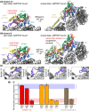
Figure 4.. Modulation of the kinesin mechanochemical cycle for microtubule depolymerization.
(a) Kinesin-13 (KLP10A, PDBid: 6B0L) adopts an open state on the straight microtubule when bound to AMPPNP (left panel). On curved tubulin protofilaments, kinesin-13 (KLP10A, PDBid: 6B0C and KIF2A, PDBid: 6BBN, the latter illustrated) adopts a closed state when bound to AMPPNP (right panel). (b) Kinesin-8 (CaKip3, PDBid: 7TQX) adopts an open state on the straight microtubule when bound to AMPPNP (left panel). On curved tubulin protofilaments, kinesin-8 (CaKip3, PDBid: 7TR3) adopts a closed state when bound to AMPPNP (right panel). Minus-end kinesin-8 neighbor shown in faded colors to highlight cooperative contacts. (c) Close-up views of loop-2 from separate kinesin families to illustrate different loop-2 structures and tubulin contacts. From left to right: KLP10A — PDBid: 6B0L, CaKip3 — PDBid: 7TQX, Pb kinesin 8b — PDBid: 7Z2A, KIF14 — PDBid: 6WWI. (d) Nucleotide-binding pocket openness measurements for high-resolution structures of kinesin-13 and kinesin-8 motors. Measurements performed using same residues as Figures 1 and 3.

Similar articles

Cited by

References

    1. Schliwa, M. and Woehlke, G. (2003) Molecular motors. Nature 422, 759–765 10.1038/nature01601 - DOI - PubMed
    1. Verhey, K.J., Cochran, J.C. and Walczak, C.E. (2015) The Kinesin Superfamily. In Kinesins and Cancer (Kozielski, F.S.B.F., ed.), pp. 1–26, Springer Netherlands, Dordrecht
    1. Lawrence, C.J., Dawe, R.K., Christie, K.R., Cleveland, D.W., Dawson, S.C., Endow, S.A.et al. (2004) A standardized kinesin nomenclature. J. Cell Biol. 167, 19–22 10.1083/jcb.200408113 - DOI - PMC - PubMed
    1. Wickstead, B., Gull, K. and Richards, T.A. (2010) Patterns of kinesin evolution reveal a complex ancestral eukaryote with a multifunctional cytoskeleton. BMC Evol. Biol. 10, 110 10.1186/1471-2148-10-110 - DOI - PMC - PubMed
    1. Pandey, H., Popov, M., Goldstein-Levitin, A. and Gheber, L. (2021) Mechanisms by which kinesin-5 motors perform their multiple intracellular functions. Int. J. Mol. Sci. 22, 6420 10.3390/ijms22126420 - DOI - PMC - PubMed

Publication types