. 2023 Nov;290(22):5322-5339.
doi: 10.1111/febs.16925. Epub 2023 Aug 17.

Cardiomyocyte external mechanical unloading activates modifications of α-actinin differently from sarcomere-originated unloading

Affiliations

Cardiomyocyte external mechanical unloading activates modifications of α-actinin differently from sarcomere-originated unloading

Christopher Solís et al. FEBS J. 2023 Nov.

Abstract

Loss of myocardial mass in a neonatal rat cardiomyocyte culture is studied to determine whether there is a distinguishable cellular response based on the origin of mechano-signals. The approach herein compares the sarcomeric assembly and disassembly processes in heart cells by imposing mechano-signals at the interface with the extracellular matrix (extrinsic) and at the level of the myofilaments (intrinsic). Experiments compared the effects of imposed internal (inside/out) and external (outside/in) loading and unloading on modifications in neonatal rat cardiomyocytes. Unloading of the cellular substrate by myosin inhibition (1 μm mavacamten), or cessation of cyclic strain (1 Hz, 10% strain) after preconditioning, led to significant disassembly of sarcomeric α-actinin by 6 h. In myosin inhibition, this was accompanied by redistribution of intracellular poly-ubiquitin K48 to the cellular periphery relative to the poly-ubiquitin K48 reservoir at the I-band. Moreover, loading and unloading of the cellular substrate led to a three-fold increase in post-translational modifications (PTMs) when compared to the myosin-specific activation or inhibition. Specifically, phosphorylation increased with loading while ubiquitination increased with unloading, which may involve extracellular signal-regulated kinase 1/2 and focal adhesion kinase activation. The identified PTMs, including ubiquitination, acetylation, and phosphorylation, are proposed to modify internal domains in α-actinin to increase its propensity to bind F-actin. These results demonstrate a link between mechanical feedback and sarcomere protein homeostasis via PTMs of α-actinin that exemplify how cardiomyocytes exhibit differential responses to the origin of force. The implications of sarcomere regulation governed by PTMs of α-actinin are discussed with respect to cardiac atrophy and heart failure.

Keywords: HCM; HFrEF; calponin homology domain; myotrope; sarcopenia.


Conflict of interest statement

Conflict of interest

RJS is a member of the scientific advisory board of Cytokinetics and a consultant to Pfizer and Edgewise Therapeutics. The remaining authors declare no competing financial interests.

Figures

Fig. 1.
Fig. 1.
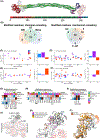
Experimental Approach to study the response of cardiac muscle cells to inside-out and outside-in forces. Two experimental paradigms of mechanical force stimulation were tested to study the effects of mechanical forces on cardiac sarcomere assembly and disassembly and the associated chemical and biophysical changes. The first approach was an intrinsic stimulus elicited by drugs that modify myosin contractility (i.e. inside-out). The second approach was an extrinsic stimulus from varying strain applied to the cardiomyocyte attachment substrate (i.e. outside-in). The inside-out approach consisted of treating NRVMs with the myosin inhibitor Mavacamten (1 μM, 6 h) or the myosin activator Omecamtiv Mecarbil (0.5 μM, 6 h). The outside-in approach consisted of 1 Hz cyclic strains for 24 h (loaded) followed by 6 h rest (unloaded) of cultured neonatal rat ventricular myocytes (NRVMs) and compared with continuously strained cultures. These two approaches were compared by quantitative immunofluorescence, targeted proteomics of α-actinin, and fluorescence recovery after photobleaching experiments (FRAP).
Fig. 2.
Fig. 2.
Sarcomere disassembly begins at the outer annulus of the cell with diminished myosin tension. (A) Untreated (UT) NRVMs were compared with NRVMs treated with OM (0.5 M) or Mava (1 μM) at 12 h after fixing and staining with α-actinin (magenta) and phalloidinrhodamine (top panels) or α-actinin (magenta) and oligo-Ub-K48 (green) antibodies (bottom panels) (scale, 20 μm). (B) Sarcomeric α-actinin content per cell area was quantified for NRVMs treated with OM and Mava over the course of 12 h. (C) The distribution of oligo-K48 ubiquitin between the non-striated annulus 5 μm from the cell edge (outer) and central sarcomeric region (inner) was quantified. (D) The oligo-Ub-K48 content of the outer relative to middle between UT, OM, and Mava (error bars indicate mean ± standard deviation; 27 measurements from independent cells per treatment, or nine quantified cells from each N = 3 independent cell culture; multicomparison tests use Tukey’s honest significance test).
Fig. 3.
Fig. 3.
Diminished myosin tension increases α-actinin and CapZ exchange dynamics. The dynamic exchange of the Z-disc proteins α-actinin and CapZ in response to chemical unloading were probed in NRVMs infected with adenoviral constructs expressing α-actinin-yellow fluorescent protein (YFP) or CapZβ1-GFP. (A) The sarcomeric (inner) and non-sarcomeric (outer) fluorescent protein pools were photobleached as in (B) while tracking the time-dependent recovery of fluorescence from dynamic exchange of proteins at the Z-discs (12 min imaging per region of interest). The FRAP recovery kinetics (kFRAP) are summarized for α-actinin-YFP (C) and CapZβ1-GFP (D) in response to treatments with OM and Mava for 6 h (error bars indicate mean ± standard deviation, 27 measurements from independent cells per treatment, or nine quantified cells from each N = 3 independent cell culture; multicomparison tests use Tukey’s honest significance test).
Fig. 4.
Fig. 4.
Sarcomere assembly due to external loading is reversed by unloading. (A) Unflexed (UF) NRVMs are compared with NRVMs treated with 24 h flexing (F), or 24 h flexing followed by 6 h rest (F-UF) after fixing and staining with α-actinin and oligo-Ub-K48 antibodies (scale, 20 μm). (B) α-actinin content per cell area shows significant gain in sarcomeric α-actinin after F (P < 0.001) and significant loss of sarcomeric α-actinin after the F-UF treatment (P < 0.01). (C) The proportion of the oligo-Ub-K48 content in the outer 5 μm annulus is not significantly different between UF, F, and F-UF outer relative to inner regions (error bars indicate mean ± standard deviation; 27 measurements from independent cells per treatment, or nine quantified cells from each N = 3 independent cell culture; multicomparison tests use Tukey’s honest significance test).
Fig. 5.
Fig. 5.
Sarcomeric oligo-K48-linked ubiquitin localizes at the I-band and increases with unloading from myosin inhibition. Super-resolution fluorescence immunostains depict oligo-Ub-K48 distribution (green) relative to (A) α-actinin, (B) myosin, and (C) cMyBP-C (magenta). The dashed squares indicate the location of the magnified images on the right (Scale bar of enlarged and magnified images is 10 and 2 μm respectively). White arrows 10 μm long show direction and fluorescence intensity profile of ubiquitin-K48 (green) and the selected sarcomeric proteins (magenta). (D) Quantification of the I-band oligo-Ub-K48 relative to the A-band oligo-Ub-K48 fixed at 6 h after treatment (error bars indicate mean ± standard deviation; 27 measurements from independent cells per treatment, or 9 quantified cells from each N = 3 independent cell culture; multicomparison tests use Tukey’s honest significance test).
Fig. 6.
Fig. 6.
Post-translational content of α-actinin reveals alterations of actin-binding interface and protein degradation sites. (A) Crystal structure of α-actinin-2 (PDB: 4D1E) Displaying color-coded domains and their arrangement across the full sequence (ABD, red; neck region, yellow; spectrin-like repeats or rod regions, green; CaMDs composed of EF-hand motifs, magenta and purple). (B) The mechanical intervention (outside-in: F, F-UF) causes more post-translational modification changes compared with the chemical intervention (inside-out: Mava, OM). (C–F) Log2 fold changes in acetylation, ubiquitination, and phosphorylation are displayed across the rat α-actinin-2 linear sequence (NCBI ref. seq.: NP_001163796.1). (C) OM and (D) Mava Log2 fold change relative to UT are presented across the linear sequence and summarized in frequency histograms. (E) F and (F) F-UF a Log2 fold change relative to UF are presented across the linear sequence and summarized in frequency histograms. Heatmap analysis of (G) modified residues found in all the treatments, (H) modified residues only found in the outside-in dataset, and (I) residues only found in the inside-out dataset. Top lines show modification of each residue and predicted degron sites. (J) Homology model of the actin binding domain (ABD) of α-actinin-2 (template, PDB ID 7R94) illustrates the spatial arrangement of CH1 and CH2 domains relative to actin, and (K) a close view depicting modified residues from G–I. (L) ABD models of actin-free (template, PDB ID 4D1E) and actin-bound (template, PDB ID 7R94) α-actinin-2 show relative displacement of CH1 and CH2 domains by arrow and spatial rearrangements of S237. Models were prepared for illustration in UCSF Chimera (version 1.6, build 42360).
Fig. 7.
Fig. 7.
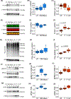
Phosphorylation, ubiquitination, ERK, and focal adhesion kinase (FAK) activity are differentially sensitive to internal and external loading and unloading. (A) Representative Acetyl-lysine and α-actinin western blots of NRVM cell lysates under untreated (UT), OM, Mava, unflexed (UF), flexed (F), and flexed-unflexed (F-UF) conditions. (B, C) Quantification of the acetyl-lysine content relative to α-actinin. (D) Representative phosphoserine western blot overlays with α-actinin for the same samples as in (A). (E, F) Quantification of the phosphoserine content relative to α-actinin. (G) Representative oligo-Ub-K48 and α-actinin western blots for the same samples as in (A). (H, I) Quantification of the oligo-Ub-K48 content relative to α-actinin. (J) Representative pERK1/2, total ERK1/2 western blots for the same samples as in (A). (K, L) Quantification of the pERK1/2 content relative to total ERK1/2. (M) Representative pFAK, and total FAK western blots for the same samples as in (A). (N, O) Quantification of pFAK relative to the total FAK. H2B is used as a loading control. N = 4 independent cell cultures for all the treatments; multicomparison tests use Tukey’s honest significance test.
Fig. 8.
Fig. 8.
Control of sarcomere assembly via regulation of α-actinin stabilization of sarcomeres in response to loading and unloading. (A) Sarcomere growth is supported by the increased cross-linking activity of structural proteins like α-actinin-2 that bundle the thin filaments forming the Z-discs, shown in an unloaded (left) or loaded sarcomere (right). (B) α-actinin-2 is shown at the molecular scale as loosely bound to actin (left). Tight binding is promoted by increasing loading through both mechanical flexing or myosin activators with similar PTMs phosphorylation (P) and acetylation (Ac). Signaling pathways are proposed to modify α-actinin at the interphase between the CH1 and CH2 domains to increase CH1 availability to bind F-actin. However, the ubiquitination (Ub) is only greater with recovery after flexing; it is not seen with myosin inhibition at the level of the sarcomere.

Similar articles

Cited by

References

    1. Maggioni MA, Castiglioni P, Merati G, Brauns K, Gunga H-C, Mendt S, Opatz OS, Rundfeldt LC, Steinach M, Werner A et al. (2018) High-intensity exercise mitigates cardiovascular deconditioning during long-duration bed rest. Front Physiol 9, 1553. - PMC - PubMed
    1. Sy MR, Keefe JA, Sutton J & Wehrens XHT (2022) Cardiac function, structural, and electrical remodeling by microgravity exposure. Am J Physiol Heart Circ Physiol 324, H1–H13. - PMC - PubMed
    1. Hoffmann B, Dehkordi P, Khosrow-Khavar F, Goswami N, Blaber AP & Tavakolian K (2022) Mechanical deconditioning of the heart due to long-term bed rest as observed on seismocardiogram morphology. NPJ Microgravity 8, 25. - PMC - PubMed
    1. Kim GH, Uriel N & Burkhoff D (2018) Reverse remodelling and myocardial recovery in heart failure. Nat Rev Cardiol 15, 83–96. - PubMed
    1. Kyriakopoulos CP, Kapelios CJ, Stauder EL, Taleb I, Hamouche R, Sideris K, Koliopoulou AG, Bonios MJ & Drakos SG (2022) LVAD as a bridge to remission from advanced heart failure: current data and opportunities for improvement. J Clin Med 11, 3542. - PMC - PubMed

Publication types

Associated data

LinkOut - more resources