. 2023 Aug 1;9(1):279.
doi: 10.1038/s41420-023-01563-2.

Bellidifolin ameliorates isoprenaline-induced cardiac hypertrophy by the Nox4/ROS signalling pathway through inhibiting BRD4

Affiliations

Bellidifolin ameliorates isoprenaline-induced cardiac hypertrophy by the Nox4/ROS signalling pathway through inhibiting BRD4

Dingyan Zhou et al. Cell Death Discov. .

Abstract

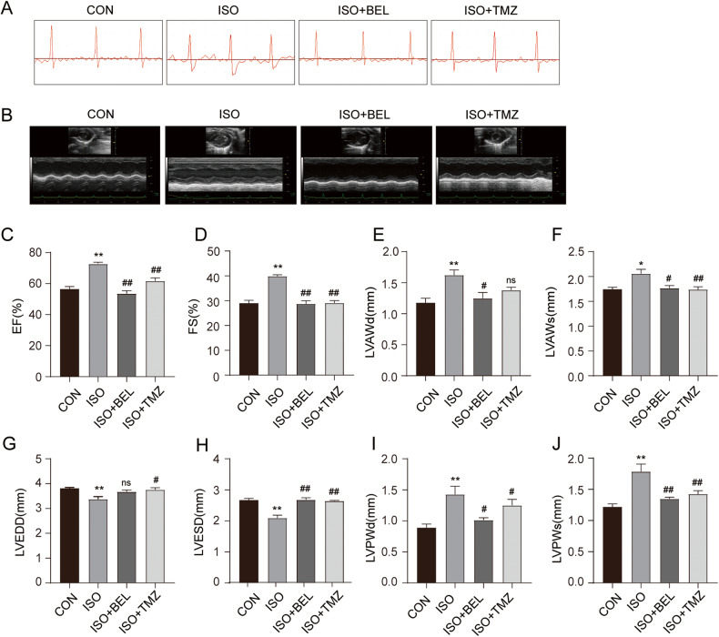
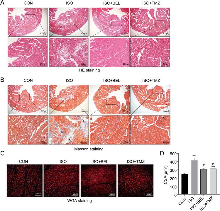
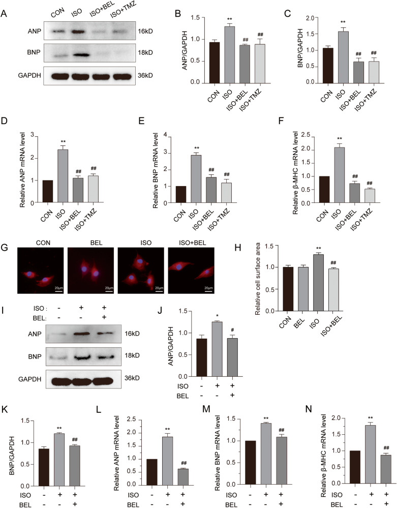
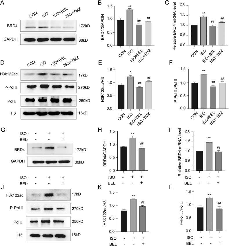
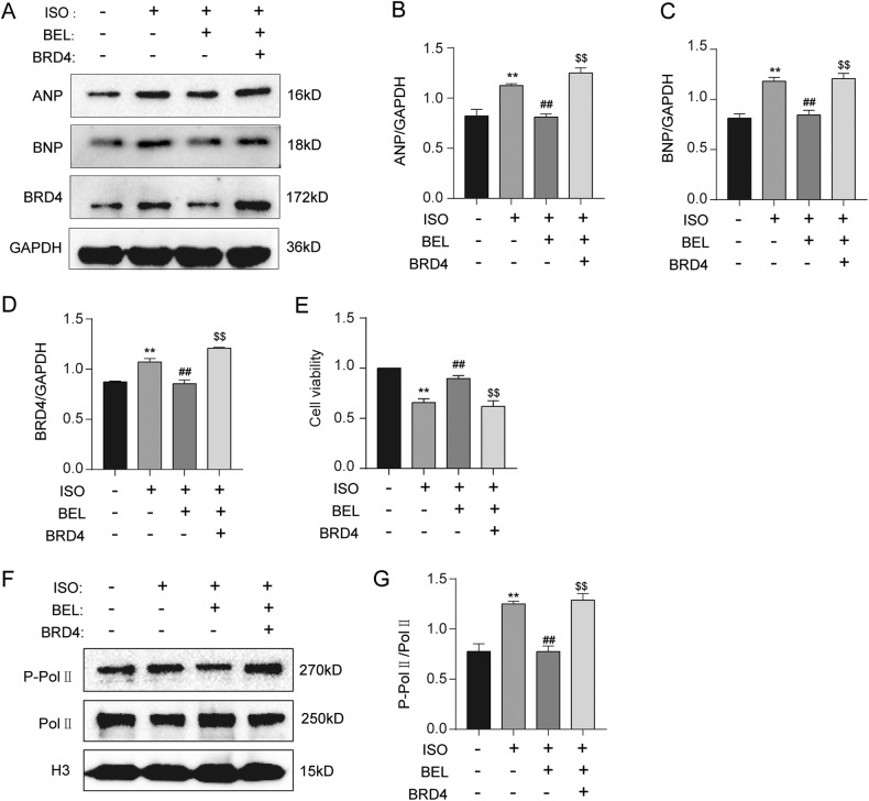
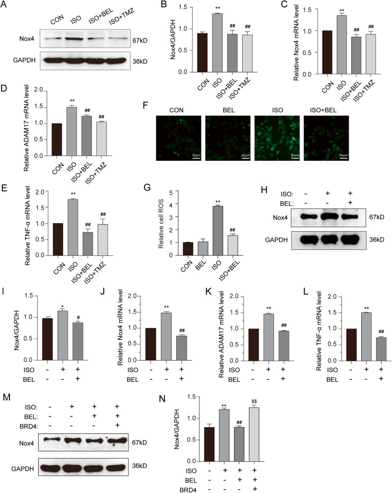
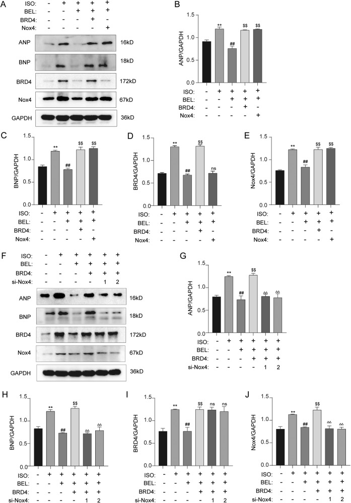
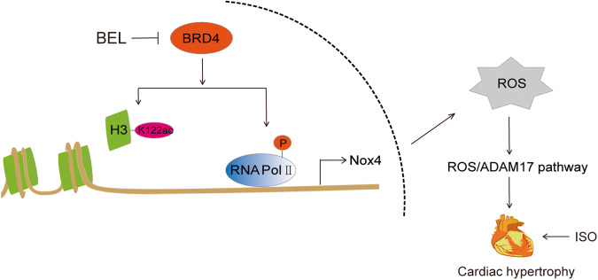
To date, there is no effective therapy for pathological cardiac hypertrophy, which can ultimately lead to heart failure. Bellidifolin (BEL) is an active xanthone component of Gentianella acuta (G. acuta) with a protective function for the heart. However, the role and mechanism of BEL action in cardiac hypertrophy remain unknown. In this study, the mouse model of cardiac hypertrophy was established by isoprenaline (ISO) induction with or without BEL treatment. The results showed that BEL alleviated cardiac dysfunction and pathological changes induced by ISO in the mice. The expression of cardiac hypertrophy marker genes, including ANP, BNP, and β-MHC, were inhibited by BEL both in mice and in H9C2 cells. Furthermore, BEL repressed the epigenetic regulator bromodomain-containing protein 4 (BRD4) to reduce the ISO-induced acetylation of H3K122 and phosphorylation of RNA Pol II. The Nox4/ROS/ADAM17 signalling pathway was also inhibited by BEL in a BRD4 dependent manner. Thus, BEL alleviated cardiac hypertrophy and cardiac dysfunction via the BRD4/Nox4/ROS axes during ISO-induced cardiac hypertrophy. These findings clarify the function and molecular mechanism of BEL action in the therapeutic intervention of cardiac hypertrophy.


Conflict of interest statement

The authors declare no competing interests.

Figures

Fig. 1
Fig. 1. BEL improved cardiac function in ISO-treated mice.
A Representative images of ECG tracings in the different groups. B Representative images of ultrasound in different groups. CJ Changes in EF, FS, LVAWd, LVAWs, LVEDD, LVESD, LVPWd, and LVPWs in the different groups (n = 6). Data are shown as the mean ± SEM. Significance: *p < 0.05 vs CON group; **p < 0.01 vs CON group; # p < 0.05 vs ISO group; ## p < 0.01 vs ISO group; ns no significant difference.
Fig. 2
Fig. 2. BEL alleviated cardiac histopathological change and cardiomyocyte hypertrophy in ISO-treated mice.
A Representative images of HE staining of left ventricular tissue in different groups. B Representative images of Masson trichrome staining of left ventricular tissue in different groups. C Representative images of WGA staining of left ventricular tissue in different groups. D Bar graph showing the changes in cardiomyocyte cross-sectional area. Data are shown as the mean ± SEM. Significance: *p < 0.05 vs CON group; **p < 0.01 vs CON group; #p < 0.05 vs ISO group; ##p < 0.01 vs ISO group.
Fig. 3
Fig. 3. BEL reversed cardiomyocyte hypertrophy induced by ISO in vitro and in vivo.
AC Protein expression levels of ANP and BNP in cardiac tissues are detected via western blotting and quantitative analysis of the relative protein expression of ANP and BNP. GAPDH is used as a loading control. (n = 3). DF The mRNA levels of ANP, BNP, and β-MHC in mice are measured by Quantified RT-PCR in each group, and relative mRNA levels normalise to GAPDH (n = 3). GH The cell surface area was measured by rhodamine-phalloidin staining in H9C2 cells. IK The protein level of ANP and BNP were determined in H9C2 cells by Western blot (n = 3). LN The mRNA levels of ANP, BNP, and β-MHC are measured by Real-time PCR (n = 3). Data are shown as the mean ± SEM. Significance: *p < 0.05 vs CON group; **p < 0.01 vs CON group; #p < 0.05 vs ISO group; ##p < 0.01 vs ISO group.
Fig. 4
Fig. 4. BEL decreased BRD4 protein and its functions induced by ISO.
A, B The protein levels of BRD4 were determined by Western blot in cardiac tissues. GAPDH was used as a loading control (n = 3). C The mRNA levels of BRD4 were measured by qRT-PCR in cardiac tissues, and relative mRNA levels normalise to GAPDH (n = 3). DF Western blot analysis was conducted to determine the phosphorylation of RNA Pol II, total expression and acetylation of K122 in cardiac tissues. H3 was used as a loading control (n = 3). GH The protein levels of BRD4 were determined by Western blot in H9C2 cells. GAPDH was used as a loading control (n = 3). I The mRNA levels of BRD4 were measured by qRT-PCR, and relative mRNA levels normalise to GAPDH (n = 3). JL Western blot was conducted to determine the phosphorylation of RNA Pol II, total H3 expression and acetylation of K122 in H9C2 cells (n = 3). Data are shown as the mean ± SEM. Significance: *p < 0.05 vs CON group; **p < 0.01 vs CON group; #p < 0.05 vs ISO group; ##p < 0.01 vs ISO group; ns no significant difference.
Fig. 5
Fig. 5. BEL inhibited ISO-induced elevation of BRD4 and its function.
AD Western blot analysis was performed to measure ANP, BNP and BRD4 expression levels in cells (n = 3). E CCK-8 assay was applied to investigate the cell viability in H9C2 cells. F, G Western blot analysis was performed to measure Pol II phosphorylation levels in H9C2 cells (n = 3). Data are shown as the mean ± SEM. Significance: *p < 0.05 vs CON group; **p < 0.01 vs CON group; #p < 0.05 vs ISO group; ##p < 0.01 vs ISO group; $p < 0.05 vs ISO + BEL group ; $$p < 0.01 vs ISO + BEL group.
Fig. 6
Fig. 6. BEL inhibited the Nox4/ROS/ADAM17 pathway in ISO-induced cardiac hypertrophy.
A, B The protein levels of Nox4 were determined by Western blot in cardiac tissues. GAPDH was used as a loading control (n = 3). C The mRNA levels of Nox4 were measured by qRT-PCR in cardiac tissues, and relative mRNA levels normalise to GAPDH (n = 3). D, E The mRNA levels of ADAM17 and TNF-a were measured by qRT-PCR in cardiac tissues, and relative mRNA levels normalise to GAPDH (n = 3). F, G DCFH-DA was used to measure the intracellular ROS. Confocal images showing ROS levels in H9C2 cells. H, I The protein levels of Nox4 were determined by Western blot in H9C2 cells. GAPDH was used as a loading control (n = 3). J The mRNA levels of Nox4 were measured by qRT-PCR in H9C2 cells, and relative mRNA levels normalise to GAPDH (n = 3). K, L The mRNA levels of ADAM17 and TNF-a were measured by qRT-PCR in H9C2 cells, and relative mRNA levels normalise to GAPDH (n = 3). M, N H9C2 cells were infected with adenovirus overexpressing BRD4 with or without BEL and ISO treatment. The protein levels of Nox4 were determined by Western blot (n = 3). Data are shown as the mean ± SEM. Significance: *p < 0.05 vs CON group; **p < 0.01 vs CON group; #p < 0.05 vs ISO group; ##p < 0.01 vs ISO group; $p < 0.05 vs ISO + BEL group ; $$p < 0.01 vs ISO + BEL group.
Fig. 7
Fig. 7. BEL inhibited ISO-induced cardiac hypertrophy via BRD4/NOX4.
AE H9C2 cells were transfected with Nox4 lentiviral and infected with adenovirus overexpressing BRD4 with or without BEL and ISO treatment. The protein levels of ANP, BNP, BRD4, and Nox4 were determined by Western blot. GAPDH was used as a loading control (n = 3). FJ H9C2 cells were transfected with si-Nox4 and infected with adenovirus overexpressing BRD4. Finally, cells were treated either with or without BEL and ISO. The protein levels of ANP, BNP, BRD4 and Nox4 were determined by Western blot. GAPDH was used as a loading control (n = 3). Data are shown as the mean ± SEM. Significance: *p < 0.05 vs CON group; **p < 0.01 vs CON group; #p < 0.05 vs ISO group; ##p < 0.01 vs ISO group; $p < 0.05 vs ISO + BEL group ; $$p < 0.01 vs ISO + BEL group; Δp < 0.05 vs ISO + BEL + BRD4 group; ΔΔp < 0.01 vs ISO + BEL + BRD4 group ; ns no significant difference.
Fig. 8
Fig. 8. Schematic representation for BEL-mediated BRD4/Nox4/ROS signalling inhibiting during ISO-induced cardiac hypertrophy.
BEL repressed BRD4 to reduce the ISO-induced acetylation of H3K122 and phosphorylation of RNA Pol II. Thus, the Nox4/ROS/ADAM17 signalling pathway was inhibited by BEL in a BRD4 dependent manner. Cardiac hypertrophy marker genes, including ANP, BNP, and β-MHC, were inhibited by BEL both in mice and in H9C2 cells. BEL alleviated cardiac hypertrophy and cardiac dysfunction via the BRD4/Nox4/ROS axes during ISO-induced cardiac hypertrophy.

Similar articles

Cited by

References

    1. Virani SS, Alonso A, Aparicio HJ, Benjamin EJ, Bittencourt MS, Callaway CW, et al. Heart Disease and Stroke Statistics—2021 update: a report from the American Heart Association. Circulation. 2021;143:e254–e743. doi: 10.1161/CIR.0000000000000950. - DOI - PubMed
    1. Tham YK, Bernardo BC, Ooi JY, Weeks KL, McMullen JR. Pathophysiology of cardiac hypertrophy and heart failure: signaling pathways and novel therapeutic targets. Arch Toxicol. 2015;89:1401–38. doi: 10.1007/s00204-015-1477-x. - DOI - PubMed
    1. Shimizu I, Minamino T. Physiological and pathological cardiac hypertrophy. J Mol Cell Cardiol. 2016;97:245–62. doi: 10.1016/j.yjmcc.2016.06.001. - DOI - PubMed
    1. Rohini A, Agrawal N, Koyani CN, Singh R. Molecular targets and regulators of cardiac hypertrophy. Pharm Res. 2010;61:269–80. doi: 10.1016/j.phrs.2009.11.012. - DOI - PubMed
    1. Liu CF, Tang WHW. Epigenetics in cardiac hypertrophy and heart failure. JACC Basic Transl Sci. 2019;4:976–93. doi: 10.1016/j.jacbts.2019.05.011. - DOI - PMC - PubMed