. 2023 Jul 31;13(1):139.
doi: 10.1186/s13578-023-01087-3.

Exploring the cellular and molecular differences between ovarian clear cell carcinoma and high-grade serous carcinoma using single-cell RNA sequencing and GEO gene expression signatures

Affiliations

Exploring the cellular and molecular differences between ovarian clear cell carcinoma and high-grade serous carcinoma using single-cell RNA sequencing and GEO gene expression signatures

Dan Guo et al. Cell Biosci. .

Abstract

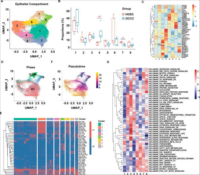
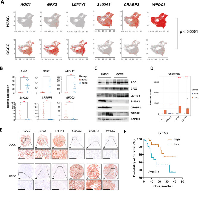
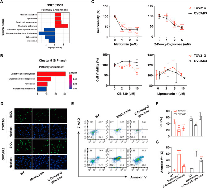
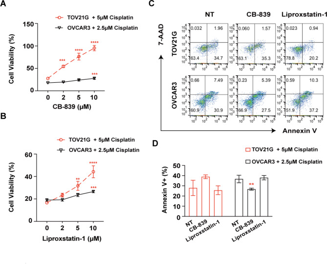
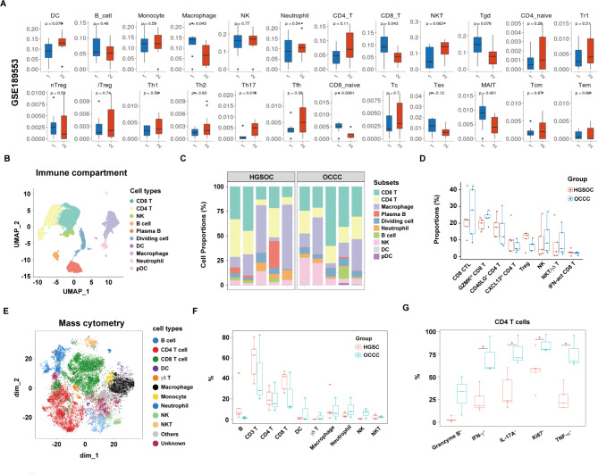
The two most prevalent subtypes of epithelial ovarian carcinoma (EOC) are ovarian clear cell carcinoma (OCCC) and high-grade serous ovarian carcinoma (HGSC). Patients with OCCC have a poor prognosis than those with HGSC due to chemoresistance, implying the need for novel treatment target. In this study, we applied single-cell RNA sequencing (scRNA-seq) together with bulk RNA-seq data from the GEO (Gene Expression Omnibus) database (the GSE189553 dataset) to characterize and compare tumor heterogeneity and cell-level evolution between OCCC and HGSC samples. To begin, we found that the smaller proportion of an epithelial OCCC cell subset in the G2/M phase might explain OCCC chemoresistance. Second, we identified a possible pathogenic OCCC epithelial cell subcluster that overexpresses LEFTY1. Third, novel biomarkers separating OCCC from HGSC were discovered and subsequently validated on a wide scale using immunohistochemistry. Amine oxidase copper containing 1 (AOC1) was preferentially expressed in OCCC over HGSC, while S100 calcium-binding protein A2 (S100A2) was detected less frequently in OCCC than in HGSC. In addition, we discovered that metabolic pathways were enriched in the epithelial compartment of the OCCC samples. In vitro experiments verified that inhibition of oxidative phosphorylation or glycolysis pathways exerted direct antitumor effects on both OCCC and HGSC cells, while targeting glutamine metabolism or ferroptosis greatly attenuated chemosensitivity only in OCCC cells. Finally, to determine whether there were any variations in immune cell subsets between OCCC and HGSC, data from scRNA-seq and mass cytometry were pooled for analysis. In summary, our work provides the first holistic insights into the cellular and molecular distinctions between OCCC and HGSC and is a valuable source for discovering new targets to leverage in clinical treatments to improve the poor prognosis of patients with OCCC.

Keywords: Bulk RNA-seq; HGSC; OCCC; Single-cell RNA-seq.


Conflict of interest statement

The authors declare that they have no competing interests.

Figures

Fig. 1
Fig. 1
A higher proportion of LEFTY1 + epithelial subset cells and a lower proportion of epithelial subset cells in the G2/M phase were observed in OCCC samples than in HGSC samples. (A) UMAP plot displaying eight epithelial cell subpopulations. Each dot represents a single cell (n = 39,722). (B) The box plot shows the comparison of each epithelial cell percentage in the OCCC and HGSC groups. (C) Heatmap showing the average expression of the top 5 most highly expressed markers among epithelial cell subsets. (D) UMAP plot displaying the epithelial cell subsets in each cell cycle phase. (E) Heatmap showing regulon activity as analyzed by SCENIC. A “regulon” refers to the regulatory network of TFs and their target genes. “On” indicates active regulons; “Off” indicates inactive regulons. (F) Pseudotime reconstruction and development of epithelial cell subsets inferred from Monocle 3. (G) Pathway analysis of each epithelial cluster. P values were calculated by two-sided Wilcoxon test. *: p < 0.05, **: p < 0.01
Fig. 2
Fig. 2
Detection and validation of new biomarkers in OCCC patients. (A) Comparisons of AOC1, GPX3, LEFTY1, S100A, CRABP2 and WFDC2 expression levels as shown in a UMAP plot. Each dot represents a single cell. (B)AOC1, GPX3, LEFTY1, S100A, CRABP2 and WFDC2 expression levels in OCCC (n = 8) and HGSC (n = 8) patients, as determined by real-time PCR. Each dot represents a single individual. (C) Western blot results of AOC1, GPX3, LEFTY1, S100A, CRABP2 and WFDC2 expression in OCCC (n = 4) and HGSC (n = 4) patients. (D) Comparisons of AOC1, GPX3, LEFTY1 and CRABP2 expression in the GSE189553 dataset. (E) Representative images of AOC1, GPX3, LEFTY1, S100A, CRABP2 and WFDC2 in the TMA of OCCC and HGSC samples after immunohistochemical staining. The scale bar is 250 μm. (F) The progression-free survival (PFS) analysis results based on GPX3 level in OCCC patients. P values were calculated by two-sided Wilcoxon test. *: p < 0.05, **: p < 0.01, ***: p < 0.001, ****: p < 0.0001
Fig. 3
Fig. 3
Glucose metabolism pathways are activated in OCCC cells and might be promising targets for both OCCC and HGSC treatments as indicated by in vitro experiments. (A) Bar plots showing the differentially expressed genes (DEGs) in the GSE189553 dataset with enriched Kyoto Encyclopedia of Genes and Genomes (KEGG) pathways. Red: OCCC; blue: HGSC. (B) Bar plots showing the differentially expressed gene (DEG)-enriched Kyoto Encyclopedia of Genes and Genomes (KEGG) pathways in Cluster 5 consisting of OCCC and HGSC patient cells. (C) TOV21G and OVCAR3 cells were treated with the indicated concentrations of CB-839, liproxstatin-1, metformin, and 2-deoxy-D-glucose, and cell viability was measured by CCK8 assay 72 h after treatment. (D) TOV21G/OVCAR3 cells were treated with metformin (5 mM) and 2-deoxy-D-glucose (5 mM) for 48 h. Representative images of EdU-positive cells by immunofluorescence. (E) TOV21G/OVCAR3 cells were treated with metformin (5 mM) and 2-deoxy-D-glucose (5 mM) for 72 h. Representative images of flow cytometry are presented. (F) Summary of the percentage of EdU-positive cells. (G) The percentage of Annexin V-positive cells was determined by flow cytometry. NT: no treatment. All assays were carried out in triplicate, and the data are presented as the mean ± S.D. * p < 0.05, ** p < 0.01, *** p < 0.001, **** p < 0.0001
Fig. 4
Fig. 4
Inhibition of glutathione metabolism or ferroptosis reverses cisplatin-induced death of OCCC cells, as determined in vitro. (A) TOV21G or OVCAR3 cells were treated with the indicated concentrations of CB-839 with cisplatin. Cell viability was measured by CCK8 assay 72 h after treatment. (B) TOV21G or OVCAR3 cells were treated with the indicated concentrations of liproxstatin-1 with cisplatin. Cell viability was measured by CCK8 assay 72 h after treatment. (C-D) TOV21G/OVCAR3 cells were treated with CB-839 (10 𝛍M) and liproxstatin-1 (10 𝛍M) with cisplatin for 72 h. Representative images and summary of the percentage of Annexin V-positive cells as determined by flow cytometry NT: no treatment. All assays were carried out in triplicate, and the data are presented as the mean ± S.D. * p < 0.05, ** p < 0.01, *** p < 0.001, **** p < 0.0001
Fig. 5
Fig. 5
Comparison of immune cell heterogeneity in OCCC and HGSC samples. (A) Comparisons of immune cell frequency and T-cell subset frequency between OCCC [1] and HGSC [2] in the GSE189553 dataset as determined by ImmunCellAI analysis. DC: Dendritic cell; B: B cell; NK: natural killer cell; NKT: natural killer T cell; nTreg: natural regulatory T cell; iTreg: induced regulatory T cell; Th: T helper; Tfh: follicular helper T; MAIT: mucosal-associated invariant T; Tcm: central memory T; Tem: effector memory T. (B) UMAP plot displaying ten immune subpopulations (n = 48,213). (C) The distribution and proportion of ten immune subsets in each sample of the HGSC and OCCC groups from scRNA-seq data. (D) Box plots showing a comparison of the percentage of each T/NK cluster between the HGSC and OCCC groups. (E) t-SNE plot showing 12 clusters of TILs from mass cytometry data. Each dot represents a single cell (n = 48,213). (F) Box plots showing the comparison of the percentages of each TIL cluster between the HGSC and OCCC groups. (G) Box plots showing the comparison of different marker expression levels in CD4 T cells in the HGSC and OCCC groups. Each dot represents a single cell. P values were calculated by two-sided Wilcoxon test. * p < 0.05

Similar articles

Cited by

References

    1. Siegel RL, Miller KD, Jemal A. Cancer statistics, 2020. CA Cancer J Clin. 2020;70(1):7–30. doi: 10.3322/caac.21590. - DOI - PubMed
    1. Torre LA, Trabert B, DeSantis CE, Miller KD, Samimi G, Runowicz CD, et al. Ovarian cancer statistics, 2018. CA Cancer J Clin. 2018;68(4):284–96. doi: 10.3322/caac.21456. - DOI - PMC - PubMed
    1. Zhu C, Zhu J, Qian L, Liu H, Shen Z, Wu D, et al. Clinical characteristics and prognosis of ovarian clear cell carcinoma: a 10-year retrospective study. BMC Cancer. 2021;21(1):322. doi: 10.1186/s12885-021-08061-7. - DOI - PMC - PubMed
    1. Chan JK, Teoh D, Hu JM, Shin JY, Osann K, Kapp DS. Do clear cell ovarian carcinomas have poorer prognosis compared to other epithelial cell types? A study of 1411 clear cell ovarian cancers. Gynecol Oncol. 2008;109(3):370–6. doi: 10.1016/j.ygyno.2008.02.006. - DOI - PubMed
    1. Kao YC, Lin MC, Lin WC, Jeng YM, Mao TL. Utility of hepatocyte nuclear factor-1β as a diagnostic marker in ovarian carcinomas with clear cells. Histopathology. 2012;61(5):760–8. doi: 10.1111/j.1365-2559.2012.04267.x. - DOI - PubMed