. 2023 Jul 31;14(1):4591.
doi: 10.1038/s41467-023-40217-w.

The HSV-1 ICP22 protein selectively impairs histone repositioning upon Pol II transcription downstream of genes

Affiliations

The HSV-1 ICP22 protein selectively impairs histone repositioning upon Pol II transcription downstream of genes

Lara Djakovic et al. Nat Commun. .

Abstract

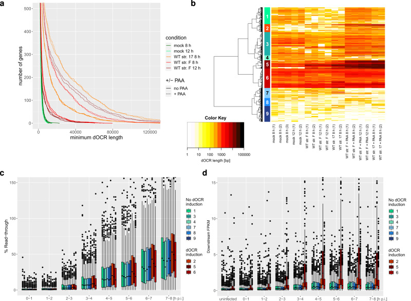
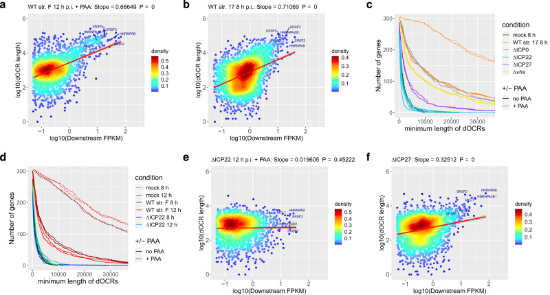
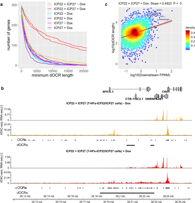
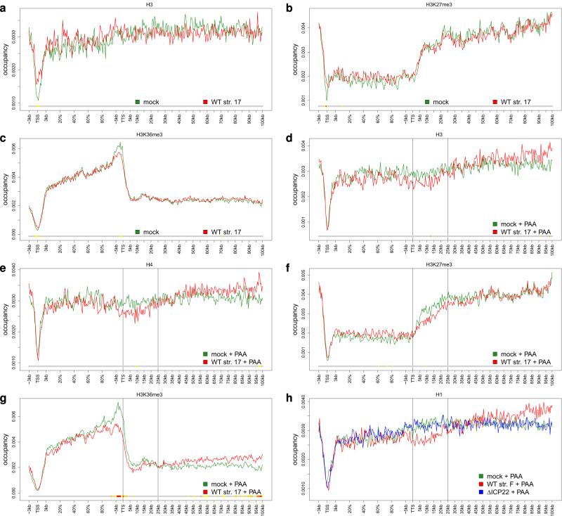
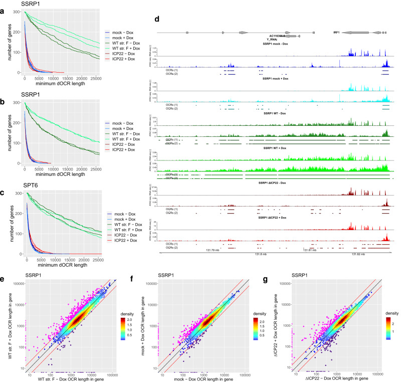
Herpes simplex virus 1 (HSV-1) infection and stress responses disrupt transcription termination by RNA Polymerase II (Pol II). In HSV-1 infection, but not upon salt or heat stress, this is accompanied by a dramatic increase in chromatin accessibility downstream of genes. Here, we show that the HSV-1 immediate-early protein ICP22 is both necessary and sufficient to induce downstream open chromatin regions (dOCRs) when transcription termination is disrupted by the viral ICP27 protein. This is accompanied by a marked ICP22-dependent loss of histones downstream of affected genes consistent with impaired histone repositioning in the wake of Pol II. Efficient knock-down of the ICP22-interacting histone chaperone FACT is not sufficient to induce dOCRs in ΔICP22 infection but increases dOCR induction in wild-type HSV-1 infection. Interestingly, this is accompanied by a marked increase in chromatin accessibility within gene bodies. We propose a model in which allosteric changes in Pol II composition downstream of genes and ICP22-mediated interference with FACT activity explain the differential impairment of histone repositioning downstream of genes in the wake of Pol II in HSV-1 infection.


Conflict of interest statement

The authors declare no competing interests.

Figures

Fig. 1
Fig. 1. Induction of dOCRs in HSV-1 infection is associated with downstream transcriptional activity.
a Number of genes with dOCR length greater than the value on the x-axis in mock and HSV-1 WT strain 17 infections with or without PAA treatment (combined data of 2 biological replicates). To avoid having to define a threshold on whether a particular dOCR length is considered as dOCR induction, we visualized dOCR lengths in each condition for all analyzed 4162 genes without read-in transcription in HSV-1 infection (excluding those with a dOCR length = 0). This depicts whether the number of genes with longer dOCRs is generally increased in the respective condition. The y-axis was limited to 500 to highlight differences in the number of genes with long dOCRs between mock and HSV-1 infection. b Hierarchical clustering analysis (Euclidean distances, Ward’s clustering criterion) of log10(dOCR length) for all analyzed genes (i.e., 4162 genes without read-in transcription in HSV-1 infection) of the samples shown in (a). To define clusters, the cutoff on the clustering dendrogram was chosen such that three groups of genes visually identified as showing dOCR induction in the heatmap resulted in separate clusters. Identified clusters are numbered from top to bottom as indicated and marked by colored rectangles. Shades of red indicate clusters with dOCR induction and shades of blue clusters without dOCR induction. c, d Boxplots showing the distribution of read-through transcription (c) and downstream FPKM (d) for the 9 clusters (n = 609, 290, 851, 176, 305, 701, 367, 289, and 574 genes for clusters 1–9, respectively) from (b). Bounds of boxes are the first and third quartiles for each condition. The center (median) is shown by the horizontal line in the box. Whiskers extend to 1.5 times the inter-quartile range. Outliers are shown as small circles, and minimum and maximum values are the lowest and highest circles, respectively. Read-through values and downstream FPKM were calculated as described in Methods from previously published 4sU-seq data (average of n = 2 biological replicates). Read-through for mock infection was defined as zero and is thus not shown. Source data are provided as a Source Data file.
Fig. 2
Fig. 2. ICP22 is required for the induction of dOCRs.
a, b Scatter plots correlating downstream FPKM against dOCR length (average of two replicates) in total RNA for WT strain F (a) and in 4sU-RNA for WT strain 17 (b) for all analyzed genes with a downstream FPKM ≥ 0.05. Colors indicate the density of points from high (red) to low (blue). The red line indicates a linear fit of log10(dOCR length) against log10(downstream FPKM). The slope of the fit and p-values for the slope of the linear regression estimate being ≠ 0 (two-sided test) were calculated using the lm function in R and are indicated on top of each figure. The error bands around the red line indicate the 95% confidence level interval for predictions from the lm linear model. Example genes with high induction of dOCRs in HSV-1 infection are highlighted. c, d Number of genes in Cluster 5 from Fig. 1b for which dOCRs reach at least a length greater than the value indicated on the x-axis for mock, WT strain 17 (c), WT strain F (d), ΔICP0 (c), ΔICP22 (c, d), ΔICP27 (c) and Δvhs infection (c). To avoid having to define a threshold on whether or not a particular dOCR length for a gene is considered dOCR induction, we visualize dOCR lengths in each condition for all 305 Cluster 5 genes (excluding only those with a dOCR length = 0 in a particular condition). This depicts whether the number of genes with longer dOCRs was generally increased or not in the respective experimental condition. Results are shown separately for two biological replicates. All infections in d were performed with and without PAA, while in c PAA treatment was only performed for WT strain 17 and ΔICP22 infection (as indicated by solid (no PAA) or dashed (+PAA) lines). e, f Scatter plots as in (a, b) correlating downstream FPKM against dOCR length in total RNA for 12 h p.i. ΔICP22 infection +PAA (e) and in 4sU-RNA for ΔICP27 infection (f). Scatter plots for other analyzed conditions are shown in Supplementary Figs. 3 and 7. Source data are provided as a Source Data file.
Fig. 3
Fig. 3. ICP22 is sufficient to induce dOCRs upon ICP27-induced read-through transcription.
a Number of genes in Cluster 5 from Fig. 1b for which dOCRs reach at least a length greater than the value indicated on the x-axis in T-HF-ICP22 cells, T-HF-ICP27 cells, and T-HF-ICP22/ICP27 cells ± Dox treatment. b Example gene (HNRNPA2B1) showing induction of dOCRs after Dox-induced ICP22 and ICP27 expression in T-HF-ICP22/ICP27 cells. Tracks show total RNA-seq (strand-specific) and Omni-ATAC-seq (non-strand-specific) read coverage (normalized to a total number of mapped human reads; averaged between replicates). Below each Omni-ATAC-seq track, the figure shows open chromatin regions (OCRs) identified with F-Seq as well as the dOCR regions calculated from the OCRs as described in Methods. For simplification, OCRs and dOCRs are shown only for the first replicate. Gene annotation is indicated at the top. Boxes represent exons, lines introns, and gene direction is indicated by arrowheads. Genomic coordinates are shown at the bottom. c Scatter plot correlating downstream FPKM in total RNA against dOCR length (average of two replicates) for Dox-induced combined ICP22 and ICP27 expression (T-HF-ICP22/ICP27 cells + Dox). Shown are all analyzed genes with a downstream FPKM ≥ 0.05. Colors indicate the density of points from high (red) to low (blue). The red line indicates a linear fit of log10(dOCR length) against log10(downstream FPKM). The slope of the fit and p-values for the slope of the linear regression estimate being ≠ 0 were calculated using the lm function in R and are indicated on top of each figure. The error bands around the red line indicate the 95% confidence level interval for predictions from the lm linear model. Example genes with high induction of dOCRs in HSV-1 infection are highlighted. The corresponding scatter plot for T-HF-ICP22/ICP27 cells without Dox treatment is shown in Supplementary Fig. 8f. Source data are provided as a Source Data file.
Fig. 4
Fig. 4. Alterations in histone distribution associated with the induction of dOCRs.
ag Metagene plots (see Methods) showing the distribution of (a, d) H3, (e) H4, (b, f) H3K27me3, and (c, g) H3K36me3 for genes with strong induction of dOCRs, i.e., Cluster 5 genes from Fig. 1b, in mock and WT strain 17 infections without (ac) and with PAA treatment (dg). Metagene plots for H1 in mock and WT strain 17 infections with PAA treatment for Cluster 5 and corresponding metagene plots for genes without induction of dOCRs are shown in Supplementary Fig. 11a–f. The color track at the bottom of each subfigure indicates the significance of paired two-sided Wilcoxon tests comparing the normalized transcript coverages of genes for each position between mock and WT infection. P‐values are adjusted for multiple testing with the Bonferroni method within each subfigure; color code: red = adj. P-value ≤ 105; orange = adj. P-value ≤ 103; yellow: adj. P-value ≤ 0.05. h Metagene plots showing the distribution of H1 in mock, WT strain F and ΔICP22 infection for genes with strong dOCR induction, i.e., Cluster 5 genes. P-values for pairwise comparisons between mock and WT strain F infection, WT strain F and ΔICP22 infection, and mock and ΔICP22 infection, respectively, were calculated as for (ag) and are shown in Supplementary Fig. 12b–d. Source data are provided as a Source Data file.
Fig. 5
Fig. 5. Depletion of FACT increases chromatin accessibility in an ICP22-dependent manner.
ac Number of genes in Cluster 5 from Fig. 1b that exhibit dOCRs with at least a length greater than the value indicated on the x-axis in mock, WT strain F and ΔICP22 infection with or without Dox-induced knockdown of SSRP1 a without and b with down-sampling of reads or c knockdown of SPT6 without down-sampling. Results for SPT6 with down-sampling of reads are shown in Supplementary Fig. 13e. d Example gene (IRF1) showing increased chromatin accessibility within the gene body in SSRP1-depleted cells in HSV-1 infection. Tracks show total RNA-seq (strand-specific) and ATAC-seq (non-strand-specific) read coverage (normalized to a total number of mapped human reads; averaged between replicates) in mock, WT and ΔICP22 infection without and with Dox-induced SSRP1 depletion. Identified OCRs for both replicates are shown separately below the read coverage tracks. Gene annotation is indicated at the top. Boxes represent exons and lines introns, and gene direction is indicated by arrowheads. Genomic coordinates are shown at the bottom. eg Scatter plots comparing the total length of open chromatin regions (OCRs) within gene bodies with and without Dox-induced knock-down of SSRP1 in e WT, f mock, and g ΔICP22 infection for all 4162 analyzed genes without read-in transcription. Colors indicate the density of points from high (red) to low (blue). Genes with ≥2-fold increased and reduced OCR lengths within the gene body are marked in magenta and violet, respectively. Red lines indicate a 2-fold change, and the black line depicts the diagonal. Source data are provided as a Source Data file.

Similar articles

Cited by

References

    1. Hennig T, Djakovic L, Dölken L, Whisnant AW. A review of the multipronged attack of herpes simplex virus 1 on the host transcriptional machinery. Viruses. 2021;13:1836. doi: 10.3390/v13091836. - DOI - PMC - PubMed
    1. Rivas HG, Schmaling SK, Gaglia MM. Shutoff of host gene expression in influenza A virus and herpesviruses: similar mechanisms and common themes. Viruses. 2016;8:102. doi: 10.3390/v8040102. - DOI - PMC - PubMed
    1. Rutkowski AJ, et al. Widespread disruption of host transcription termination in HSV-1 infection. Nat. Commun. 2015;6:7126. doi: 10.1038/ncomms8126. - DOI - PMC - PubMed
    1. Wang X, et al. Herpes simplex virus blocks host transcription termination via the bimodal activities of ICP27. Nat. Commun. 2020;11:293. doi: 10.1038/s41467-019-14109-x. - DOI - PMC - PubMed
    1. Hennig T, et al. HSV-1-induced disruption of transcription termination resembles a cellular stress response but selectively increases chromatin accessibility downstream of genes. PLOS Pathog. 2018;14:e1006954. doi: 10.1371/journal.ppat.1006954. - DOI - PMC - PubMed

Publication types