. 2023 Jul 12;24(14):11379.
doi: 10.3390/ijms241411379.

Assessing the Impact of Polyethylene Nano/Microplastic Exposure on Human Vaginal Keratinocytes

Affiliations

Assessing the Impact of Polyethylene Nano/Microplastic Exposure on Human Vaginal Keratinocytes

Paola Pontecorvi et al. Int J Mol Sci. .

Abstract

The global rise of single-use throw-away plastic products has elicited a massive increase in the nano/microplastics (N/MPLs) exposure burden in humans. Recently, it has been demonstrated that disposable period products may release N/MPLs with usage, which represents a potential threat to women's health which has not been scientifically addressed yet. By using polyethyl ene (PE) particles (200 nm to 9 μm), we showed that acute exposure to a high concentration of N/MPLs induced cell toxicity in vaginal keratinocytes after effective cellular uptake, as viability and apoptosis data suggest, along with transmission electron microscopy (TEM) observations. The internalised N/MPLs altered the expression of junctional and adherence proteins and the organisation of the actin cortex, influencing the level of genes involved in oxidative stress signalling pathways and that of miRNAs related to epithelial barrier function. When the exposure to PE N/MPLs was discontinued or became chronic, cells were able to recover from the negative effects on viability and differentiation/proliferation gene expression in a few days. However, in all cases, PE N/MPL exposure prompted a sustained alteration of DNA methyltransferase and DNA demethylase expression, which might impact epigenetic regulation processes, leading to accelerated cell ageing and inflammation, or the occurrence of malignant transformation.

Keywords: microplastics; nano/microparticles uptake; nanoplastics; period products; polyethylene; vaginal keratinocytes.


Conflict of interest statement

The authors declare no conflict of interest.

Figures
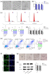
Figure 1
Figure 1
Evaluation of vaginal keratinocyte morphology and viability after acute PE N/MPL exposure. (a) Representative images of VK2 E6/E7 treated for 48 h with Tween20 (Veh; 0.0025%) or PE N/MPLs at low and high concentrations (25 and 250 μg/mL). Pictures were taken at 40× magnification. Scale bars are 50 μm. (b) Graphical representation of the percentage of viable vaginal keratinocytes after 48, 72, and 120 h vehicle or PE N/MPL exposure obtained with a Trypan blue dye exclusion assay. (c) Representative plots of FACS cell cycle analysis at 48 h and percentage histograms at 48, 72, and 120 h exposure to vehicle or PE N/MPLs. Arrowheads in FACS plots indicate the ideal position of G1 and G2 peaks. (d) Histograms showing gene expression levels of cell cycle and apoptosis markers in cells treated for 48 h with PE N/MPLs compared to vehicle. (e) Representative dot plots and histograms displaying the percentage of apoptotic cells upon vehicle and PE particle exposure for 72 h. Blue corresponds to areas of lower cell density, red and orange are areas of high cell density, green and yellow indicate mid-range density areas. (f) Illustrative IF pictures for Lamin B (green, left) and DAPI (blue, right) staining of VK2 E6/E7 treated with vehicle or PE N/MPLs 25–250 μg/mL for 72 h. Pictures were taken at 40× magnification. Scale bars are 50 μm. (g) Representative pictures of β-galactosidase staining of VK2 E6/E7 exposed to vehicle or PE 25 and 250 μg/mL for 72 h. H2O2 treatment was used as a positive control. Pictures were taken at 40× magnification. Scale bars are 50 μm. (h) Illustrative Western blot and densitometric analysis for the autophagy markers LC3 B and p62 in VK2 E6/E7 exposed to vehicle or PE 25 and 250 μg/mL for 48 h (β-Actin was used as loading control) (Figure S2). For all the experiments, statistical analysis was conducted by unpaired, two-tailed Student’s t-test (n > 3) with “ns” non-significant, * p ≤ 0.05 and ** p ≤ 0.005.
Figure 2
Figure 2
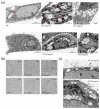
Assessment of cellular uptake of PE N/MPLs by vaginal keratinocytes upon acute exposure. (a) TEM analysis of VK2 E6/E7 cells after 72 h of treatment with 25 and 250 μg/mL of PE N/MPLs or vehicle. Upper panel: From left to right, VK2 E6/E7 cells exposed to vehicle versus 25 μg/mL of PE N/MPLs. In the latter, extracellular accumulation of N/MPLs (asterisks) is evident between cells, in proximity to surface microvilli and intercellular junctions (red arrows). Lower panel: From left to right, VK2 E6/E7 cells exposed to vehicle versus 250 μg/mL of PE N/MPLs. In the latter, the N/MPLs (asterisks) are principally localised within cytoplasmic endocytic structures, often distributed around the nucleus. Two different vehicle pictures are present in the panel, since we performed the experiment with two distinct vehicle volumes matching the two PE N/MPLs concentrations. (b) Optical microscopy analysis of VK2 E6/E7 cells exposed for 72 h to vehicle versus PE N/MPLs coated with 0.2% human keratin in aqueous solution. (c) Ultrastructural analysis of the uptake of PE N/MPLs (250 μg/mL) coated with 0.2% human keratin in aqueous solution. TEM micrographs show PE N/MPLs (asterisks) deposited on the plasma membrane of VK2 E6/E7 cell, with initial uptake in forming vacuoles (top), and internalisation within peripheral and perinuclear endosome-like organelles (bottom). The NP mean diameter, reported with coloured lines in the figure (bottom), is consistent with that of the N/MPLs used in the experiment. PM: plasma membrane; N: nucleus. Scale bar: 1 μm.
Figure 3
Figure 3
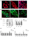
Analysis of the effects of PE N/MPL acute exposure on VK2 E6/E7 cytoskeleton and cell stress pathways. (a) Representative IF acquisitions showing actin cytoskeleton of vaginal keratinocytes treated with vehicle or PE N/MPLs at 25–250 μg/mL for 72 h. Actin is stained in red with DyLight™ 554 Phalloidin, and nuclei are stained in blue with DAPI. Arrowheads indicate gaps in the cytoskeletal net. Pictures were taken at 40× magnification. Scale bars are 50 μm. (b) Illustrative IF pictures showing β-Catenin localisation in VK2 E6/E7 exposed to vehicle or PE N/MPLs 25–250 μg/mL for 72 h. β-Catenin is stained in green with Alexa Fluor™ 488, while nuclei are stained in blue with DAPI. Acquisitions were obtained at 40× magnification. Scale bars are 50 μm. (c) Representative Western blot and corresponding densitometry for β-Catenin, E-cadherin, and Pan Keratin expression in vaginal keratinocytes exposed to vehicle or PE N/MPLs at 25–250 μg/mL for 72 h. β-Actin was used as a loading control (Figure S2). (d) RT-qPCR graphs showing relative expression of stress and redox homeostasis genes in VK2 E6/E7 exposed for 48 h to vehicle or PE 25–250 μg/mL. GAPDH mRNA level was used as endogenous control. (e) Histograms displaying epithelial barrier function-related microRNA expression in vaginal keratinocytes treated with vehicle or low and high PE particle concentrations for 48 h. U6 snRNA levels were employed as endogenous control. All the statistical analyses were conducted by unpaired, two-tailed Student’s t-test (n = 3) with “ns” non-significant, * p ≤ 0.05 and ** p ≤ 0.005.
Figure 4
Figure 4
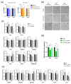
Assessing the impact of discontinuous PE N/MPL exposure on cell viability, morphology, proliferation, inflammation, and epigenetic regulation in vaginal keratinocytes. (a) Graphs showing the percentage of viable cells upon 48 h of exposure to low and high concentrations of PE particles and 144 h post-wash-out obtained via Trypan blue and MTT assays. (b) Representative pictures of VK2 E6/E7 exposed to vehicle or PE N/MPLs for 48 h and 144 h after wash-out taken throughout the experimental points at 40× magnification. Scale bars are 50 μm. (c) Histograms for RT-qPCR analysis displaying a relative expression of apoptosis and cell cycle markers, and proliferation/differentiation-associated genes in VK2 E6/E7 exposed to vehicle or PE 25–250 μg/mL for 48 h and after 144 h wash-out. (d) Graphs showing relative fold regulation for VEGF concentrations detected in cell culture supernatants at 48 h of exposure to PE 25–250 μg/mL or vehicle and at 144 h from wash-out. (e) Relative gene expression of epigenetic regulation enzymes in vaginal keratinocytes exposed to PE 25–250 μg/mL or vehicle for 48 h and at 144 h post-wash-out. All the statistical analyses were conducted by unpaired, two-tailed Student’s t-test (n ≥ 3) with “ns” non-significant, * p ≤ 0.05 and ** p ≤ 0.005.
Figure 5
Figure 5
Assessment of the impact of PE N/MPL chronic exposure on vaginal keratinocytes viability, morphology, proliferation, inflammation, epigenetic regulation, and transformation potential. (a) Diagrams showing the results of the Trypan blue assay with the percentage of viable cells for VK2 E6/E7 chronically exposed to low and high concentrations of PE N/MPLs or vehicle for 21 days. (b) Illustrative images of vaginal keratinocytes treated with vehicle or PE 25–250 μg/mL at day 3 to 21 of exposure. Pictures were taken at 40× magnification. Scale bars are 50 μm. (c) Graphs for RT-qPCR analysis displaying a relative expression of proliferation/differentiation-associated genes in VK2 E6/E7 chronically exposed to vehicle or PE 25–250 μg/mL for 21 days. (d) Histograms showing relative fold regulation for VEGF concentrations detected in VK2 E6/E7 cell culture supernatants at 3 to 21 days of exposure to PE 25–250 μg/mL or vehicle. (e) Relative gene expression of epigenetic regulation enzymes in vaginal keratinocytes exposed to PE 25–250 μg/mL or vehicle for 21 days. (f) Representative images of colony formation assays of VK2 E6/E7 chronically exposed to PE 25 μg/mL or vehicle for one month and two weeks. Staining was performed with Crystal violet. Relative absorbance for solubilised Crystal violet was reported on a diagram. Statistical analyses were conducted by unpaired, two-tailed Student’s t-test (n = 3) with “ns” non-significant, * p ≤ 0.05 and ** p ≤ 0.005.

Similar articles

Cited by

References

    1. Llorca M., Farré M. Current Insights into Potential Effects of Micro-Nanoplastics on Human Health by in-vitro Tests. Front. Toxicol. 2021;3:752140. doi: 10.3389/ftox.2021.752140. - DOI - PMC - PubMed
    1. Andrady A.L., Neal M.A. Applications and societal benefits of plastics. Philos. Trans. R. Soc. B Biol. Sci. 2009;364:1977–1984. doi: 10.1098/rstb.2008.0304. - DOI - PMC - PubMed
    1. Hartmann N.B., Hüffer T., Thompson R.C., Hassellöv M., Verschoor A., Daugaard A.E., Rist S., Karlsson T., Brennholt N., Cole M., et al. Are We Speaking the Same Language? Recommendations for a Definition and Categorization Framework for Plastic Debris. Environ. Sci. Technol. 2019;53:1039–1047. doi: 10.1021/acs.est.8b05297. - DOI - PubMed
    1. Park H., Park B. Review of Microplastic Distribution, Toxicity, Analysis Methods, and Removal Technologies. Water. 2021;13:2736. doi: 10.3390/w13192736. - DOI
    1. Carbery M., O’Connor W., Palanisami T. Trophic transfer of microplastics and mixed contaminants in the marine food web and implications for human health. Environ. Int. 2018;115:400–409. doi: 10.1016/j.envint.2018.03.007. - DOI - PubMed

LinkOut - more resources