Review
. 2023 Jun 24;13(7):1035.
doi: 10.3390/biom13071035.

Voltage-Gated Proton Channels in the Tree of Life

Affiliations
Review

Voltage-Gated Proton Channels in the Tree of Life

Gustavo Chaves et al. Biomolecules. .

Abstract

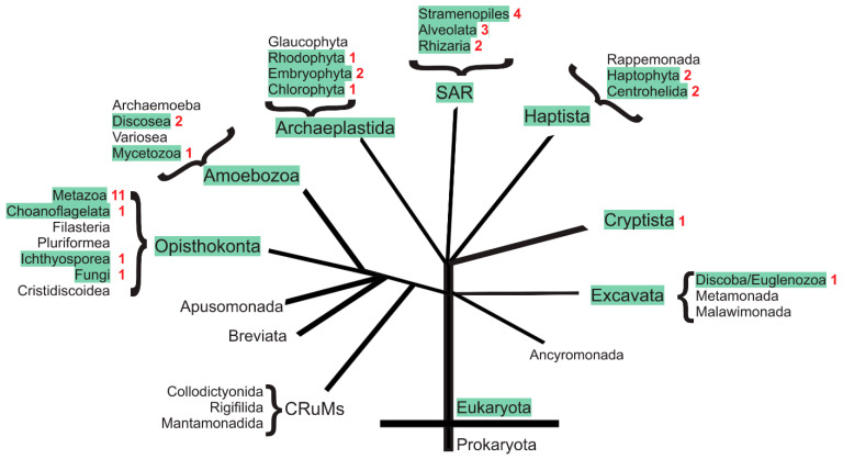
With a single gene encoding HV1 channel, proton channel diversity is particularly low in mammals compared to other members of the superfamily of voltage-gated ion channels. Nonetheless, mammalian HV1 channels are expressed in many different tissues and cell types where they exert various functions. In the first part of this review, we regard novel aspects of the functional expression of HV1 channels in mammals by differentially comparing their involvement in (1) close conjunction with the NADPH oxidase complex responsible for the respiratory burst of phagocytes, and (2) in respiratory burst independent functions such as pH homeostasis or acid extrusion. In the second part, we dissect expression of HV channels within the eukaryotic tree of life, revealing the immense diversity of the channel in other phylae, such as mollusks or dinoflagellates, where several genes encoding HV channels can be found within a single species. In the last part, a comprehensive overview of the biophysical properties of a set of twenty different HV channels characterized electrophysiologically, from Mammalia to unicellular protists, is given.

Keywords: Aplysia; Ecdysozoa; Lophotrochozoa; NADPH oxidase; coccolithophores; insect; mollusks; pH-dependent gating; voltage-gated proton channel; voltage-sensing.


Conflict of interest statement

The authors declare no conflict of interest.

Figures

Figure 1
Figure 1
Simplified scheme of the respiratory burst in phagocytes. The activity of the NADPH oxidase reduces intracellular pH and depolarizes the plasma membrane. The voltage-gated proton channel HV1 compensates for the charge by conducting protons out of the cell. Additionally, the outflux of protons counteracts the decreased pH. Therefore, HV1 and NADPH oxidase are perfect synergistic partners during the respiratory burst.
Figure 2
Figure 2
Architecture of the voltage-sensing and conduction pore domains of HV, KV, and NaV channels. As examples, a homology model of the human HV1, the crystal structure of the KV1.2 (PDB: 3LUT [108]), and the bacterial NaVAB channels (PDB 3RVY [109]) are shown. The voltage-sensing domain of all voltage-gated channels consists of four transmembrane helices (S1–S4). In HV channels, the voltage-sensing domain is also the ion conduction pore. In KV, NaV, and also CaV channels, the ion conduction pore is formed by a tetrameric assembly of two further transmembrane helices S5 and S6. The voltage-sensing arginines, three in HV, four in NaV, and five plus a lysine in KV are shown as sticks. Upward, resp. downward movement of the S4 (orange) helix is coupled to the activation, resp. deactivation, of the channels upon membrane depolarization, resp. hyperpolarization. A phenylalanine, shown as sticks, in the hydrophobic gasket that separates the intracellular and extracellular vestibules of the channels is conserved among voltage-gated ion channels.
Figure 3
Figure 3
HV channels within the eukaryotic tree of life. Phylae and Subphylae containing at least one species harboring an HV channel gene are marked in green. The maximal number of different HV channel genes per species is indicated in red.
Figure 4
Figure 4
Hv channels in Metazoa. (A) Tree of metazoan life. The maximal number of different HV channel genes per species is indicated in red. (B) Maximal number of HV channels genes per species in different phylae.
Figure 5
Figure 5
Single Vrev measurements of the HV4 channel from Crassostrea gigas are plotted against the EH predicted value at different proton gradients (ΔpH = pHo – pHi). The dotted line shows equality between Vrev and EH. The inset depicts an example of a Vrev experimental determination by the tail currents method in a whole-cell patch clamp configuration at ΔpH = 0.5. Repolarization of the cell membrane after a depolarizing pulse was conducted in Δ10 mV/step from −60 mV to −20 mV) with a holding potential of −60 mV. The arrow indicates the point where H+ currents reverse (Vrev), here ~33 mV. Taken from [125].
Figure 6
Figure 6
ΔpH-gating of the human HV1 channel from molecular dynamics simulations. The ΔpH-dependent gating from a deactivated (left, brown) to an activated (right, blue) conformation moves the three gating charges R1 to R3 in S4 (orange ribbon) outwards, passing the second arginine R2 through the hydrophobic gasket (HG). Changes of the location and orientation of the gating charges in the two conformations result in different interaction networks both in the internal and in the external vestibules, as illustrated for the selectivity filter aspartate (D112 in hHV1, here in red) that interact with R1 in the deactivated but with R2 in the activated conformation. For more details, please see [176].
Figure 7
Figure 7
pH-dependent gating of HV channels in different organisms. The evaluation of the pH-dependent gating of HV channels can be described by a linear function of the form Vthres = slope·Vrev + offset from data obtained at different ΔpH conditions. (Upper panel, left) Families of H+ currents elicited by two different proton channels from Aplysia californica at symmetrical pH = 6.5. A zoom-in of dashed areas for each family is shown on the right side. The membrane potential where currents are first detected (Vthres) differs for both channels. AcHV1 activates positive to Vrev (here at +10 mV) and H+ currents are then outwardly directed (A). In contrast, AcHV2 activates negative to Vrev (here at −30 mV), permitting H+ influx (B). (Upper panel, right) pH-dependent gating of AcHV1 (orange line) and AcHV2 (blue line) represented according to Equation (3). Both channels present identical pH-sensing with the same slope (Vthres/Vrev) but have distinct offsets. The dotted line represents equality between Vthres and Vrev and delimits the equilibrium between outward and inward driving forces. Data over and below the dotted line indicate outwardly and inwardly directed H+ driving force, respectively. While AcHV1 extrudes H+ alkalinizing the cytosol, AcHV2 conducts inward currents that acidify the cell. From [124]. (Lower panel, left) Vthres vs. ΔpH (pHo − pHi) plot determined for some functionally tested HV channels from different species in a ΔpH ranging from −1 to +1. The pH-dependent gating of native HV channels of 15 cell types (from distinct organisms including rat, hamster, mouse, human, frog, and snail) described by [192] is displayed for comparison (black solid triangles). The dotted line shows equality between Vrev and Vthres. Most of HV channels are proton extruders but there are few notable exceptions, i.e., the channels from the dinoflagellate K. veneficum (kHV1), the fungi A. oryzae (AoHV1), the stick insect E. tiaratum (EtHV1), or the type-2 channel from sea hare A. californica (AcHV2). (Lower panel, right) the pH-dependent gating on the external pHo for several HV channels is represented as ΔgH/ΔpH. The values indicate the shift of the conductance–voltage curves expected once pHo is changed in one unit while maintaining pHi constant. The dotted line shows the 40 mV/unit of pH thumb rule describing the pH-dependent gating for most of HV channels [192]. The channels are grouped by organism in insects (red), mollusks (light blue), dinoflagellates (green), ascidians (light cyan), fungi (orange), mammals (blue), fishes (grey), corals (dark green), sea urchins (pink), coccolithophores (dark cyan), and 15 native HV from different species. Np: Nicoletia phytophila, Et: Extatosoma tiaratum, Cg: Crassostrea gigas, Ac: Aplysia californica, Ht: Helisoma trivolvis, Kv: Karlodinium veneficum, Lp: Lingulodinium polyedrum, Ci: Ciona intestinalis, Ao: Aspergillus oryzae, Sl: Suillus luteus, h: homo sapiens, m: Mus musculus, Rn: Rattus norvegicus, Dr: Danio rerio, Am: Acropora millepora, Sp: Strongylocentrotus purpuratus, Eh: Emiliania huxleyi, Cp: Coccolithus pelagicus ssp braarudii.

Similar articles

Cited by

References

    1. Thomas R.C., Meech R.W. Hydrogen ion currents and intracellular pH in depolarized voltage-clamped snail neurones. Nature. 1982;299:826–828. doi: 10.1038/299826a0. - DOI - PubMed
    1. Fogel M., Hastings J.W. Bioluminescence: Mechanism and Mode of Control of Scintillon Activity. Proc. Natl. Acad. Sci. USA. 1972;69:690–693. doi: 10.1073/pnas.69.3.690. - DOI - PMC - PubMed
    1. Henderson L.M., Chappell J.B., Jones O.T.G. The superoxide-generating NADPH oxidase of human neutrophils is electrogenic and associated with an H+ channel. Biochem. J. 1987;246:325–329. doi: 10.1042/bj2460325. - DOI - PMC - PubMed
    1. Baldridge C.W., Gerard R.W. The extra respiration of phagocytosis. Am. J. Physiol. Content. 1932;103:235–236. doi: 10.1152/ajplegacy.1932.103.1.235. - DOI
    1. DeCoursey T. Hydrogen ion currents in rat alveolar epithelial cells. Biophys. J. 1991;60:1243–1253. doi: 10.1016/S0006-3495(91)82158-0. - DOI - PMC - PubMed

Grants and funding

This research received no external funding.

LinkOut - more resources