. 2023 Sep 22;51(17):9166-9182.
doi: 10.1093/nar/gkad631.

The HDAC6-RNF168 axis regulates H2A/H2A.X ubiquitination to enable double-strand break repair

Affiliations

The HDAC6-RNF168 axis regulates H2A/H2A.X ubiquitination to enable double-strand break repair

Lingyu Qiu et al. Nucleic Acids Res. .

Abstract

Histone deacetylase 6 (HDAC6) mediates DNA damage signaling by regulating the mismatch repair and nucleotide excision repair pathways. Whether HDAC6 also mediates DNA double-strand break (DSB) repair is unclear. Here, we report that HDAC6 negatively regulates DSB repair in an enzyme activity-independent manner. In unstressed cells, HDAC6 interacts with H2A/H2A.X to prevent its interaction with the E3 ligase RNF168. Upon sensing DSBs, RNF168 rapidly ubiquitinates HDAC6 at lysine 116, leading to HDAC6 proteasomal degradation and a restored interaction between RNF168 and H2A/H2A.X. H2A/H2A.X is ubiquitinated by RNF168, precipitating the recruitment of DSB repair factors (including 53BP1 and BRCA1) to chromatin and subsequent DNA repair. These findings reveal novel regulatory machinery based on an HDAC6-RNF168 axis that regulates the H2A/H2A.X ubiquitination status. Interfering with this axis might be leveraged to disrupt a key mechanism of cancer cell resistance to genotoxic damage and form a potential therapeutic strategy for cancer.


Figures

Graphical Abstract
Graphical Abstract
Figure 1.
Figure 1.
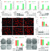
HDAC6 is involved in DSB repair. (A) pEJ5-GFP U2OS or DR-U2OS cells were transfected with HDAC6 or negative control (NC) siRNAs and subjected to NHEJ and HR assays, separately. (B) pEJ5-GFP U2OS or DR-U2OS cells were transfected with Myc-HDAC6 or empty vector and subjected to NHEJ and HR assays, separately. (C, D) HeLa cells were transfected with HDAC6 or NC siRNAs for 48 h, treated with 10 μM VP16 for 2 h, and washed three times with fresh medium before recovery for the indicated times. All samples were then subjected to a comet assay. Representative images (C) and statistical analyses (D) are shown. (E, F) HeLa cells were transfected with Flag-HDAC6 plasmids for 48 h. After 3 Gy irradiation, the cells were counted and seeded for the colony formation assays. For the control (Ctr) group, 500 cells were seeded in each plate. For the IR-treated group, 5000 cells were seeded in each plate. Representative images (E) and statistical analyses (F) are shown. (G, H) HDAC6 WT or HDAC6 KO HCT116 cells underwent 3 Gy irradiation (IR) and were counted and seeded for colony formation assays. For the control (Ctr) group, 500 cells were seeded in each plate. For the IR-treated group, 5000 cells were seeded in each plate. Representative images (G) and statistical analyses (H) are shown. All data represent the means ± SD (n = 3, *P< 0.05, **P< 0.01, ***P< 0.001).
Figure 2.
Figure 2.
HDAC6 interacts with H2A/H2A.X in response to DSBs, inhibiting H2A/H2A.X ubiquitination. (A) HeLa cells were transfected with Flag-HDAC6 or an empty plasmid for 48 h. Chromatin fractions were subjected to immunoprecipitation with an anti-Flag antibody. Western blots of indicated proteins are shown. (B) Whole cell lysates from HeLa cells were extracted and immunoprecipitated using an anti-HDAC6 (up) or an anti-H2A.X (down) antibody. Rabbit IgG was used as a negative control. Western blotting was performed with the indicated antibodies. (C) GST or GST-H2A.X fusion proteins were expressed in bacteria, purified, and then incubated with His-HDAC6 protein. Western blotting was performed to detect HDAC6 protein levels and Coomassie Brilliant Blue (CBB) staining was performed to detect GST or GST-H2A.X levels. The asterisk indicates the corresponding protein bands. (D) HCT116 cells were co-transfected with Flag-H2A.X, Myc-HDAC6, or an empty plasmid for 48 h. Flag-H2A.X was immunoprecipitated from whole cell lysates and eluted for western blotting with the indicated antibodies (FK2: anti-ubiquitin conjugate antibody, Pan-me: anti-pan methyl Lysine antibody, p-S/T: anti-Phospho-(Ser/Thr) antibody, Pan-ac: anti-acetyl lysine antibody) to detect post-translational modifications. (E) HDAC6 WT or HDAC6 KO HCT116 cells were transfected with Flag-H2A.X and HA-ub, with or without Myc-HDAC6 for 48 h, before they were exposed to IR (10 Gy) and released for 1 h. Flag-H2A.X proteins were immunoprecipitated from the whole cell lysates and eluted for western blotting to detect changes in ubiquitination status.
Figure 3.
Figure 3.
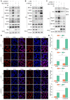
HDAC6 is associated with the H2A/H2A.X ubiquitination signaling cascade in DSB repair. (A–E) All cells were irradiated with 10 Gy followed by release for 1 h, after transfection with the indicated plasmids or siRNAs. (A) HeLa cells were transfected with Myc-HDAC6 or an empty plasmid for 48 h. Whole cell lysate and chromatin fractions were subjected to western blotting. (B) HeLa cells were transfected with HDAC6 or negative control (NC) siRNAs for 48 h. Whole cell lysate and chromatin fractions were subjected to western blotting. (C) HDAC6 KO HCT116 cells were transfected with Myc-HDAC6 or empty plasmid for 48 h. Whole cell lysate and chromatin fractions were subjected to immunoblotting. (D, E) HeLa cells were transfected with HDAC6 or negative control (NC) siRNAs for 48 h. The cells were then fixed, and the samples were labeled with the indicated antibodies. Representative images (D) and statistical analyses (E) are shown. The data represent the means ± SD (n = 5, *P< 0.05, **P< 0.01, ***P< 0.001).
Figure 4.
Figure 4.
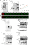
HDAC6 regulates the H2A/H2A.X ubiquitination signaling cascade in an RNF168-dependent manner during DSB repair. (A) HeLa cells were transfected with Flag-HDAC6 or an empty plasmid for 48 h. Whole cell lysates were subjected to immunoprecipitation with an anti-Flag antibody before western blotting. (B) HeLa cells were transfected with Myc-HDAC6 or empty plasmid for 48 h and exposed to 10 Gy irradiation (IR) and released for 1 h. Chromatin fractions were subjected to immunoprecipitation with an anti-H2A.X antibody followed by western blotting. Rabbit IgG was used as a negative control. (C, D) HeLa cells were co-transfected with GFP-RNF168 and mCherry-HDAC6 or an empty plasmid for 48 h and then subjected to laser micro-IR. Images were captured every 10 s for 200 s (C), and the IR path signal intensity was quantified (D). (E) Recombinant His-HDAC6 and GST-RNF168 were subjected to in vitro ubiquitination assays in the presence of ATP, E1 (UBE1), E2 (UbcH5c), ub (ubiquitin) and H2A/H2B dimer as indicated. Western blotting was performed with the indicated antibodies. (F, G) HeLa cells were transfected with the indicated plasmids for 48 h and then exposed to 10 Gy irradiation and released for 1 h. Whole cell lysate and chromatin fractions were analyzed by western blotting with the indicated antibodies. WT: wild-type; ΔDC1: DAC1 domain deleted; ΔDC2: DAC2 domain deleted; ΔZnF: ZnF-UBP domain deleted (F); HDAC6 catalytic mutant (Y386F/Y782F) (G). (H) HeLa cells were co-transfected with Flag-H2A.X, HA-ub, with Myc-HDAC6 WT or Myc-HDAC6 2YF (catalytic mutant) for 48 h and then exposed to 10 Gy irradiation and released for 1 h. The Flag-H2A.X proteins were immunoprecipitated from the whole cell lysates and eluted to detect the ubiquitination changes by western blotting with the indicated antibodies. All data represent the means ± SD.
Figure 5.
Figure 5.
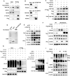
Nuclear HDAC6 is displaced from chromatin and degraded via the proteasome in response to DSBs. (AB) HeLa cells were exposed to an increasing dose of irradiation (IR) and released after 1 h or treated with 10 μM etoposide (VP16) for the indicated time. Then, whole cell lysate (β-actin), nuclear (Lamin-B1), and chromatin (H3) fractions were isolated and analyzed by western blotting. (C) HeLa cells were treated with 10 μM MG132 for 3 h or 50 μM CHQ for 12 h, then exposed to 10 Gy IR and released for 1 h before analysis of the whole cell lysate and nuclear fractions by western blotting. (D–F) HeLa cells were co-transfected with Flag-HDAC6 and HA-ub (D), Flag-HDAC6 and different HA-ubiquitin constructs (WT: wild type ubiquitin, K48R: K48-mutant ubiquitin, K63R: K63-mutant ubiquitin) (E) or Flag-HDAC6 wild-type (WT) or mutant (K116R) (F) for 48 h. Then, the cells were treated with 10 μM MG132 for 2 h, exposed to 10 Gy IR, and released for 1 h. The nuclear fractions were then subjected to immunoprecipitation with an anti-Flag antibody and analyzed by western blotting.
Figure 6.
Figure 6.
RNF168 directly interacts with HDAC6 and mediates DSB-induced nuclear HDAC6 degradation. (A) HeLa cells were transfected with Flag-HDAC6 or an empty plasmid for 48 h. Whole cell lysates were subjected to immunoprecipitation with an anti-Flag antibody and analyzed by western blotting. (B) Whole cell lysates from HeLa cells were extracted and subjected to immunoprecipitation using an anti-RNF168 (up) or an anti-HDAC6 (down) antibody and subsequent analysis by western blotting. Rabbit IgG served as a negative control. (C) HeLa cells were transfected with Flag-HDAC6 or an empty plasmid for 48 h. Then, the cells were treated with 10 μM MG132 for 2 h, exposed to 10 Gy irradiation (IR), and released for the indicated times. The nuclear fractions were subjected to immunoprecipitation with an anti-Flag antibody and analyzed by western blotting. (D) GST or GST-RNF168 fusion proteins were incubated with His-HDAC6 protein. Western blotting was performed to detect HDAC6 protein levels, and Coomassie Brilliant Blue (CBB) staining was performed to detect GST or GST-RNF168 levels (indicated by the asterisk). (E) HeLa cells were transfected with RNF168 or negative control (NC) siRNAs for 48 h then exposed to 10 Gy IR and released for 1 h before the whole cell lysate and nuclear fractions were analyzed by western blotting. (F) RNF168-WT or RNF168 KO HCT116 cells were transfected with Flag-RNF168 or empty plasmids for 48 h, then exposed to 10 Gy IR and released after 1 h before the whole cell lysate and nuclear fractions were analyzed by western blotting. (G) HCT116 cells were transfected with RNF168 or negative control (NC) siRNAs for 12 h, then co-transfected with the indicated plasmids for 48 h. Then, the cells were treated with 10 μM MG132 for 2 h and exposed to 10 Gy IR and released for 1 h before the nuclear fractions were subjected to immunoprecipitation with an anti-Flag antibody and analyzed by western blotting. (H) Recombinant His-HDAC6 and GST-RNF168 were subjected to in vitro ubiquitination assays performed in a reactive system containing E1 (UBE1), E2 (UBE2D3), HDAC6 (substrate), and ubiquitin with increasing RNF168 (E3 ligase). The reactions were analyzed by western blotting. (I) HDAC6 KO cells were transfected with Flag-HDAC6 wild-type (WT) or mutant (K116R) for 48 h, then exposed to 10 Gy IR and released after 1 h before the whole cell lysate and chromatin fractions were analyzed by western blotting.
Figure 7
Figure 7
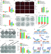
RNF168-mediated nuclear HDAC6 degradation is beneficial for DSB repair and cell survival. (A, B) pEJ5-GFP U2OS (A) and DR-U2OS (B) cells were transfected with Flag-HDAC6 wild-type (WT) or mutants (K116R or 2YF) for 48 h and subjected to NHEJ (A) and HR (B) assays, respectively. (C, D) HeLa cells were transfected with Flag-HDAC6 wild-type (WT) or mutants (K116R or 2YF) for 48 h, exposed to 10 Gy irradiation (IR), and then allowed to recover under normal conditions for the indicated times. All samples were then analyzed by comet assay. Representative images (C) and data analysis (D) are shown. (E, F) HDAC6-WT, HDAC6 KO, HDAC6 KO re-transfected Flag-HDAC6(WT), and Flag-HDAC6(K116R) stably expressing cells were exposed to 3 Gy IR and released for 8 h. The cells were then treated with colcemid (0.03 μg/ml) for an additional 6 h before chromosomal abnormality analysis. Representative images (E) and data analysis (F) are shown. (G, H) HeLa cells were transfected with Flag-HDAC6 wild-type (WT) or mutants (K116R or 2YF) for 48 h and then exposed to 3 Gy IR. The cells were counted and seeded for colony formation assays. For the control (Ctr) group, 500 cells were seeded in each plate, while for the IR-treated group, 5000 cells were seeded in each plate. (I, J) HDAC6-WT, HDAC6 KO, HDAC6 KO re-transfected Flag-HDAC6 (WT), and HDAC6 KO re-transfected Flag-HDAC6 (K116R) stably expressing cells were exposed to 3 Gy IR before being counted and seeded for colony formation assays. For the control (Ctr) group, 500 cells were seeded in each plate, while for the IR-treated group, 5000 cells were seeded in each plate. (K) A model of the role played by HDAC6 in DSB repair. All data represent the means ± SD (n = 60 for E–F, I–J; n = 3 for A–D, G–H; *P< 0.05, **P< 0.01, ***P< 0.001).

Similar articles

Cited by

References

    1. Lord C.J., Ashworth A.. The DNA damage response and cancer therapy. Nature. 2012; 481:287–294. - PubMed
    1. Ciccia A., Elledge S.J.. The DNA damage response: making it safe to play with knives. Mol. Cell. 2010; 40:179–204. - PMC - PubMed
    1. Panier S., Durocher D. Push back to respond better: regulatory inhibition of the DNA double-strand break response. Nat. Rev. Mol. Cell Biol. 2013; 14:661–672. - PubMed
    1. Jackson S.P., Bartek J.. The DNA-damage response in human biology and disease. Nature. 2009; 461:1071–1078. - PMC - PubMed
    1. Zhang K., Ning Y., Kong F., Chen X., Cai Y.. Genome instability in pathogenesis of tuberculosis. Genome Instab. Dis. 2021; 2:331–338.

Publication types