. 2023 Sep;50(3):169.
doi: 10.3892/or.2023.8606. Epub 2023 Jul 28.

Girdin regulates both migration and angiogenesis in pancreatic cancer cell lines

Affiliations

Girdin regulates both migration and angiogenesis in pancreatic cancer cell lines

Yuichi Hayashi et al. Oncol Rep. 2023 Sep.

Abstract

Girdin, an actin‑binding protein, is reportedly involved in the invasion and angiogenesis of various cancers. It has been suggested that the flavonoid Scutellarin (SCU) inhibits Girdin signaling. In the present study, the function and therapeutic applications of Girdin in pancreatic cancer (PaCa) were investigated. Immunohistochemical staining of Girdin in resected PaCa specimens from the Department of Gastroenterological Surgery, Nagoya City University Graduate School of Medical Science showed that high Girdin expression was associated with poor overall survival and relapse‑free survival, as well as with T factor, indicating invasion into the surrounding tissues. On the other hand, Girdin was highly expressed in almost all PaCa cell lines, and the migration ability of Girdin‑knockdown cell lines was decreased even under epidermal growth factor (EGF) stimulation. In addition, SCU suppressed PaCa cell migration by inhibiting the phosphorylation of Girdin. The expression and production of vascular endothelial growth factor A (VEGF‑A) was significantly decreased in Girdin‑knockdown cell lines. Furthermore, in Matrigel tube formation assays performed using culture supernatant, the lumen‑forming ability of vascular endothelial cells was also decreased in Girdin‑knockdown cell lines. However, SCU treatment did not significantly alter the expression or production of VEGF‑A. These results suggested that Girdin is involved in EGF signaling‑mediated migration of PaCa cells, that SCU inhibits PaCa invasion by suppressing Girdin activity, and that Girdin is also involved in angiogenesis via an activation pathway different from the action site of SCU. Girdin may be a prognostic biomarker, and the development of a novel molecular‑targeted drugs for Girdin may improve the prognosis of PaCa in the future.

Keywords: cancer angiogenesis; cancer migration; girdin; pancreatic cancer; scutellarin.


Conflict of interest statement

The authors declare that they have no competing interests.

Figures
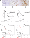
Figure 1.
Figure 1.
Relationship between Girdin expression in PaCa specimens and its prognosis. (A) IHC of tissues resected from patients with PaCa at Nagoya City University Graduate School of Medical Sciences. Paraffin-embedded PaCa tissues were analyzed for Girdin expression by IHC using an anti-Girdin antibody. In the PaCa tissues, there were differences in the staining intensity for Girdin, and they were classified into four types according to the intensity (−, +, ++ and +++). The number of cases in each of the four staining intensity categories was follows: -, 6 cases; +, 23 cases; ++, 38 cases; and +++, 23 cases. (B) Kaplan-Meier curves showing OS RFS in 90 PaCa patients at Nagoya City University Graduate School of Medical Sciences. The tissues in both survival groups were classified into the weakly stained (n=29) and the strongly stained (n=61) according to the staining intensity. (i.e. ‘-’ and ‘+’ represent weak staining, ‘++’ and ‘+++’ represent strong staining). (C) Kaplan-Meier curves showing OS and RFS of 177 and 69 PaCa patients, respectively, from the database in the Kaplan-Meier plotter website (https://kmplot.com/analysis/). PaCa, pancreatic cancer; IHC, Immunohistochemistry; OS, overall survival; RFS, relapse-free survival.
Figure 2.
Figure 2.
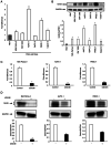
Expression and knockdown of Girdin in PaCa cell line. (A) Expression of Girdin mRNA in normal pancreatic and PaCa cell lines. The reverse transcription-quantitative PCR experiments were calculated using a comparative quantitative method with calibration curves. The vertical axis represents the ratio of Girdin expression to that in the human pancreatic ductal epithelial cell lines. (B) Western blotting was performed using Girdin and GAPDH antibodies. The density of bands detected was measured, and the ratio of Girdin to GAPDH was plotted. (C) Transfection of PaCa cell lines with negative control siRNA or Girdin siRNA. Girdin was knocked down by transfection of Girdin siRNA for MIA PaCa-2, AsPC-1 and PANC-1. (D) The knockdown of Girdin in these cell lines was confirmed by western blotting. The data are presented as the mean ± SEM. All experiments were performed in triplicate and repeated three times. *P<0.01 [A: One-way ANOVA with Dunnett's multiple comparison test. B: Student's t-test (unpaired), two-tailed]. PaCa, pancreatic cancer; siRNA, small interfering RNA.
Figure 3.
Figure 3.
PaCa cells migration is enhanced by EGF stimulation and decreased by suppression of Girdin activation. (A) In vitro cell migration assays in Boyden double chambers. The Transwell assays were performed as described in Materials and Methods. The data are presented as the mean ± SEM. All of the experiments were performed in triplicate and repeated three times. (B) Immunocytochemistry and IF with anti-phosphate-specific Girdin antibody. MIA PaCa-2 cells were cultured for 2 h in the presence or absence of EGF, followed by fixation and IF staining. Anti-β-actin mouse and anti-Girdin (phospho Y1764) rabbit antibodies were used as primary antibodies. Anti-mouse goat IgG (Alexa Fluor® 488; green) and anti-rabbit goat IgG (Cy3; red) were used as secondary antibodies, and DAPI was used for nuclear staining (blue). *P<0.01 (one-way ANOVA with Bonferroni's multiple comparison test, two-tailed). IF, immunofluorescence; si-, small interfering.
Figure 4.
Figure 4.
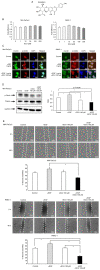
SCU suppresses PaCa cell migration by inhibiting Girdin phosphorylation. (A) The chemical structure of SCU. SCU is a flavonoid with a molecular weight of 462.36 g/mol. (B) The toxicological effects of SCU on PaCa cells were evaluated using the WST-1 assay. SCU was administered to MIA PaCa-2 and PANC-1 at various concentrations and incubated for 72 h, followed by addition of WST-1 reagent and measurement of absorbance. The values are expressed relatively to the control. (C) Immunocytochemistry/Immunofluorescence was used to evaluate the level of phosphorylated Girdin in MIA PaCa-2 and PANC-1 cells with or without EGF and 100 µM SCU. The cells were incubated with each reagent for 30 min and then stained. White arrows indicate the location of the lamellipodia enhanced by EGF stimulation. In addition, western blotting was used to quantify phosphorylated Girdin (Y1764). (D) The changes in Girdin and p-Girdin expression with SCU treatment were evaluated by western blotting. The ratio of p-Girdin to total Girdin (T-Girdin) is shown in the graph. The expression of p-Girdin was enhanced by EGF (1 ng/ml) stimulation, whereas p-Girdin expression was significantly suppressed under SCU treatment, even under EGF stimulation. (E) The effects of SCU on the migration ability of PaCa cells was evaluated by wound healing assay. MIA PaCa-2 and PANC-1 cells were seeded in 24-well plates at 100,000 cells per well, respectively. On the following day, after cell settlement, scratches were made in a monolayer using a pipette tip. Each reagent was added to medium containing 2% FBS, and the cells were incubated for 48 h and then observed under a phase contrast microscope. The length of the gaps at three random locations were measured and averaged. The data are presented as the mean ± SEM. All experiments were performed in triplicate and repeated three times. *P<0.01 (B: One-way ANOVA with Dunnett's multiple comparison test. D and E: One-way ANOVA with Bonferroni's multiple comparison test, two-tailed). SCU, scutellarin; PaCa, pancreatic cancer; p-, phosphorylated.
Figure 5.
Figure 5.
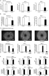
Girdin regulates angiogenic activity by upregulating VEGF-A gene expression. (A and B) Evaluation of VEGF-A (A) expression and (B) secretion in PaCa cell lines (MIA PaCa-2, AsPC-1 and PANC-1) with knockdown of Girdin. (A) RT-qPCR experiments were calculated using a comparative quantitative method with calibration curves. The vertical axis of the figure shows the ratio of VEGF-A expression to that of the respective control group. (B) After knockdown of Girdin in each cell line, the cells were cultured in medium containing 2% FBS for 72 h, and the supernatant was collected to measure VEGF-A by ELISA. The vertical axis of the figure shows the absolute concentration of VEGF-A. (C) The HUVEC tube formation assay was used to evaluate the angiogenic ability in vitro. The PaCa cell lines after knockdown of Girdin were cultured in 2% FBS-containing medium for 72 h, and then the cell supernatant was mixed with the same volume of 2% FBS-containing medium to make conditioned medium (CM). In the present study, the EA.hy926 cell line was used as immortalized HUVECs. EA.hy926 cells were cultured in the prepared conditioned medium for 16 h on Matrigel, and the number of endotubes was counted by phase-contrast microscopy. (D) Effect of SCU on the angiogenic ability of PaCa cell lines. MIA PaCa-2, AsPC-1 and PANC-1 cells were treated with 1 ng/ml EGF and 100 µM SCU in 2% FBS-containing medium to verify the alteration of VEGF-A expression and secretion. For RT-qPCR, cDNA prepared 2 h after the addition of reagents was used; for ELISA, the cell supernatant collected 72 h after the addition of reagents was used. The data are presented as the mean ± SEM. All experiments were performed in triplicate and repeated three times. *P<0.01 (A and B: Student's t-test. C and D: One-way ANOVA with Bonferroni's multiple comparison test, two-tailed). PaCa, pancreatic cancer; SCU, scutellarin; RT-qPCR, reverse transcription-quantitative PCR; HUVEC, human umbilical vein endothelial cell.
Figure 6.
Figure 6.
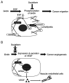
Working model of Girdin for migration and angiogenesis in PaCa cells. (A) A working model of the involvement of Girdin in the migratory ability of PaCa and the inhibitory effect of SCU. Girdin, which binds to actin fibers, is likely activated through phosphorylation via the EGF signaling pathway and involved in the migration ability of PaCa. SCU suppressed the migration ability of PaCa by inhibiting the activation of Girdin through EGF signaling. (B) A working model for the involvement of Girdin in the angiogenic ability of PaCa. Girdin appears to enhance angiogenesis of human umbilical vein endothelial cells by enhancing transcription and secretion of VEGF-A. SCU, which inhibits the phosphorylation of Girdin, had no effects on the expression of VEGF-A. Therefore, Girdin was suggested to be involved in the expression of VEGF-A through a different pathway. PaCa, pancreatic cancer; SCU, scutellarin.

Similar articles

Cited by

References

    1. Siegel RL, Miller KD, Fuchs HE, Jemal A. Cancer statistics, 2022. CA Cancer J Clin. 2022;72:7–33. doi: 10.3322/caac.21708. - DOI - PubMed
    1. Matsuo Y, Sawai H, Ochi N, Yasuda A, Sakamoto M, Takahashi H, Funahashi H, Takeyama H, Guha S. Proteasome inhibitor MG132 inhibits angiogenesis in pancreatic cancer by blocking NF-kappaB activity. Dig Dis Sci. 2010;55:1167–1176. doi: 10.1007/s10620-009-0814-4. - DOI - PubMed
    1. Enomoto A, Murakami H, Asai N, Morone N, Watanabe T, Kawai K, Murakumo Y, Usukura J, Kaibuchi K, Takahashi M. Akt/PKB regulates actin organization and cell motility via Girdin/APE. Dev Cell. 2005;9:389–402. doi: 10.1016/j.devcel.2005.08.001. - DOI - PubMed
    1. Omori K, Asai M, Kuga D, Ushida K, Izuchi T, Mii S, Enomoto A, Asai N, Nagino M, Takahashi M. Girdin is phosphorylated on tyrosine 1798 when associated with structures required for migration. Biochem Biophys Res Commun. 2015;458:934–940. doi: 10.1016/j.bbrc.2015.02.065. - DOI - PubMed
    1. Yamamura Y, Asai N, Enomoto A, Kato T, Mii S, Kondo Y, Ushida K, Niimi K, Tsunoda N, Nagino M, et al. Akt-Girdin signaling in cancer-associated fibroblasts contributes to tumor progression. Cancer Res. 2015;75:813–823. doi: 10.1158/0008-5472.CAN-14-1317. - DOI - PubMed

MeSH terms

Substances

Grants and funding

Funding: No funding was received.