[Preprint]. 2023 Jul 19:2023.07.18.549504.
doi: 10.1101/2023.07.18.549504.

ACKR3-arrestin2/3 complexes reveal molecular consequences of GRK-dependent barcoding

Affiliations

ACKR3-arrestin2/3 complexes reveal molecular consequences of GRK-dependent barcoding

Qiuyan Chen et al. bioRxiv. .

Abstract

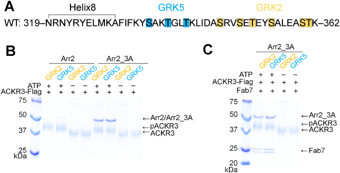
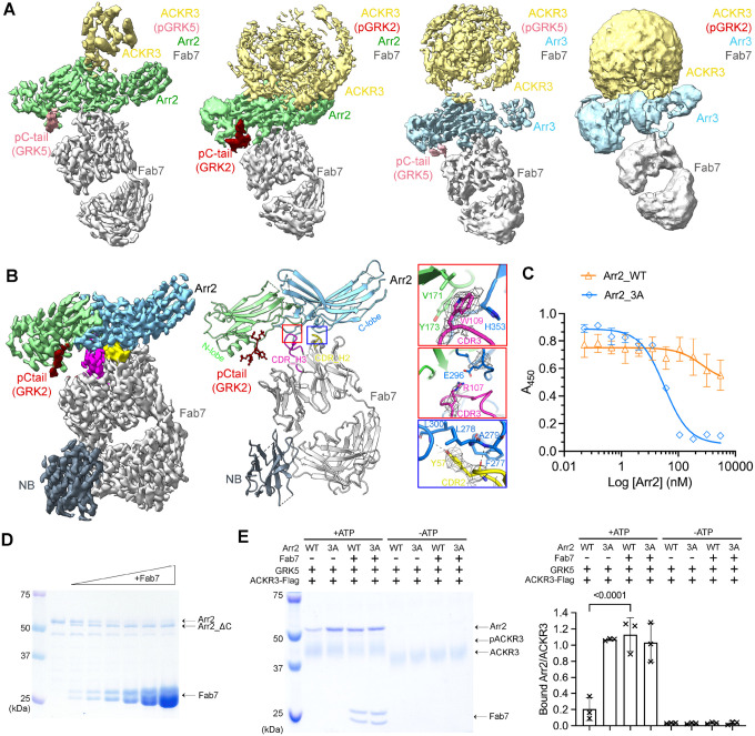
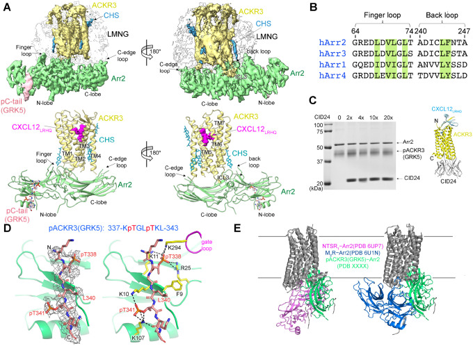
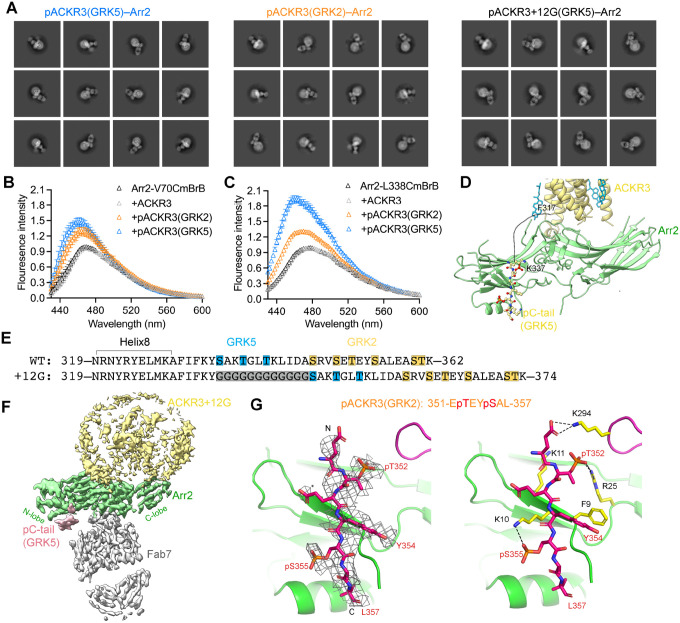
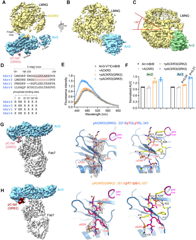
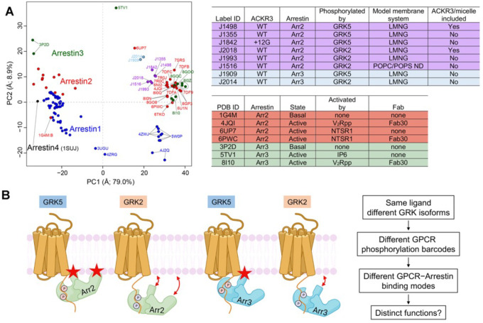
Atypical chemokine receptor 3 (ACKR3, also known as CXCR7) is a scavenger receptor that regulates extracellular levels of the chemokine CXCL12 to maintain responsiveness of its partner, the G protein-coupled receptor (GPCR), CXCR4. ACKR3 is notable because it does not couple to G proteins and instead is completely biased towards arrestins. Our previous studies revealed that GRK2 and GRK5 install distinct distributions of phosphates (or "barcodes") on the ACKR3 carboxy terminal tail, but how these unique barcodes drive different cellular outcomes is not understood. It is also not known if arrestin2 (Arr2) and 3 (Arr3) bind to these barcodes in distinct ways. Here we report cryo-electron microscopy structures of Arr2 and Arr3 in complex with ACKR3 phosphorylated by either GRK2 or GRK5. Unexpectedly, the finger loops of Arr2 and 3 directly insert into the detergent/membrane instead of the transmembrane core of ACKR3, in contrast to previously reported "core" GPCR-arrestin complexes. The distance between the phosphorylation barcode and the receptor transmembrane core regulates the interaction mode of arrestin, alternating between a tighter complex for GRK5 sites and heterogenous primarily "tail only" complexes for GRK2 sites. Arr2 and 3 bind at different angles relative to the core of ACKR3, likely due to differences in membrane/micelle anchoring at their C-edge loops. Our structural investigations were facilitated by Fab7, a novel Fab that binds both Arr2 and 3 in their activated states irrespective of receptor or phosphorylation status, rendering it a potentially useful tool to aid structure determination of any native GPCR-arrestin complex. The structures provide unprecedented insight into how different phosphorylation barcodes and arrestin isoforms can globally affect the configuration of receptor-arrestin complexes. These differences may promote unique downstream intracellular interactions and cellular responses. Our structures also suggest that the 100% bias of ACKR3 for arrestins is driven by the ability of arrestins, but not G proteins, to bind GRK-phosphorylated ACKR3 even when excluded from the receptor cytoplasmic binding pocket.


Figures

Figure 1.
Figure 1.. Arrestin binding to ACKR3 depends on GRK2/5 phosphorylation.
(A) Sequence of the ACKR3 C-tail with the observed GRK2 phosphorylation sites highlighted in orange and the unique sites observed for GRK5 in blue. (B) A flag pulldown assay shows that comparable amounts of Arr2_3A, but not WT Arr2, binds to ACKR3 phosphorylated by GRK2 or GRK5. The interaction is abolished when ACKR3 is not phosphorylated. (C) Flag pulldown assays show that Fab7 coelutes with pACKR3(GRK2)–Arr2_3A and pACKR3(GRK5)–Arr2_3A. This interaction is dependent on GRK activity because non-phosphorylated ACKR3 does not pulldown Arr2_3A or Fab7.
Figure 2.
Figure 2.. Structural and functional characterization of Fab7, a new arrestin conformational sensor.
(A) Sharpened maps of the 3.2 Å pACKR3(GRK5)–Arr2–Fab7, the 3.5 Å pACKR3(GRK2)–Arr2–Fab7, the 3.9 Å pACKR3(GRK5)–Arr3–Fab7, and the 7.3 Å pACKR3(GRK2)–Arr3–Fab7 complexes. ACKR3 in all four complexes is solubilized in LMNG/CHS detergent micelles. (B) Sharpened map and model of the 3.0 Å pACKR3(GRK2)–Arr2–Fab7–NB in POPC/POPS nanodiscs (PDB entry XXXX). The density of the ACKR3 TM domain and the nanodisc is not evident. Insets show interfacial details discussed in the text. (C) ELISA analysis of Fab7 competition assay reveals that preactivated Arr2 (IC50 ~35 nM) competes for Fab7 binding more efficiently than Arr2 WT (IC50 > 5 μM). Error bars represent S.D. from three technical replicates. (D) Limited trypsin digestion of Arr2 WT in the presence of increasing concentrations of Fab7. (E) A Flag pulldown assay shows that Fab7 significantly promotes Arr2 WT binding to pACKR3(GRK5), but it does not increase Arr2 WT or 3A binding non-phosphorylated ACKR3. One representative gel is shown. The ratios between the density of bound Arr2 and that of bound ACKR3 are compared using one-way ANOVA followed by a Dunnett’s multiple comparison test (P < 0.0001). Error bars represent S.D. from three technical replicates.
Figure 3.
Figure 3.. The interface between pACKR3(GRK5) and Arr2 is supported by a novel and several conventional interactions.
(A) Sharpened maps and models of pACKR3(GRK5)–Arr2 from the pACKR3(GRK5)–Arr2–Fab7 complex (PDB entry XXXX) with Fab7 density omitted. (B) Sequence alignment of the arrestin finger and back loops. Conserved hydrophobic residues involved in receptor and detergent/membrane binding are highlighted in green. (C) A flag pulldown assay in the absence or presence of increasing concentrations of CID24 shows no competition with Arr2 binding. CID24 binds to the cytoplasmic cleft of ACKR3 (PDB entry 7SK6) and thus blocks the access to the TM core. (D) Interactions of the pACKR3(GRK5) C-tail with the Arr2 N-lobe in the pACKR3(GRK5)–Arr2–Fab7 complex (PDB entry XXXX). Electron density for the pACKR3(GRK5) phospho-peptide is shown as a wire cage contoured at 12σ. Phosphate interactions below 4 Å are shown as black dash lines. (E) Comparison of Arr2 from the pACKR3(GRK5) structure with the NTSR1 (PDB entry 6UP7) and the M2R (PDB entry 6U1N) complexes after alignment of the receptor TM cores.
Figure 4.
Figure 4.. GRK barcoding dictates distinct arrestin binding modes to ACKR3.
(A) 2D class averages for pACKR3(GRK5)–Arr2–Fab7, pACKR3(GRK2)–Arr2–Fab7 and pACKR3+12G(GRK5)–Arr2–Fab7. (B, C) Fluorescence spectra of Arr2-V70CmBrB (B) and Arr2-L338CmBrB (C) alone (black), or in the presence of non-phosphorylated (grey), GRK2 (orange) or GRK5 (blue) phosphorylated ACKR3. Error bars represent S.E. from three technical replicates. (D) An 18-residue disordered linker from ACKR3 TM7 to the beginning of the GRK5 barcode is shown as a dashed line. The model shown is that of the pACKR3(GRK5)–Arr2–Fab7 complex (PDB entry XXXX). (E) Twelve glycine residues were inserted between Helix8 and the C-tail of ACKR3 (ACKR3+12G) to extend the GRK5 phosphorylation sites to where the GRK2 phosphorylation sites begin. (F) Sharpened map of the 3.3Å pACKR3+12G(GRK5)–Arr2–Fab7 complex highlights a more heterogenous interface between ACKR3+12G and Arr2. (G) Interactions of the ACKR3 C-tail phosphorylated by GRK2 with Arr2 N-lobe in the pACKR3(GRK2)–Arr2–Fab7 complex (PDB entry XXXX). The electron density of ACKR3 GRK2 phospho-peptide is shown as a wire cage contoured at 10σ. Phosphate contacts below 4 Å are shown as black dashed lines.
Figure 5.
Figure 5.. Arr3 binds to ACKR3 in a unique way compared to Arr2, but with similar responses to barcoding by different GRK isoforms.
(A, B) Sharpened map of pACKR3(GRK5)–Arr3 from the pACKR3(GRK5)–Arr3–Fab7 complex with Fab7 omitted. (C) Sharpened map of pACKR3(GRK5)–Arr2 from the pACKR3(GRK5)–Arr2–Fab7 complex with Fab7 omitted. Arr2 is positioned at the same angle as Arr3 in (B) to highlight the ~30° difference in orientation with respect to detergent micelle. (D) Sequence alignment of arrestin C-edge loops and receptor phosphates binding sites. The C-edge loop which is unique in Arr2 is highlighted in pink. (E) Fluorescence spectra of Arr3-V71CmBrB alone (black), or in the presence of non-phosphorylated ACKR3 (grey), pACKR3(GRK2) (orange) or pACKR3(GRK5) (blue). Error bars represent S.D. from three technical replicates. (F) Area under curve (AUC) value from the data in 4B and 5C normalized to Arr2 and Arr3, respectively, allows a direct comparison between Arr2 and Arr3. The AUC value obtained in the presence of GRK2 or GRK5 phosphorylated ACKR3 was compared between Arr2 and Arr3 using t test and p value is shown. (G) Sharpened map of pACKR3(GRK5)–Arr3–Fab7. Interactions of the ACKR3 C-tail phosphorylated by GRK5 with the Arr3 N-lobe in the pACKR3(GRK5)–Arr3–Fab7 complex. Electron density of the pACKR3(GRK5) phospho-peptide is shown as a wire cage contoured at 10σ. Distances below 4Å are shown as black dash line. (H) Sharpened map of pACKR3(GRK2)–Arr3–Fab7. Interactions of the ACKR3 C-tail phosphorylated by GRK5 with the Arr3 N-lobe in the pACKR3(GRK2)–Arr3–Fab7 complex. Electron density of the pACKR3(GRK2) phospho-peptide is shown as a wire cage contoured at 12σ. Distances below 4 Å are shown as black dash line.
Figure 6.
Figure 6.. PCA reveals the conformational landscape of arrestins and their complexes with ACKR3.
(A) Conformational map derived from all previously deposited arrestin structures with new structures of arrestin–Fab7 complexes from this paper (light blue and purple circles) superposed. Blue, red, green, and black circles otherwise correspond to structures that include Arr1, Arr2, Arr3, and Arr4, respectively. Structures with Fab7 (light blue and purple circles) or Fab30 (red and green and PC1>15, on right) fall in distinct clusters. Detailed information on the models used for PCA is provided in Table S2. The PC1 axis corresponds to the well-established twist between the N- and C-lobes of arrestin characteristic of activation (Movie S1), whereas the PC2 axis corresponds to an activation-independent “wag” of the C-lobe relative to the N-lobe (Movie S2). The “ACKR3/micelle included” column refers to whether the solubilized receptor was included in the reconstruction (i.e., nanodisc (ND) or LMNG micelle). (B) The distinct configurations of ACKR3–arrestin complexes mediated by different GRK barcodes and different arrestin isoforms identified in this paper may be generally applicable to other 7TM receptors and trigger distinct cellular outcomes. Stars indicate the position of the finger and C-edge loops (Arr2 only) as they engage the membrane. The GRK2 barcode in the C tail of ACKR3 is further from the receptor core than that of GRK5, yielding in our experiments a larger proportion of “tail-mode” complexes. In the case of ACKR3, its 100% bias towards arrestin seems to be entirely driven by GRK phosphorylation and not receptor interactions with arrestin.

Similar articles

References

    1. Balkwill F. (2004). The significance of cancer cell expression of the chemokine receptor CXCR4. Semin Cancer Biol 14, 171–179. - PubMed
    1. Beyett T.S., Chen Q., Labudde E.J., Krampen J., Sharma P.V., and Tesmer J.J.G. (2020). Characterization of a hyperphosphorylated variant of G protein-coupled receptor kinase 5 expressed in E. coli. Protein Expr Purif 168, 105547. - PMC - PubMed
    1. Bloch J.S., Mukherjee S., Kowal J., Filippova E.V., Niederer M., Pardon E., Steyaert J., Kossiakoff A.A., and Locher K.P. (2021). Development of a universal nanobody-binding Fab module for fiducial-assisted cryo-EM studies of membrane proteins. Proc Natl Acad Sci U S A 118. - PMC - PubMed
    1. Bous J., Fouillen A., Orcel H., Trapani S., Cong X., Fontanel S., Saint-Paul J., Lai-Kee-Him J., Urbach S., Sibille N., et al. (2022). Structure of the vasopressin hormone-V2 receptor-beta-arrestin1 ternary complex. Sci Adv 8, eabo7761. - PMC - PubMed
    1. Burns J.M., Summers B.C., Wang Y., Melikian A., Berahovich R., Miao Z., Penfold M.E., Sunshine M.J., Littman D.R., Kuo C.J., et al. (2006). A novel chemokine receptor for SDF-1 and I-TAC involved in cell survival, cell adhesion, and tumor development. J Exp Med 203, 2201–2213. - PMC - PubMed

Publication types