. 2023 Sep 7;31(9):1086-1099.e6.
doi: 10.1016/j.str.2023.06.014. Epub 2023 Jul 19.

Conformational exchange at a C2H2 zinc-binding site facilitates redox sensing by the PML protein

Affiliations

Conformational exchange at a C2H2 zinc-binding site facilitates redox sensing by the PML protein

Thomas A Bregnard et al. Structure. .

Abstract

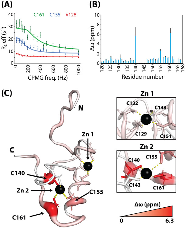
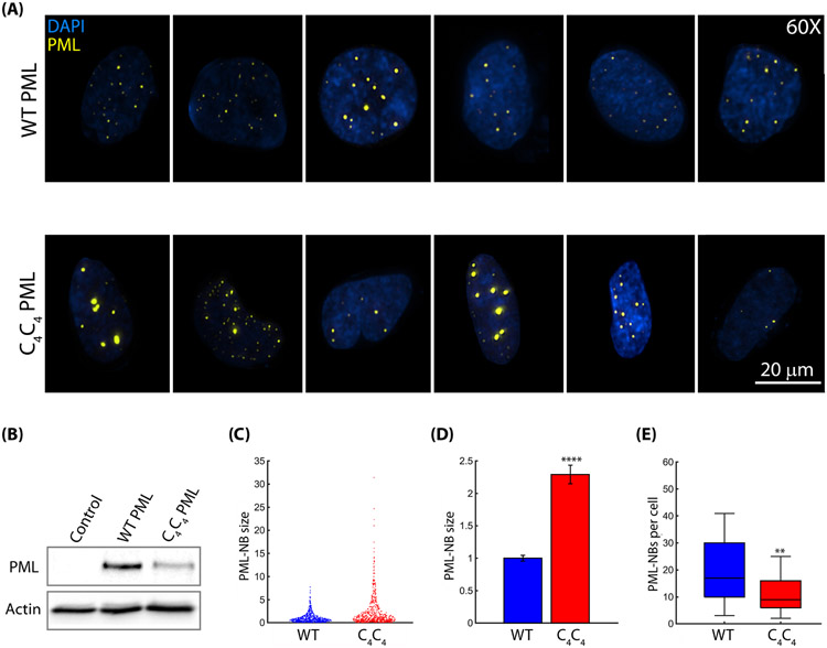
The promyelocytic leukemia protein, PML, plays a vital role in the cellular response to oxidative stress; however, the molecular mechanism of its action remains poorly understood. Here, we identify redox-sensitive sites of PML. A molecule of PML is cysteine-rich and contains three zinc-binding domains including RING, B-box1, and B-box2. Using in vitro assays, we have compared the sensitivity of the isolated RING and B-box1 domains and shown that B-box1 is more sensitive to oxidation. NMR studies of PML dynamics showed that one of the Zn-coordination sites within the B-box1 undergoes significant conformational exchange, revealing a hotspot for exposure of reactive cysteines. In agreement with the in vitro data, enhancement of the B-box1 Zn-coordination dynamics led to more efficient recruitment of PML into PML nuclear bodies in cells. Overall, our results suggest that the increased sensitivity of B-box1 to oxidative stress makes this domain an important redox-sensing component of PML.

Keywords: Carr-Purcell-Meiboom-Gill (CPMG); PML nuclear bodies; conformational change; molecular dynamics; nuclear magnetic resonance (NMR); oxidative stress; promyelocytic leukemia (PML); relaxation dispersion; zinc finger.


Conflict of interest statement

Declaration of interests The authors declare that they have no conflicts of interest with the contents of this article.

Figures

Figure 1:
Figure 1:. Treatment with the oxidizing agent H2O2 induces multimerization of RING and B-box1.
(A) Diagram of the TRIM of PML. Numbers above each domain indicate amino acid sequence boundaries. The types of zinc-coordinating sites are indicated; RING, B-box1, and B-box2 each coordinate two zinc ions through a set of four cysteine and/or histidine residues per zinc. (B) In vitro treatment of isolated RING (top) and B-box1 (bottom) domains with a range of concentrations of H2O2. Non-reduced gels (left) indicate dose-dependent multimerization of both RING and B-box1 in response to H2O2. Reduced gels (right) display little multimerization, suggesting that multimerization of both domains is through intermolecular disulfide bonds. Arrows to the right indicate the monomeric band for each domain: 6.5 kDa for RING, 6 kDa for B-box1. (C) PAR assay to detect zinc release. Both RING and B-box1 release zinc in response to H2O2 treatment in a dose-dependent fashion. Asterisks indicate statistically significant differences in zinc release between RING and B-box1 at the indicated concentration of H2O2. *, p < 0.05; **, p < 0.01; ***, p < 0.001; ****, p < 0.0001. (D) Quantification of soluble protein after H2O2 treatment. Purified RING and B-box1 domains were treated as in (B). After treatment, the precipitated protein was removed by centrifugation, and the remaining soluble protein was quantified by absorbance at 280 nm. Whereas RING remains highly soluble, B-box1 precipitates out of solution following exposure to high concentrations of H2O2. Asterisks indicate statistically significant differences in soluble protein relative to the 0 mM H2O2 control; p values are the same as in part (C). Error bars represent standard deviations derived from three experimental repeats.
Figure 2:
Figure 2:. PML B-box1 and RING display limited pico-nanosecond timescale motions.
(A) Per-residue S2 values for PML B-box1. Uncertainties were estimated using the covariance matrix method Local flexibility, indicated by lower S2, is limited almost exclusively to the N terminus and the immediate C terminus. (B) Structural representation of flexible regions of B-box1. More flexible residues appear thicker and red, while rigid portions of the domain are thinner and blue. The N-terminus and C-terminus are labeled. Insets at the right show close-up views of the zinc-coordinating residues. Dashed lines connect the atoms involved in coordination to each zinc atom (black spheres). (C) Per-residue S2 values for PML RING. Local flexibility is observed for residues 49-50 at N-terminus and residues 98-104 at the immediate C-terminus. Uncertainties were estimated using the covariance matrix method (D) Structural representation of flexible regions of RING.
Figure 3:
Figure 3:. C2H2 Zn-coordinating site of B-box1 undergoes conformational exchange.
(A) Representative 15N relaxation dispersion profiles of selected residues. Errors were estimated on the basis of repeat measurements at two CPMG fields, and a minimum error of 4% was used. (B) Amide 15N NMR chemical shift differences (Δω) between the major and minor conformations of B-box1. Uncertainties were estimated using the covariance matrix method (C) Values from (B) mapped onto the structure of the B-box1. The termini and the zinc atoms are labeled. Side chains of zinc-coordinating residues are shown as sticks. Residues with the largest Δω are labeled. Insets depict close-up views of the two zinc-coordinating sites.
Figure 4:
Figure 4:. B-box1 dimerization.
(A) Overlay of 15N-HSQC spectra of B-box1 at decreasing protein concentrations. The spectra are color-coded from red to cyan as indicated in the legend. The inset shows an enlarged area of the spectrum outlined by the square. (B) A bar graph of concertation-dependent chemical shift perturbations Δω. (C) Concertation-dependent chemical shift perturbations mapped on the structure of B-box1 (PDB ID: 2MVW). The Δω are color coded as a gradient from white (smallest) to cyan (largest) and identify the dimerization interface. (D) A graph of total Δω as a function of B-box1 concentration used to estimate the KD of dimerization. Δωtotal was calculated as a sum of individual backbone amide Δω. (E) Concertation-dependent multimer composition of B-box1 assessed by AUC c(s) distribution plots and used to estimate KD of dimerization.
Figure 5:
Figure 5:. Zinc-coordinating residues at the Zn 2 site of the B-box1 C4C4 mutant undergo conformational exchange.
(A) Representative 15N relaxation dispersion profiles of the B-box1 C4C4 mutant. Errors were estimated on the basis of repeat measurements at two CPMG fields , and a minimum error of 4% was used (B) Amide 15N NMR chemical shift differences (Δω) between the major and minor conformations of B-box1 C4C4 shown as bar graphs. Uncertainties were estimated using the covariance matrix method . (C) Δω from panel (B) mapped on a structural model of C4C4. More intense red color and thicker ribbon indicate larger Δω. The N- and C-termini, and zinc atoms are labeled. Side chains of zinc-coordinating residues are shown as sticks. Insets depict close-up views of the two zinc-coordinating sites. Note that data could not be obtained for residues 141-146.
Figure 6:
Figure 6:. Treatment with H2O2 induces multimerization and zinc release by a C4C4 mutant of B-box1.
(A) PAR assay to detect zinc release. C4C4 releases zinc more readily than WT B-box1 at concentrations below the reaction saturation point. Asterisks indicate statistically significant differences in zinc release between WT B-box1 and C4C4 at the indicated concentration of H2O2. *, p < 0.05; **, p < 0.01; ***, p < 0.001; ****, p < 0.0001. Data for WT B-box1 are reproduced from Figure 1C. Error bars represent standard deviations derived from three experimental repeats. (B) Normalized c(s) distributions of C4C4 at 0.1mM (top) and 1mM (bottom). The monomeric species is set to a height of 1 in each distribution. Numbers represent the predicted size of the primary peak of each protein in Da. (C) In vitro treatment of isolated C4C4 with a range of concentrations of H2O2. C4C4 multimerizes in a H2O2 dose-dependent fashion (left) but exists as a monomer under reducing conditions (right). Arrows to the right indicate the position of the 6kDa monomeric band. Bands at 28 and 34kDa correspond to traces of GST and GST-tagged domains, respectively, that remained after size-exclusion chromatography. (D) Quantification of soluble protein after H2O2 treatment. C4C4 remains nearly 100% soluble after centrifugation. Error bars represent standard deviations derived from three experimental repeats. (E) Impact of H155C and H161C mutations on Zn 2 coordination in C4C4. Point mutations were modeled on the B-box1 NMR structure (PDB ID: 2MVW) using PyMol. Mutation of H155 to cysteine increases the hypothetical coordination bond length, which likely alters local folding of C4C4 around the Zn 2 site.
Figure 7:
Figure 7:. PML-NB formation in cells expressing WT and oxidation-sensitive C4C4 mutant of PML.
(A) Six representative live cell images of PML−/− U2OS cells transfected with either wild type (top panel) or C4C4 mutant (bottom panel) of EYFP-tagged PML. Images are shown as an overlay of the DAPI (blue) and YFP (yellow) channels. A 20 μm scale bar is included. (B) Western blot of the PML expressed in PML−/− U2OS cells transfected with either WT or C4C4 mutant of EYFP-tagged PML (isoform I). Untransfected cells were used as a negative control. Actin served as a loading control. (C) Scatter plot of individual PML-NB sizes in cells transfected with WT PMLI (blue) and C4C4 mutant PMLI (red). PML-NB sizes were normalized to the mean WT size. (D) The plot of the mean PML-NB size in cells transfected with WT PMLI (blue) and C4C4 mutant PMLI (red). Error bars represent +/− SEM, n = 623. NB size was normalized to the mean WT size. Two-tailed t-test ****P<0.0001 (E) Boxplot of the number of PML NBs per cell for both wild type and C4C4 mutant of PMLI, n = 48. Two-tailed t-test **P=0.0013.
Figure 8:
Figure 8:. A model for the impacts of oxidation on PML multimerization.
Under reducing conditions, the isolated RING and B-box1 domains are monomeric. B-box1 exchanges between a major conformation that coordinates Zn 2 through four cysteine and histidine residues and a minor conformation in which one or both cysteine side chains are transiently exposed. Oxidative stress enables disulfide bond formation by cysteine side chains in the C2H2 site of B-box1, which leads to B-box1 multimerization. RING does not lose either zinc atom or form large multimers, although it can dimerize through a surface-exposed cysteine side chain (not shown). Intense oxidative stress promotes the release of zinc from RING and B-box1, which leads to extensive multimerization of both domains.

Similar articles

Cited by

References

    1. Lallemand-Breitenbach V, and de Thé H (2010). PML nuclear bodies. Cold Spring Harbor perspectives in biology 2, a000661. 10.1101/cshperspect.a000661. - DOI - PMC - PubMed
    1. Sahin U, Lallemand-Breitenbach V, and de Thé H (2014). PML nuclear bodies: regulation, function and therapeutic perspectives. The Journal of pathology 234, 289–291. 10.1002/path.4426. - DOI - PubMed
    1. Guan D, and Kao H-Y (2015). The function, regulation and therapeutic implications of the tumor suppressor protein, PML. Cell & Bioscience 5, 60. 10.1186/s13578-015-0051-9. - DOI - PMC - PubMed
    1. Lallemand-Breitenbach V, Zhu J, Puvion F, Koken M, Honoré N, Doubeikovsky A, Duprez E, Pandolfi PP, Puvion E, Freemont P, and de Thé H (2001). Role of promyelocytic leukemia (PML) sumolation in nuclear body formation, 11S proteasome recruitment, and As2O3 - induced PML or PML/retinoic acid receptor alpha degradation. The Journal of experimental medicine 193, 1361–1371. 10.1084/JEM.193.12.1361. - DOI - PMC - PubMed
    1. Luciani JJ, Depetris D, Usson Y, Metzler-Guillemain C, Mignon-Ravix C, Mitchell MJ, Megarbane A, Sarda P, Sirma H, Moncla A, et al. (2006). PML nuclear bodies are highly organised DNA-protein structures with a function in heterochromatin remodelling at the G2 phase. Journal of cell science 119, 2518–2531. 10.1242/jcs.02965. - DOI - PubMed

Publication types

MeSH terms

LinkOut - more resources