. 2023 Jul 18;13(1):11596.
doi: 10.1038/s41598-023-38156-z.

Mechanism and therapeutic implications of pomalidomide-induced immune surface marker upregulation in EBV-positive lymphomas

Affiliations

Mechanism and therapeutic implications of pomalidomide-induced immune surface marker upregulation in EBV-positive lymphomas

Hannah K Jaeger et al. Sci Rep. .

Abstract

Epstein-Barr virus (EBV) downregulates immune surface markers to avoid immune recognition. Pomalidomide (Pom) was previously shown to increase immune surface marker expression in EBV-infected tumor cells. We explored the mechanism by which Pom leads to these effects in EBV-infected cells. Pom increased B7-2/CD86 mRNA, protein, and surface expression in EBV-infected cells but this was virtually eliminated in EBV-infected cells made resistant to Pom-induced cytostatic effects. This indicates that Pom initiates the upregulation of these markers by interacting with its target, cereblon. Interestingly, Pom increased the proinflammatory cytokines IP-10 and MIP-1∝/β in EBV infected cells, supporting a possible role for the phosphoinositide 3-kinase (PI3K)/AKT pathway in Pom's effects. Idelalisib, an inhibitor of the delta subunit of PI3 Kinase, blocked AKT-Ser phosphorylation and Pom-induced B7-2 surface expression. PU.1 is a downstream target for AKT that is expressed in EBV-infected cells. Pom treatment led to an increase in PU.1 binding to the B7-2 promoter based on ChIP analysis. Thus, our data indicates Pom acts through cereblon leading to degradation of Ikaros and activation of the PI3K/AKT/PU.1 pathway resulting in upregulation of B7-2 mRNA and protein expression. The increased immune recognition in addition to the increases in proinflammatory cytokines upon Pom treatment suggests Pom may be useful in the treatment of EBV-positive lymphomas.


Conflict of interest statement

Drs. Yarchoan and Davis are co-inventors on US Patent 10,001,483 entitled “Methods for the treatment of Kaposi’s sarcoma or KSHV induced lymphoma using immunomodulatory compounds, and uses of biomarkers”. In is our understanding that foreign patents have also been filed for this invention. An immediate family member of R. Yarchoan is a co-inventor on patents or patent applications related to internalization of target receptors, epigenetic analysis, and ephrin tyrosine kinase inhibitors. These inventions were all made as full-time employees of the US government under 45 Code of Federal Regulations Part 7. All rights, title, and interest to these patents have been or should by law be assigned to the U.S. Department of Health and Human Services. The government conveys a portion of the royalties it receives to its employee inventors under the Federal Technology Transfer Act of 1986 (P.L. 99–502). This research was supported in part by a CRADA between the NCI and Celgene Corporation (now Bristol Myers Squibb). Dr. Yarchoan also reports receiving drugs for clinical trials from Merck, EMD-Serano, Eli Lilly, and CTI BioPharma through CRADAs with the NCI, and he has received drug supply for laboratory research from Janssen Pharmaceuticals.

Figures

Figure 1
Figure 1
Pom-resistant cells show low cereblon expression and Pom no longer increases surface markers or T-cell activation. Live cell number for (A) WT Daudi or (B) Pom-Resistant Daudi cells after DMSO (solid) or 1 µM Pom (dashed). (C) Representative immunoblots from 3 separate experiments of cytoplasmic extracts for cereblon and nuclear extracts for IKZF-1 following DMSO or 1 µM Pom treatment for 48 h. Protein was normalized to actin and the numbers under the blots represent the fold changes calculated against control (DMSO-treated) cells. (D) FACS analysis on live cells showing the mean fold changes + / − SD for B7-2 for DMSO (black bars) and Pom treated (grey bars), compared to DMSO-treated wild type (WT) cells. (E) Mean fold changes + / − SD for ICAM-1 for DMSO (black bars) and Pom treated (grey bars). Average fold change in median fluorescent intensity (MFI) compared to DMSO treated cells. Error bars represent standard deviation from 7 independent experiments (****p ≤ 0.0001, ***p ≤ 0.001, *p < 0.05, ns not significant). (F) Activation of Jurkat IL-2 reporter cells by WT and Pom-resistant cells exposed to varying amounts of Pom. WT or Pom-resistant Daudi cells were treated with DMSO vehicle, 1 or 10 µM Pom for 2 days and then co-incubated with Jurkat IL-2 reporter T-cells in the presence of varying concentrations of anti-CD3 antibody. Luminescence, a measure of T-cell activation, was measured after 6 h. Viability for BCBL-1 cells was greater than 83% for DMSO, 1 µM and 10 µM Pom. Results show the mean + / − SD from three independent experiments. (*p < 0.05, ns not significant).
Figure 2
Figure 2
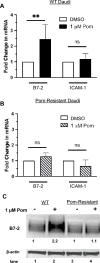
Pom increases the mRNA and protein levels for B7-2 in WT but not in Pom-resistant Daudi cells. (AB) Mean mRNA levels of B7-2 and ICAM-1 in (A) WT Daudi and (B) Pom-Resistant Daudi cells treated with DMSO (control) or Pom 1 µM after 24 h. The mRNA levels of B7-2 and ICAM-1 are normalized to 18S RNA. Error bars represent standard deviation from 7 independent experiments in WT Daudi cells and 3 independent experiments in Pom-Resistant Daudi cells. Statistically significant differences (**p ≤ 0.01, ns not significant) between control and Pom-treated cells are indicated. (C) Protein levels by immunoblot of B7-2 and β-actin from cytoplasmic lysates of WT or Pom-Resistant cells 48 h after DMSO or Pom treatment. Fold changes shown under each lane are values normalized to the actin level in the DMSO control for each sample.
Figure 3
Figure 3
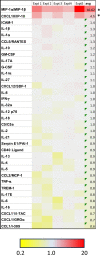
Pom significantly increases MIP-1∝/β and IP-10 in the supernatant of Daudi cells following Pom-treatment. Heat map depicting the fold changes over control for 35 different cytokines. Daudi cells were treated with 1 µM Pom for 48 h and then supernatant was collected and analyzed for changes in cytokine expression using the Proteome Profiler Human Cytokine Array Kit. Shown on the right column are the means for 5 independent experiments run in duplicate. Color range is from 0.2 to 20-fold with yellow indicating a decrease in levels, light blue indicating no change in levels and red indicating an increase in levels. Statistically significant differences between control and Pom-treated cells are indicated for those cytokines whose expression changed by at least an average of twofold (*p ≤ 0.05).
Figure 4
Figure 4
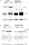
Pom increases PU.1 and B7-2 levels while decreasing Ikaros levels in WT but not in Pom-resistant cells. (A) Protein levels by immunoblot (using MES running buffer) for PU.1, B7-2, and β-actin from nuclear and cytoplasmic extracts of WT Daudi cells 48 h after DMSO or 1 µM Pom treatment. Fold changes shown under each lane are values normalized to the actin level in the DMSO control for each extract. (B) Protein levels by immunoblot using MOPS running buffer for PU.1, Ikaros, TBP and β-actin from nuclear lysates of WT and Pom-resistant Daudi cells 48 h after DMSO or Pom treatment. Fold changes shown under each lane are values normalized to TBP in the DMSO control for each cell type. Note that PU.1 runs as a doublet when using MOPS running buffer which provides better separation.
Figure 5
Figure 5
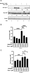
Idelalisib inhibits AKT-Ser473 phosphorylation and inhibits B7-2 surface expression induced by Pom. Daudi cells were pretreated with DMSO or PI3K δ-subunit inhibitor idelalisib for 2 h and then treated with 1 μM Pom for an additional 48 h. (A) Representative immunoblots of cytoplasmic extracts show AKT-Ser473 phosphorylation levels. Fold change of AKT phosphorylation was calculated by first normalizing to β-actin, followed by normalization to total AKT present and then to their respective DMSO controls. (BC) Surface expression levels of (B) B7-2 and (C) ICAM-1 were measured by flow cytometry using PerCP/Cy5.5-conjugated anti-B7-or anti-ICAM-1 antibodies in Daudi cells treated with Pom (1 μM) and/or idelalisib (1 or 10 μM). Graphs show fold changes in median fluorescence intensity (MFI) of (B) B7-2 and (C) ICAM-1 upon treatment with Pom and/or idelalisib relative to DMSO-treated cells. Shown are the means ± standard deviations of at least 4 separate experiments. Statistically significant differences (****p ≤ 0.0001, ***p ≤ 0.001, **p ≤ 0.01, *p ≤ 0.05, ns not significant) between control and Pom-treated cells are indicated. The immunoblots were performed 3 times and the result from one representative experiment is shown.
Figure 6
Figure 6
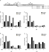
Pom increases PU.1 occupancy at the promoter of B7-2/CD-86. (A) Schematic for human B7-2 (CD86 gene) and promoter regions pA-pD. (BE) Fold enrichment levels for PU.1 in DMSO and Pom (1 µM) treated WT Daudi cells at the four promoter regions pA-pD using 4 different PU.1 antibodies (ab 1–4) and one antibody to IRF8 (ab5). The fold enrichments are adjusted to the values obtained using the IgG control antibody. Dashed line shows the IgG value to which all the antibodies were normalized. Error bars represent the standard deviation for each lane and values are normalized to the actin mRNA level in the DMSO control for each protein extract. Promoter regions are pA (− 677 to − 480), pB (− 217 to − 74), pC (− 74 to + 15) and pD (− 325 to − 258) relative to each isoform’s transcription start site.
Figure 7
Figure 7
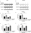
Idelalisib inhibits AKT-Ser473 phosphorylation and B7-2 surface expression induced by Pom in BL41 + and Namalwa cells. BL41 + and Namalwa cells were pretreated with DMSO or the PI3K δ-subunit inhibitor idelalisib for approximately 2 h and then treated with DMSO or 1 µM Pom for an additional 48 h. Representative immunoblots of cytoplasmic extracts show AKT phosphorylation levels with Pom treatment and/or idelalisib for (A) BL41 + cells or (B) Namalwa cells. Fold changes of AKT phosphorylation and total AKT are shown below the blot and calculated by first normalizing to β-actin. AKT phosphorylation was normalized to total AKT present after normalizing to actin. BL41 + and Namalwa cells treated as indicated in above were analyzed for surface expression levels of (C, D) B7-2 and (E, F) ICAM-1 by flow cytometry using PerCP/Cy5.5-conjugated anti-ICAM-1 or anti-B7-2 antibodies. Graphs show fold change in median fluorescent intensity (MFI) of surface expression levels measured via flow cytometry relative to DMSO-treated cells. The data are means + / − SD from 3 or more experiments and the statistically significant differences (****p ≤ 0.0001, ***p ≤ 0.001, **p ≤ 0.01, *p ≤ 0.05, ns not significant) between various treatments are indicated.

Similar articles

References

    1. Bjorklund CC, et al. Rate of CRL4(CRBN) substrate Ikaros and Aiolos degradation underlies differential activity of lenalidomide and pomalidomide in multiple myeloma cells by regulation of c-Myc and IRF4. Blood Cancer J. 2015;5:e354. doi: 10.1038/bcj.2015.66. - DOI - PMC - PubMed
    1. Chamberlain PP, et al. Structure of the human Cereblon-DDB1-lenalidomide complex reveals basis for responsiveness to thalidomide analogs. Nat. Struct. Mol. Biol. 2014;21:803–809. doi: 10.1038/nsmb.2874. - DOI - PubMed
    1. Chang X, Zhu Y, Shi C, Stewart AK. Mechanism of immunomodulatory drugs' action in the treatment of multiple myeloma. Acta Biochim. Biophys. Sin. Shanghai. 2014;46:240–253. doi: 10.1093/abbs/gmt142. - DOI - PMC - PubMed
    1. Polizzotto MN, et al. Pomalidomide for symptomatic Kaposi's Sarcoma in people with and without HIV infection: A phase I/II study. J. Clin. Oncol. 2016;34:4125–4131. doi: 10.1200/JCO.2016.69.3812. - DOI - PMC - PubMed
    1. Ramaswami R, et al. Safety, activity, and long-term outcomes of pomalidomide in the treatment of Kaposi Sarcoma among individuals with or without HIV infection. Clin. Cancer Res. 2022;28:840–850. doi: 10.1158/1078-0432.CCR-21-3364. - DOI - PMC - PubMed

Publication types

MeSH terms