. 2023 Jul 17;17(1):46.
doi: 10.1186/s13036-023-00367-3.

Study of the regulatory elements of the Ovalbumin gene promoter using CRISPR technology in chicken cells

Affiliations

Study of the regulatory elements of the Ovalbumin gene promoter using CRISPR technology in chicken cells

Sara Yousefi Taemeh et al. J Biol Eng. .

Abstract

Background: Hormone-dependent promoters are very efficient in transgene expression. Plasmid-based reporter assays have identified regulatory sequences of the Ovalbumin promoter that are involved in response to estrogen and have shown that the deletion of the steroid-dependent regulatory element (SDRE) and negative regulatory element (NRE) leads to a steroid-independent expression of a reporter. However, the functional roles of these regulatory elements within the native genomic context of the Ovalbumin promoter have not been evaluated.

Results: In this study, we show that the negative effects of the NRE element on the Ovalbumin gene can be counteracted by CRISPR interference. We also show that the CRISPR-mediated deletion of SDRE and NRE promoter elements in a non-oviduct cell can lead to the significant expression of the Ovalbumin gene. In addition, the targeted knock-in of a transgene reporter in the Ovalbumin coding region and its expression confirms that the truncated promoter of the Ovalbumin gene can be efficiently used for an estrogen-independent expression of a foreign gene.

Conclusions: The methodology applied in this paper allowed the study of promoter regulatory sequences in their native nuclear organization.

Keywords: Avian expression systems; CRISPR technology; Chicken fibroblast; Gene editing; Ovalbumin promoter; Regulatory sequences.


Conflict of interest statement

The authors declare no competing interests.

Figures

Fig. 1
Fig. 1
CRISPRi-mediated activation of the Ovalbumin promoter in DF1 cells. A The schematic representation of the promoter and coding region of the OVA gene in DF1 cells. Two regulatory elements of SDRE and NRE are shown in the distal promoter. The bottom panel shows binding sites for two guide RNAs (Silencer-gRNA and CAR-gRNA) that bind the silencer and CAR regions in the NRE element, respectively. SDRE, steroid-dependent regulatory element; NRE, negative regulatory element; CAR, COUP-adjacent repressor site; COUP, Chicken OVA upstream promoter; TATA, TATA box; TSS, transcription start site; dCas9, Catalytically dead Cas9. The enlarged inset in the lower section of panel A shows the location and orientation of PAM regions and protospacers for the two regulatory regions of ‘silencer’ and ‘CAR’. B The left panel shows agarose gel electrophoresis for analysis of the RT-PCR products which were amplified by primers P8 and P9 (for OVA, Fig. 2A and Table 1). The right panel shows agarose gel electrophoresis for analysis of the RT-PCR products which were amplified by P10 and P11 (for GAPDH, Table 1). RNA was extracted from DF1 cells which were transfected with CRISPRi vectors that target the NRE element at CAR, Silencer, both CAR and silencer sequences, and pdCas9-X as the negative control. The expected amplicon size for OVA was 179 bp, and for GAPDH was 187 bp. WT, wild-type; Magnum, hormonally-activated tissue of magnum from  a 35-week-old laying hen; M, DNA size marker; NTC, no template control. C Upregulation of the OVA mRNA in CRISPRi-modified DF1 cells was assessed by RT-qPCR. Upon transfection with CRISPRi vectors that target the NRE element at CAR, Silencer, and both CAR and silencer sequences, an increment in the OVA gene expression level was determined. The transcript levels for OVA in the hormonally-activated tissue of the magnum (from a 35-week-old laying hen) show the highest level of expression. The gene expression ratio for the OVA over GAPDH was calculated by the Pfaffl method of relative quantification [38]. For each group of CRISPRi-DF1 cells, three biological replicates were used. Each biological replicate was read as three technical replicates. The Mann–Whitney assay was used to analyze significant statistical differences between groups. The asterisk (*) indicates statistical differences with p values < 0.05
Fig. 2
Fig. 2
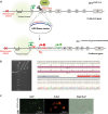
Design and validation of the targeted deletion of Ovalbumin distal promoter elements in DF1 cells. A The schematic representation of CRISPR/Cas9 mediated deletion strategy of the OVA promoter in DF1 cells. The top diagram shows the wild-type (WT) chicken OVA locus. The two guide RNA (SDRE-gRNA and NRE-gRNA) binding sites are shown. The NRE- and SDRE- gRNAs target two positions downstream of NRE (downstream of CAR) and upstream of SDRE, respectively. The bottom diagram shows the locus after CRISPR-mediated deletion of the distal OVA promoter in DF1 cells (DF1+/OVA Pro ∆ cell). The PCR primers used for the assessment of deletion (P5 to P7), and the OVA gene expression (P8 and P9, used in Figs. 1 and 3) are shown as small red arrows. B Two-step genomic PCR to confirm the deletion of the distal promoter of the OVA gene. In the first PCR (using P5 and P7 primers, Table 1), an amplicon of 1310 bp was amplified from the wild-type (WT) allele (In the first PCR, amplicon of ~ 370 bp were not detected from the promoter-deleted (DF1) alleles). In a hemi-nested PCR (using P5 and P6 primers), amplicons of 1256 bp and ~ 316 bp were amplified from the wild-type and promoter-deleted (DF1) alleles, respectively. C Alignment of the representative sequences of the wild-type (WT DF1) and promoter-deleted (DF1) sequences determined by Sanger sequencing. The gRNA-binding sites are shown in blue, and the PAM regions are shown in green letters. WT, wild-type; DF1 , DF1 cells knockout for the distal OVA promoter (DF1 +/OVA Pro ∆); NHEJ, non-homologous end-joining; ERE, estrogen-responsive enhancer element; TSSL, tissue-specific silencer-like element; SDRE, steroid-dependent regulatory element; NRE, negative regulatory element; CAR, COUP-adjacent repressor site; COUP, Chicken OVA upstream promoter; TATA, TATA box; TSS, transcription start site; P, primer. M, DNA size marker; NTC, no template control
Fig. 3
Fig. 3
Gene expression ratio for Ovalbumin transcript in DF1+/OVA Pro ∆ cells. A Agarose gel (2%) electrophoresis for analysis of the RT-PCR products amplified by primers P8 and P9 (for OVA, Fig. 1), and P10 and P11 (for GAPDH). The expected amplicon size for OVA and GAPDH are 179 bp and 187 bp, respectively. WT, wild-type; DF1 , distal OVA promoter knockout DF1 cells (DF1 +/OVA Pro ∆); M, DNA size marker; NTC, no template control; RT, reverse transcriptase. The full-length gel electrophoresis images are shown in Fig. S3. B Upregulation of the OVA mRNA in DF1 +/OVA Pro ∆cells was assessed by RT-qPCR. Upon deletion of the distal OVA promoter, an increased level of expression of the OVA gene was determined (DF1). The transcript levels of OVA for these samples (Three isogenic DF1 +/OVA Pro ∆ clones) were ~ 104-fold higher than the OVA transcript levels in the wild-type DF1 (WT DF1). The transcript levels for OVA in the hormonally-activated tissue of the magnum (from a 35-week-old laying hen) show the highest level of expression. The gene expression ratio for the OVA over GAPDH was calculated by the Pfaffl method of relative quantification [38]. The Mann–Whitney assay was used to analyze significant statistical differences between the WT-DF1 group and DF1 and magnum groups. * and ** show statistical differences with p values < 0.05 and < 0.01, respectively
Fig. 4
Fig. 4
Activation of transgene expression in DF1 +/OVA Pro ∆−Tg (promoterless dsRed) cells. A The schematic representation of CRISPR HDR mediated knockin strategy in DF1+/OVA Pro ∆ cells. The top diagram shows the donor vector that was designed to have a promoterless DsRed2 and a CMV-Puro-EGFP cassette flanked by left and right homology arms. The OVA E2 indicates the gRNA-binding site on exon 2 of the OVA (+ 174 to + 1784) gene. The bottom diagram shows the allele after CRISPR-HDR insertion of the reporter cassette (DF1 +/OVA Pro ∆−Tg (promoterless dsRed)). PCR primers (P12 and P13) were used for the assessment of the CRISPR-HDR insertion of the promoterless DsRed2 in DF1 +/OVA Pro ∆−Tg (promoterless dsRed) cells. B Genomic PCR analysis of the targeted gene knock-in DF1 +/OVA Pro ∆−Tg (promoterless dsRed) cells. For the assessment of the CRISPR-HDR insertion of the promoterless DsRed2 in DF1 ∆−Tg cells, primers (P12 and P13) were used to amplify a 2569 bp amplicon. The insertion-specific PCR products of DF1 ∆−Tg cells were sequenced by Sanger sequencing and aligned to the donor plasmid (used as a DNA repair template during transfection). C Fluorescence microscopy of DF1 ∆−Tg cells indicating DsRed2 expression under the control of the endogenous truncated OVA promoter, Magnification: 20X. DF1, DF1 cells knock-out for distal OVA promoter (DF1 +/OVA Pro ∆); DF1 ∆−Tg cells, promoterless DsRed2 knockin DF1 cells (DF1 +/OVA Pro ∆−Tg (promoterless dsRed)); HDR, homology-directed repair; M, DNA size marker; WT, wild-type; NTC, no template control
Fig. 5
Fig. 5
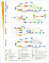
A schematic model depicting the mechanism of increased expression of the Ovalbumin gene in different cell types in steroid-dependent and –independent manners. The main induction for the expression of the OVA gene in oviduct cells is estrogen that by binding to the SDRE region overcomes the inhibitory circuits exerted by the tissue-specific silencer-like element (TSSL), and negative regulatory element (NRE). The CRISPR/CAS-mediated deletion of the regulatory sequences of the OVA distal promoter (SDRE, NRE, and the linker in between) leads to the expression of the OVA gene in DF1 cells. The CRISPR-mediated interference of regulatory sequences of the NRE element as well leads to an increased expression of the OVA gene in DF1 cells

Similar articles

Cited by

References

    1. Dehdilani N, Yousefi Taemeh S, Goshayeshi L, Dehghani H. Genetically engineered birds; pre-CRISPR and CRISPR era†. Biol Reprod. 2022;106:24–46. doi: 10.1093/biolre/ioab196. - DOI - PubMed
    1. Harvey AJ, Speksnijder G, Baugh LR, Morris JA, Ivarie R. Expression of exogenous protein in the egg white of transgenic chickens. Nat Biotechnol. 2002;20:396–399. doi: 10.1038/nbt0402-396. - DOI - PubMed
    1. Sato N, Matsuda K, Sakuma C, Foster DN, Oppenheim RW, Yaginuma H. Regulated gene expression in the chicken embryo by using replication-competent retroviral vectors. J Virol. 2002;76:1980–1985. doi: 10.1128/JVI.76.4.1980-1985.2002. - DOI - PMC - PubMed
    1. Thaisuchat H, Baumann M, Pontiller J, Hesse F, Ernst W. Identification of a novel temperature sensitive promoter in CHO cells. BMC Biotechnol. 2011;11:51. doi: 10.1186/1472-6750-11-51. - DOI - PMC - PubMed
    1. Liu Z, Tyo KEJ, Martínez JL, Petranovic D, Nielsen J. Different expression systems for production of recombinant proteins in Saccharomyces cerevisiae. Biotechnol Bioeng. 2012;109:1259–1268. doi: 10.1002/bit.24409. - DOI - PMC - PubMed

LinkOut - more resources