Review
. 2023 Jul 7;24(13):11213.
doi: 10.3390/ijms241311213.

Identification of Regulatory Molecular "Hot Spots" for LH/PLOD Collagen Glycosyltransferase Activity

Affiliations
Review

Identification of Regulatory Molecular "Hot Spots" for LH/PLOD Collagen Glycosyltransferase Activity

Daiana Mattoteia et al. Int J Mol Sci. .

Abstract

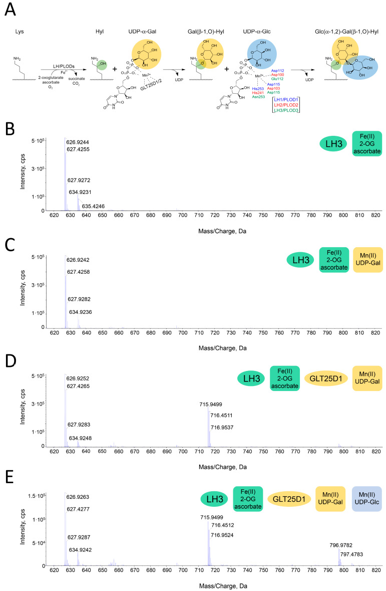
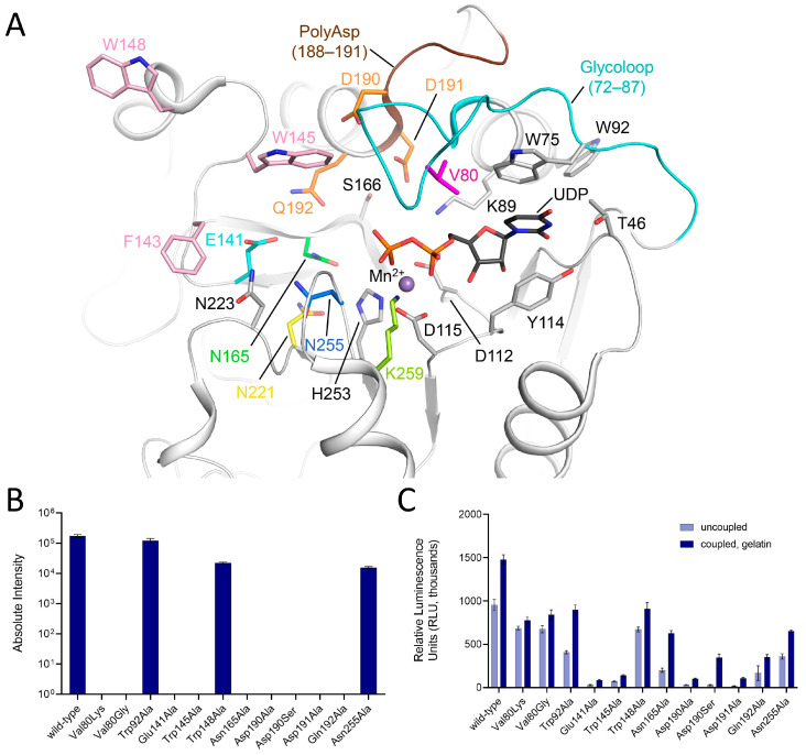
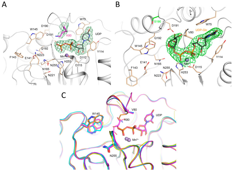
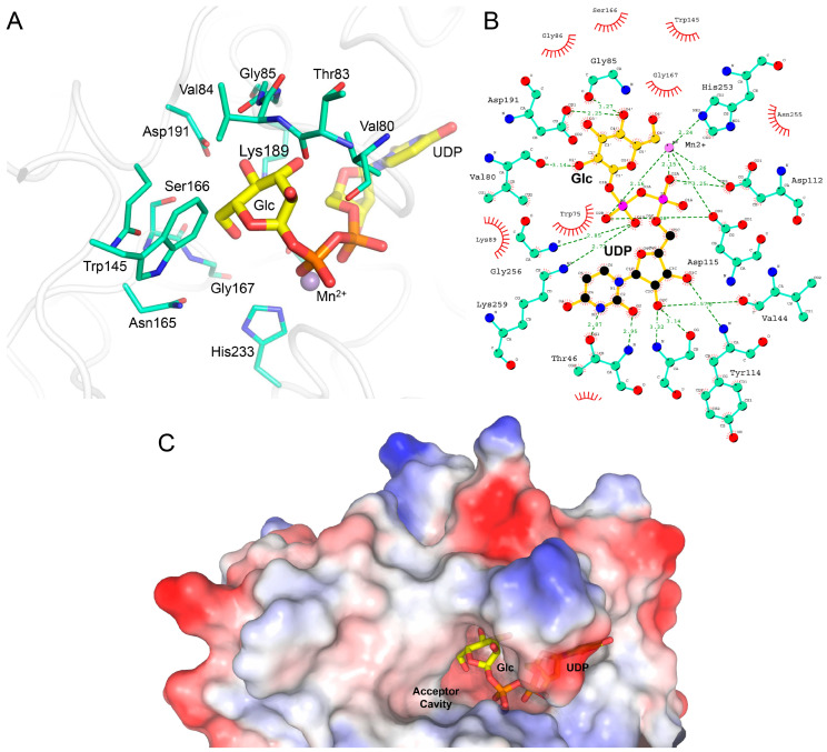
Hydroxylysine glycosylations are post-translational modifications (PTMs) essential for the maturation and homeostasis of fibrillar and non-fibrillar collagen molecules. The multifunctional collagen lysyl hydroxylase 3 (LH3/PLOD3) and the collagen galactosyltransferase GLT25D1 are the human enzymes that have been identified as being responsible for the glycosylation of collagen lysines, although a precise description of the contribution of each enzyme to these essential PTMs has not yet been provided in the literature. LH3/PLOD3 is thought to be capable of performing two chemically distinct collagen glycosyltransferase reactions using the same catalytic site: an inverting beta-1,O-galactosylation of hydroxylysines (Gal-T) and a retaining alpha-1,2-glucosylation of galactosyl hydroxylysines (Glc-T). In this work, we have combined indirect luminescence-based assays with direct mass spectrometry-based assays and molecular structure studies to demonstrate that LH3/PLOD3 only has Glc-T activity and that GLT25D1 only has Gal-T activity. Structure-guided mutagenesis confirmed that the Glc-T activity is defined by key residues in the first-shell environment of the glycosyltransferase catalytic site as well as by long-range contributions from residues within the same glycosyltransferase (GT) domain. By solving the molecular structures and characterizing the interactions and solving the molecular structures of human LH3/PLOD3 in complex with different UDP-sugar analogs, we show how these studies could provide insights for LH3/PLOD3 glycosyltransferase inhibitor development. Collectively, our data provide new tools for the direct investigation of collagen hydroxylysine PTMs and a comprehensive overview of the complex network of shapes, charges, and interactions that enable LH3/PLOD3 glycosyltransferase activities, expanding the molecular framework and facilitating an improved understanding and manipulation of glycosyltransferase functions in biomedical applications.

Keywords: collagen biosynthesis; collagen glycosylations; extracellular matrix; glycosyltransferase; lysyl hydroxylase; post-translational modifications.


Conflict of interest statement

Authors declare no conflict of interest.

Figures

Figure 1
Figure 1
A direct assay to probe Lys-to-Glc-Gal-Hyl conversion. The reaction schematic monitored by the assay is depicted in (A). The MS spectra show the results obtained by incubating a synthetic GIKGIKGIKGIK peptide (MW 1254 Da) with enzymes and cofactors, as shown in the figure legends. All peaks identified are doubly charged, resulting in nominal masses corresponding to half of the expected MW. (B) Using LH3/PLOD3 and LH activity cofactors (i.e., 2-OG and Fe2+), MS peaks corresponding to a singly hydroyxylated Lys on the peptide (i.e., 635 Da) appear. (C) The addition of Gal-T activity cofactors (i.e., UDP-Gal and Mn2+) to the same mixture as in (B) does not yield additional MS peaks. (D) When the mixture in (C) is incubated with GLT25D1, the MS peaks corresponding to Gal-Hyl are found (i.e., 716 Da). (E) When the same mixture as in (D) also contains UDP-Glc, the peaks corresponding to Glc-Gal-Hyl appear (i.e., 797 Da).
Figure 2
Figure 2
Structural and functional features of the LH3/PLOD3 glycosyltransferase (GT) domain. (A) Cartoon representation of the LH3/PLOD3 GT domain (PDB ID: 6FXR) showing the key residues shaping the catalytic site as sticks. The PolyAsp motif (brown) and the glycoloop (cyan) involved in the binding of UDP-sugar donor substrates are shown. The residues implicated in the catalytic activity and investigated in this works are colored, while the residues depicted in gray have already been shown to be essential in Mn2+ (purple sphere) and UDP (black sticks) coordination. (B) Summary of the evaluation of the Glc-T activity of LH3/PLOD3 mutants compared to the wild-type using MS direct assays. (C) Evaluation of the Glc-T activity of LH3/PLOD3 mutants compared to wild-type using luminescence-based indirect assays. Each graph shows the enzymatic activity detected in the absence (i.e., “uncoupled”, light blue) or presence of gelatin, which was used as the acceptor substrate. The plotted data are baseline-corrected, where the baseline was the background control. In both (B,C) panels, the error bars represent standard deviations from the averages of independent experiments (N > 3).
Figure 3
Figure 3
Structural characterization of LH3/PLOD3 mutants. (A) Crystal structure of the LH3/PLOD3 p.(Val80Lys) mutant in complex with UDP-glucose and Mn2+. Electron density is visible for the mutated lysine and the UDP portion of the donor substrate (green mesh, 2FoFc omit electron density map, contoured at 1.3 σ). Catalytic residues shaping the enzyme cavity are shown as sticks; Mn2+ is shown as a purple sphere. Consistent with what was observed in the crystal structure of wild-type LH3/PLOD3, the glucose moiety of the donor substrate is not visible in the experimental electron density. (B) Crystal structure of the LH3/PLOD3 p.(Asp190Ser) mutant in complex with UDP-glucose and Mn2+. Electron density is visible for the mutated Serine and for the entire donor substrate, including the sugar moiety (green mesh, 2FoFc omit electron density map, contoured at 1.3 σ). Colors and representations as in (A). (C) Superposition of wild-type, p.(Val80Lys), and p.(Asp190Ser) LH3/PLOD3 available crystal structures in substrate-free (cyan for wild-type, yellow for p.(Val80Lys), respectively) and with UDP-glucose bound (marine for wild-type, orange for p.(Val80Lys), magenta for p.(Asp190Ser), respectively) states. Notably, the conformations adopted by the side chain of Trp145 upon ligand binding are consistent in the wild-type and in the mutant enzyme. As the glycoloop is flexible in substrate-free structures, the side chains of Val/Lys80 are only visible in the in UDP-sugar-bound structures.
Figure 4
Figure 4
Binding mode of the glucose moiety of the UDP-Glc donor substrate observed in the crystal structure of LH3/PLOD3 p.(Asp190Ser). (A) Highlight of the amino acid network surrounding the Glc moiety of the donor substratein the crystal structure. UDP-Glc is shown as thick yellow sticks, whereas amino acids found at less than 5 Å distance from the Glc moiety are shown as thin blue/green sticks. (B) Overview of the interaction network surrounding the UDP-Glc donor substrate in the co-crystal structure with LH3/PLOD3 p.(Asp190Ser). Colors are as in (A). Figure made with LIGPLOT+ [36]. (C) The conformation adopted by the Glc moiety of the UDP-Glc substrate in the glycosyltransferase catalytic site of LH3/PLOD3 p.(Asp190Ser) leaves an empty cavity that is geometrically and sizably compatible with the Gal moiety of the acceptor substrate. Shown is a surface rendering of the GT domain of LH3/PLOD3 p.(Asp190Ser) colored by electrostatic potential, with highlights of the UDP-Glc donor substrate shown as sticks.
Figure 5
Figure 5
Ser178 in LH1/PLOD1, corresponding to Asp190 in LH3/PLOD3, is a key residue for Glc-T activity for both enzyme isoforms. (A) Direct MS-based assays comparing the signal associated with Glc-Gal-Hyl using wild-type and Ser178Asp LH1/PLOD1 variants. (B) Evaluation of the Glc-T activity of LH1 wild-type and Ser178Asp using luminescence-based indirect assays. The analysis of coupled and uncoupled enzymatic activities is as in Figure 2C. In both panels, the error bars represent standard deviations from the averages of independent experiments (N > 3).
Figure 6
Figure 6
Characterization of UDP-sugar analogs. (A) Thermal stability of LH3 wild-type (solid green) using differential scanning fluorimetry (DSF) in the presence of various Mn2+ and several UDP-sugars. A prominent stabilization effect is achieved in the presence of the biological donor substrates UDP-galactose (solid blue), UDP-Glucose (solid purple), and free UDP (solid black). A milder stabilization effect is also obtained with UDP-xylose (red dash) and UDP-glucuronic acid (green dash). (B) Luminescence-based competition assays evaluating the binding of increasing concentrations of UDP-GlcA or UDP-Xyl to wild-type LH3/PLOD3 in the presence of either UDP-Gal (left) or UDP-Glc (right) and acceptor substrates (i.e., gelatin). (C) Crystal structure of LH3 wild-type in complex with Mn2+ and UDP-glucuronic acid shows clear electron density for UDP (2FoFc omit electron density maps, green mesh, contour level 1.2 σ). The glucuronic acid (shown in yellow) can be modelled even if with the partial electron density. (D) Crystal structure of the LH3 Val80Lys mutant in complex with Mn2+ and UDP-glucuronic acid. While the UDP backbone can be modelled in the electron density (black sticks) (2FoFc omit electron density maps, green mesh, contour level 1.2 σ), in this case, no electron density is present for the glucuronic acid (shown in yellow). In addition, the portion of the glycoloop containing the mutated lysine is flexible from residue 79 to 83 (shown as cyan spheres). (E) Crystal structure of LH3 wild-type in complex with Mn2+ and UDP-xylose. Similar to UDP-GlcA, UDP shows clear electron density (2FoFc omit electron density maps, green mesh, contour level 1.2 σ), whereas partial density is shown for the xylose moiety (shown in pink).

Similar articles

Cited by

References

    1. Myllyharju J., Kivirikko K.I. Collagens, modifying enzymes and their mutations in humans, flies and worms. Trends Genet. 2004;20:33–43. doi: 10.1016/j.tig.2003.11.004. - DOI - PubMed
    1. Luther K.B., Hulsmeier A.J., Schegg B., Deuber S.A., Raoult D., Hennet T. Mimivirus collagen is modified by bifunctional lysyl hydroxylase and glycosyltransferase enzyme. J. Biol. Chem. 2011;286:43701–43709. doi: 10.1074/jbc.M111.309096. - DOI - PMC - PubMed
    1. Cummings R.D. The repertoire of glycan determinants in the human glycome. Mol. Biosyst. 2009;5:1087–1104. doi: 10.1039/b907931a. - DOI - PubMed
    1. Spiro R.G. Characterization and quantitative determination of the hydroxylysine-linked carbohydrate units of several collagens. J. Biol. Chem. 1969;244:602–612. doi: 10.1016/S0021-9258(18)94398-X. - DOI - PubMed
    1. Spiro R.G. The structure of the disaccharide unit of the renal glomerular basement membrane. J. Biol. Chem. 1967;242:4813–4823. doi: 10.1016/S0021-9258(18)99529-3. - DOI - PubMed

LinkOut - more resources