. 2023 Jul 6;13(1):10974.
doi: 10.1038/s41598-023-37364-x.

Formation of nuclear CPSF6/CPSF5 biomolecular condensates upon HIV-1 entry into the nucleus is important for productive infection

Affiliations

Formation of nuclear CPSF6/CPSF5 biomolecular condensates upon HIV-1 entry into the nucleus is important for productive infection

Charlotte Luchsinger et al. Sci Rep. .

Abstract

The early events of HIV-1 infection involve the transport of the viral core into the nucleus. This event triggers the translocation of CPSF6 from paraspeckles into nuclear speckles forming puncta-like structures. Our investigations revealed that neither HIV-1 integration nor reverse transcription is required for the formation of puncta-like structures. Moreover, HIV-1 viruses without viral genome are competent for the induction of CPSF6 puncta-like structures. In agreement with the notion that HIV-1 induced CPSF6 puncta-like structures are biomolecular condensates, we showed that osmotic stress and 1,6-hexanediol induced the disassembly of CPSF6 condensates. Interestingly, replacing the osmotic stress by isotonic media re-assemble CPSF6 condensates in the cytoplasm of the cell. To test whether CPSF6 condensates were important for infection we utilized hypertonic stress, which prevents formation of CPSF6 condensates, during infection. Remarkably, preventing the formation of CPSF6 condensates inhibits the infection of wild type HIV-1 but not of HIV-1 viruses bearing the capsid changes N74D and A77V, which do not form CPSF6 condensates during infection1,2. We also investigated whether the functional partners of CPSF6 are recruited to the condensates upon infection. Our experiments revealed that CPSF5, but not CPSF7, co-localized with CPSF6 upon HIV-1 infection. We found condensates containing CPSF6/CPSF5 in human T cells and human primary macrophages upon HIV-1 infection. Additionally, we observed that the integration cofactor LEDGF/p75 changes distribution upon HIV-1 infection and surrounds the CPSF6/CPSF5 condensates. Overall, our work demonstrated that CPSF6 and CPSF5 are forming biomolecular condensates that are important for infection of wild type HIV-1 viruses.


Conflict of interest statement

The authors declare no competing interests.

Figures

Figure 1
Figure 1
Viral mutations, inhibitors, and restriction factors affect the recruitment of CPSF6 to NS. (A) To induce the formation of CPSF6 condensates, A549 cells were challenged with HIV-1-Luc viruses at an MOI of ~ 2 for 24 h. Subsequently, cells were fixed, permeabilized, and immunolabeled using specific antibodies directed against SC35 (green) and CPSF6 (red). Secondary antibodies were Cy5-conjugated donkey anti-mouse IgG and Alexa-594-conjugated donkey anti-rabbit IgG, respectively. For Nuclei were stained with DAPI (blue). Cells were examined by fluorescence microscopy. Merging the red and green channels generated the lower left image; yellow indicates the overlapping location of the red and green channels. Merging all channels generated the lower right image. (B) To evaluate the effect of viral mutations and inhibitors in the formation of CPSF6 condensates, we infected A549 cells with wild-type or the indicated mutant HIV-1-GFP viruses for 24 h using an MOI of ~ 2 when possible or normalized by p24. A549 cells were also infected with wild-type HIV-1-GFP with reverse transcription inhibitors 10 μM nevirapine (Nev) and 10 μM zidovudine (AZT), the integration inhibitor 10 μM raltegravir (Ral), or 5 μM cyclosporin A (CsA), as indicated. Cells were fixed, permeabilized, and immunolabeled with rabbit anti-CPSF6 and mouse anti-SC35 antibodies. The secondary antibodies were Alexa-594-conjugated donkey anti-rabbit IgG and Cy5-conjugated donkey anti-mouse IgG. The percentage of cells containing CPSF6 in nuclear speckles was determined by visual inspection of 200 cells per sample in three independent experiments. In parallel infectivity was assayed by measuring the % of GFP-positive cells for in three independent experiments (lower panels). (C) To evaluate the effect of restriction factors in the recruitment of CPSF6 to NS, we infected A549 cells stably expressing the restriction factors TRIM5αrh or TRIMCyp with HIV-1-GPF at an MOI of ~ 2 for 24 h. The control experiment used the empty vector LPCX. Cells were fixed, permeabilized, and immunolabeled, as in (B). The percentage of cells containing CPSF6 in NS was determined as in (B). Similarly, infectivity was assayed by measuring the percentage of GFP-positive cells (lower panel). (D) To determine the stability of CPSF6 condensates, we infected A549 cells with HIV-1-GFP at an MOI of ~ 2 for 24-, 48-, 72-, or 96-h as indicated. Uninfected cells were used as control (mock). At the indicated times, cells were fixed, permeabilized, and immunolabeled, as in (B). The percentage of cells containing CPSF6 condensates in the nucleus (N) or in the cytosol (C) was determined by visual inspection of 80 cells per sample and normalized to the total amount of cells in three independent experiments (n = 240 cells). Data represent the mean ± standard deviation. **p ≤ 0.05, **p ≤ 0.0, ***p ≤ 0.001, ****p ≤ 0.0001; ns, not statistically significant; unpaired two-tailed t test.
Figure 2
Figure 2
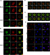
Osmotic stress and 1,6-hexanediol affect the stability of CPSF6 condensates. The formation of CPSF6 condensates was induced by infection of A549 cells by HIV-1-GFP at an MOI of ~ 2 for 24 h (A, I, and Q). Uninfected cells were used as the control (mock) (E, M, and U). Subsequently, infected, and uninfected cells were subjected to hypotonic stress for 5 min (B and F); hypertonic stress (200 mM NaCl-supplemented DMEM) for 15 min (J and N); or 3% 1,6-hexanediol for 3 min (R and V). After hypotonic, hypertonic, or 1,6-hexanediol treatment, the cells were incubated in isotonic medium (complete DMEM) for 10 min (C and G), 30 min (J and O), or 10 min (S and W), respectively. Cells were fixed, permeabilized, and immunolabeled with rabbit polyclonal antibody against CPSF6. The secondary antibody was Alexa-594-conjugated donkey anti-rabbit IgG. Nuclei were stained with DAPI. Stained cells were examined by fluorescence microscopy. The percentage of cells containing CPSF6 condensates in the nucleus (N) or in the cytosol (C) in the hypotonic (D and H), hypertonic (L and P), or 1,6-hexanediol (T and X) treatment was determined by visual inspection of 80 cells per sample in three independent experiments (n = 240 cells). Data represent the mean ± standard deviation. *p ≤ 0.05, **p ≤ 0.0, ***p ≤ 0.001, ****p ≤ 0.0001; ns, not statistically significant; unpaired two-tailed t test. Scale bar = 10 μm.
Figure 3
Figure 3
GS-CA-1 stabilizes the HIV-1-induced CPSF6 condensates in the nucleus upon hypotonic stress in A549 cells. To induce the formation of CPSF6 condensates, we infected A549 cells with HIV-1-GFP at an MOI of ~ 2 for 24 h (A, F, and K). Infected cells were incubated with DMSO (as the vehicle control) (B), 10 nM GS-CA-1 (G), or 10 μM PF74 (L) for 30 min. Cells were subsequently subjected to hypotonic stress with DMSO (C), 10 nM GS-CA-1 (H), or 10 μM PF74 (M) for 5 min. After hypotonic stress, cells were incubated in an isotonic medium with DMSO (D), 10 nM GS-CA-1 (I), or 10 μM PF74 (N) for 10 min. Cells were fixed, permeabilized, and immunolabeled with rabbit polyclonal antibody to human CPSF6. The secondary antibody was Alexa-594-conjugated donkey anti-rabbit IgG. Nuclei were stained with DAPI. Stained cells were examined by fluorescence microscopy. The percentage of cells containing CPSF6 condensates in the nucleus (N) or in the cytosol (C) under hypotonic stress with DMSO (E), 10 nM GS-CA-1 (J), or 10 μM PF74 (O) was determined as in Fig. 2. Data represent the mean ± standard deviation. *p ≤ 0.05, **p ≤ 0.0, ***p ≤ 0.001, ****p ≤ 0.0001; ns, not statistically significant; unpaired two-tailed t test. Scale bar = 10 μm.
Figure 4
Figure 4
GS-CA-1 reassembles the HIV1-induced CPSF6 condensates in the nucleus after 1,6-hexanediol treatment in A549 cells. To induce the formation of CPSF6 condensates, we infected A549 cells with HIV-1-GFP at an MOI of ~ 2 for 24 h (A, F, and K). Infected cells were incubated with DMSO (B), 10 nM GS-CA-1 (G), or 10 μM PF74 (L) for 30 min. Cells were treated with 3% 1,6-hexanediol with DMSO (C), 10 nM GS-CA-1 (H), or 10 μM PF74 (M) for 3 min. After 3% 1,6-hexanediol treatment, cells were incubated in an isotonic medium with DMSO (D), 10 nM GS-CA-1 (I), or 10 μM PF74 (N) for 10 min. Cells were fixed, permeabilized, and immunolabeled with rabbit polyclonal antibody to CPSF6. The secondary antibody was Alexa-594-conjugated donkey anti-rabbit IgG. Nuclei were stained with DAPI. Stained cells were examined by fluorescence microscopy. The percentage of cells containing CPSF6 condensates in the nucleus (N) or in the cytosol (C) for the 1,6-hexanediol treatments with DMSO (E), 10 nM GS-CA-1 (J), or 10 μM PF74 (O) was determined by visual inspection of 80 cells per sample in three independent experiments (n = 240 cells). Data represent the mean ± standard deviation. *p ≤ 0.05, **p ≤ 0.0, ***p ≤ 0.001, ****p ≤ 0.0001; ns, not statistically significant; unpaired two-tailed t test. Scale bar = 10 μm.
Figure 5
Figure 5
Formation of CPSF6 condensates is required for wild-type HIV-1 infection. A549 cells were infected wild-type or mutant HIV-1-GFP viruses at an MOI of 2, which is enough the infect more than 90% of the cells 24 h post-infection. At 10 h post-infection, cells were incubated or not with a hypertonic medium to prevent the formation of CPSF6 condensates. At 24 h post-infection, infectivity was measured as the percentage of GFP-positive cells using a flow cytometer. Data represent the mean ± standard deviation. *p ≤ 0.05, **p ≤ 0.0, ***p ≤ 0.001, ****p ≤ 0.0001; ns, not statistically significant; unpaired two-tailed t test.
Figure 6
Figure 6
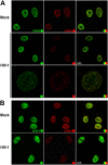
HIV-1 infection induces the formation of CPSF5 condensates that colocalize with CPSF6. A549 cells were infected or not (mock) with HIV-1-Luc at an MOI of ~ 2 for 24 h (A and B). Cells were fixed, permeabilized, and coimmunostained using rabbit polyclonal antibody to CPSF6 with mouse monoclonal antibody against CPSF5 (A) or with mouse monoclonal antibody against CPSF7 (B). Secondary antibodies were Alexa-488-conjugated donkey anti-mouse IgG (green channel) and Alexa-594-conjugated donkey anti-rabbit IgG (red channel). Stained cells were examined by fluorescence microscopy. Merging the red and green channels generated the third image in each row; yellow indicates overlapping localization of the red and green channels. Experiments were repeated at least three times, and a representative experiment is shown. Scale bar = 10 μm.
Figure 7
Figure 7
HIV-1 infection induces the formation of CPSF6 and CPSF5 condensates in human T cells and human primary macrophages. Jurkat T cells (A) and macrophages (B) were infected or not (mock) with HIV-1-GFP at an MOI of ~ 2 for 48 or 72 h, respectively. At 48hpi, 2,5 × 105 Jurkat cells were seeded on glass coverslips previously treated with poly-D-lysine for 1 h. Cells were fixed, permeabilized, and immunostained using rabbit polyclonal antibody to CPSF6, mouse monoclonal antibody against CPSF5 or mouse monoclonal antibody against CPSF7. Secondary antibody was Alexa-594-conjugated donkey anti-rabbit IgG (red channel). Nuclei were stained with DAPI (blue channel). Stained cells were examined by fluorescence microscopy. Merging the red and blue channels generated the third image in each row; magenta indicates overlapping of the red and blue channels. Experiments were repeated at least three times, and a representative experiment is shown. Scale bar = 10 μm.
Figure 8
Figure 8
HIV-1 infection induces nuclear redistribution of LEDGF/p75. A549 cells were infected or not (mock) with wild-type HIV-1-Luc (AD) or with HIV-1-GFP viruses bearing the capsid mutations N74D and A77V (E) at an MOI of ~ 2 for 24 h. Cells were fixed, permeabilized, and stained using rabbit polyclonal antibody to LEDGF/p75 (E) with mouse monoclonal antibody to CPSF6 (A and C) or with mouse monoclonal antibody to CPSF5 (B and D). Secondary antibodies were Alexa-488-conjugated donkey anti-mouse IgG (green channel) and Alexa-594-conjugated donkey anti-rabbit IgG (red channel). Nuclei were stained with DAPI (channel blue). Stained cells were examined by fluorescence microscopy. Merging the red and green channels generated the yellow color that indicates overlapping localization of these channels. Merging the red and blue channels generated the magenta color that indicates overlapping localization of these channels. For C and D z-stack images at 0.2 μm intervals were acquired. The white circle indicates an example of a condensate that contains CPSF6 (C) or CPSF5 (D) surrounded by LEDGF/p75. Scale bar = 10 μm.

Similar articles

Cited by

References

    1. Francis AC, et al. Publisher correction: HIV-1 replication complexes accumulate in nuclear speckles and integrate into speckle-associated genomic domains. Nat. Commun. 2020;11:6165. doi: 10.1038/s41467-020-20152-w. - DOI - PMC - PubMed
    1. Selyutina A, Persaud M, Lee K, KewalRamani V, Diaz-Griffero F. Nuclear import of the HIV-1 core precedes reverse transcription and uncoating. Cell Rep. 2020;32:108201. doi: 10.1016/j.celrep.2020.108201. - DOI - PMC - PubMed
    1. Dharan A, Bachmann N, Talley S, Zwikelmaier V, Campbell EM. Nuclear pore blockade reveals that HIV-1 completes reverse transcription and uncoating in the nucleus. Nat. Microbiol. 2020;5:1088–1095. doi: 10.1038/s41564-020-0735-8. - DOI - PMC - PubMed
    1. Burdick RC, et al. HIV-1 uncoats in the nucleus near sites of integration. Proc. Natl. Acad. Sci. U. S. A. 2020;117:5486–5493. doi: 10.1073/pnas.1920631117. - DOI - PMC - PubMed
    1. Rensen E, et al. Clustering and reverse transcription of HIV-1 genomes in nuclear niches of macrophages. EMBO J. 2021;40:e105247. doi: 10.15252/embj.2020105247. - DOI - PMC - PubMed

Publication types

MeSH terms

Substances