. 2023 Jul 6;19(7):e1011477.
doi: 10.1371/journal.ppat.1011477. eCollection 2023 Jul.

Changes in SUMO-modified proteins in Epstein-Barr virus infection identifies reciprocal regulation of TRIM24/28/33 complexes and the lytic switch BZLF1

Affiliations

Changes in SUMO-modified proteins in Epstein-Barr virus infection identifies reciprocal regulation of TRIM24/28/33 complexes and the lytic switch BZLF1

Carlos F De La Cruz-Herrera et al. PLoS Pathog. .

Abstract

SUMO modifications regulate the function of many proteins and are important in controlling herpesvirus infections. We performed a site-specific proteomic analysis of SUMO1- and SUMO2-modified proteins in Epstein-Barr virus (EBV) latent and lytic infection to identify proteins that change in SUMO modification status in response to EBV reactivation. Major changes were identified in all three components of the TRIM24/TRIM28/TRIM33 complex, with TRIM24 being rapidly degraded and TRIM33 being phosphorylated and SUMOylated in response to EBV lytic infection. Further experiments revealed TRIM24 and TRIM33 repress expression of the EBV BZLF1 lytic switch gene, suppressing EBV reactivation. However, BZLF1 was shown to interact with TRIM24 and TRIM33, resulting in disruption of TRIM24/TRIM28/TRIM33 complexes, degradation of TRIM24 and modification followed by degradation of TRIM33. Therefore, we have identified TRIM24 and TRIM33 as cellular antiviral defence factors against EBV lytic infection and established the mechanism by which BZLF1 disables this defence.


Conflict of interest statement

The authors have declared that no competing interests exist.

Figures

Fig 1
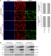
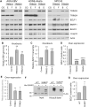
Fig 1. Inhibition of SUMOylation promotes EBV lytic infection.
A and B. AGS-EBV cells were treated with siRNA against SUMO1 or negative control siRNA, then analyzed by Western blot with antibodies against BZLF1, SUMO1 and actin (A) or fixed and stained with BZLF1 antibody for IF (B). The percentage of BZLF1 expressing cells was determined from 300 cells in three independent experiments and average values are shown on the graph. C. Western blot analysis of AGS-EBV and NPC43 cells treated with the SUMO E1 conjugating enzyme inhibitor ML792 or DMSO (-) before (Un) or the indicated hours after induction of EBV lytic reactivation with TPA/NaB. Total protein extracts were probed with antibodies against SUMO1, BZLF1, BMRF1, BALF2, Vcap18 and actin.
Fig 2
Fig 2. Characterization of cell lines used for the proteomic experiments.
A. IF of the 6His-SUMO1 or 6His-SUMO2 cells showing most express both the mCherry marker linked to SUMO and the lytic switch protein BZLF1 (detected with anti-BZLF1 antibody) after dox induction. B. Comparison of expression of EBV lytic proteins before (Un) or the indicated hours after dox-induction of the lytic cycle in AGS-EBV-Z cells with and without integrated 6His-SUMO1 or 2. Arrow and star indicate endogenous and integrated BZLF1, respectively.
Fig 3
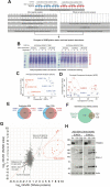
Fig 3. Overview of the proteomics experiments.
A. The label-free proteomics experimental design to monitor changes to cellular protein abundance and the SUMOylation landscape during EBV reactivation. B. Coomassie-stained gel of the whole cell extract triplicate data for total proteome analysis. C + D Principal component analysis of proteomic data for both GG-K peptide intensity and whole protein intensity for the SUMOylation analysis (C) and the total proteome analysis (D). E. Summary of substrates and sites identified based on having at least one intensity reported in at least one MS run associated with SUMO1 or SUMO2 derived samples. F. Numbers of proteins identified and quantified from the whole cell lysate analysis, and the degree of overlap with the SUMO substrates identified. G. Relationship between total protein abundance change (x-axis) and GG-K peptide abundance change (y-axis) during 24h EBV reactivation. x- and y-axes show average of SUMO1 and SUMO2 ratios. y = x line is shown in red. Sites in viral proteins have red markers, cellular proteins in grey. Selected sites are indicated. H. Examples of two proteins (SALL4 and TRIM24) found to be altered in levels upon EBV reactivation. Western blots were performed on the cell lysates used for the mass spectrometry analyses using antibodies against TRIM24 and SALL4. Positions of SALL4A (*) and SALL4B (#) are indicated.
Fig 4
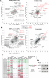
Fig 4. Proteomics data summary.
A + B. Charts indicating changes in abundance to SUMO1 (x-axis) and SUMO-2 (y-axis) GG-K peptides (left) and cellular proteins (right) derived from whole cell extracts from the 6His-SUMO1 cells (x-axis) and 6His-SUMO2 cells (y-axis) in response to 12 hours (A) or 24 hours (B) EBV reactivation. Data derived from viral proteins are indicated by red markers and cellular proteins are grey, with the shade indicating the statistical significance of the change (see key) derived from the sum of the two -log10 p-values for the x and y-axis data. Selected sites and proteins with large deviation from 0 combined with high significance are indicated. C. Tabular summary of the SUMO1 and SUMO2 site-level data and total protein level data for TRIM24, TRIM28 and TRIM33. Cells are coloured by fold change with a thick border indicating statistical significance (p<0.05). D. AGS-EBV-Z cells were treated with dox (or left untreated) with and without MG132 for 12 hours as indicated. Western blots were then performed using antibodies against TRIM24, BZLF1 and actin.
Fig 5
Fig 5. Verification of changes in SUMOylation in selected cellular and viral proteins.
A and B. Whole cell lysates from AGS-EBV-Z-6His-SUMO1 and AGS-EBV-Z-6His-SUMO2 cells before (Un) or 12 or 24 hours after dox induction were analysed by Western blotting using the indicated antibodies. Examples of cellular proteins with altered SUMOylation after EBV reactivation (A) and EBV proteins with SUMO modifications (B) are shown. Stars indicate nonmodified proteins. Arrowheads and brackets indicate SUMO conjugated proteins. C. The indicated cell lines were treated with ML792 or left untreated then reactivated with dox for 24 hours or left untreated. Whole cell lysates were then analysed by Western blotting and probed for EBV lytic proteins (BORF2, BMRF1, Vcap18) and TRIM33.
Fig 6
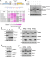
Fig 6. SUMOylation and phosphorylation of TRIM33 and verification of TRIM24 loss in NPC43 and Akata cell infections.
A. Upper panel: Schematic of TRIM33 showing domain structure and sites of SUMOylation and phosphorylation identified in the proteomics experiment. Lower Panel: Summary of MS peptide intensity data for the indicated TRIM33 modifications. Mono-SUMOylated peptides, di-SUMOylated peptides, and peptides co-modified by SUMO and phospho moieties are grouped. Intensity in each sample relative to the average intensity across all samples is indicated and coloured according to the scale shown below (values represent the average of three replicates per condition). Peptides not detected in a particular sample are shown in grey. Cell line and duration of EBV reactivation are shown in the headers. B. AGS-EBV-Z cells were treated with dox (or left untreated) with and without ML792 for 12 hours as indicated. Whole cell lysates were incubated with or without FastAP phosphatase, followed by Western blotting with TRIM33 and vinculin antibodies. C. NPC43-Z cells were treated with dox for 0, 24 or 48 hours, then whole cell lysates were analysed by Western blotting using TRIM33, TRIM24, BZLF1 and actin antibodies. The bracket marks the position of bands corresponding to SUMO-modified TRIM33. D. NPC43-Z cells were treated with dox for 12 hours (or left untreated) in the presence of DMSO (negative control), ML792 or MG132 as indicated, and the lysates were anlaysed as in C. E. Akata-Z cells were treated with dox for 0, 24 or 48 hours, then whole cell lysates were analysed by Western blotting using TRIM33, TRIM24, BZLF1 and actin antibodies. Two exposures are shown for TRIM33, with the phospho shift evident in the lighter (top) exposure and the SUMO-modified forms seen in darker exposure and marked by the bracket. F. Akata-Z cells were treated with dox for 24 hours (or left untreated) in the presence of DMSO (negative control) or ML792, and the lysates were anlaysed as in E.
Fig 7
Fig 7. TRIM33 and TRIM24 suppress EBV reactivation.
A. TRIM33 or TRIM24 was knocked out using CRISPR-Cas9 in pools of AGS-EBV, HONE-Akata and NPC43 cells with two different guide RNAs (E and F for TRIM33; B and C or D for TRIM24) and compared to a negative control guide RNA (CG) targeting the Adeno-Associated Virus Integration Site 1 (AAVS1). Western blots were performed on whole cell lysates with antibodies against TRIM33, TRIM24, four EBV proteins (BZLF1, BMRF1, BALF2, Vcap18) and actin. B and C. Total DNA and RNA were extracted from AGS-EBV cells in A and EBV genome amplification (B) and BZLF1 transcripts (C) were quantified by qPCR. D. AGS-EBV cells were transfected with plasmids expressing a negative control FLAG-tagged protein (DDX24) or FLAG-tagged TRIM33 or TRIM24, then stained for BZLF1 and FLAG and imaged by IF. For each of the transfected plasmids, the percentage of FLAG-positive cells expressing BZLF1 was determined in 100 cells in three independent experiments. E. AGS cells were co-transfected with pZp-luc (firefly luciferase) and pRL-promotorless (renilla) reporter plasmids along with FLAG-TRIM33 or FLAG-TRIM24 expression plasmid or empty plasmid control and harvested 24 hours post-transfection. Firefly and renilla luciferase levels were quantified and luciferase values were normalized to renilla for three independent experiments. F. WT and SUMO mutant (SUMOm) TRIM33 were expressed in 293T cells with or without 6His-SUMO1 and 10% of cell lysates were analysed as input samples. SUMO-modified proteins were isolated from the remaining samples on metal chelating resin (His pull down). Samples were analysed by Western blotting using anti-FLAG antibody. G. The same experiments as in D except that WT TRIM33 was compared to the TRIM33 SUMOm. All graphs show average values with standard deviation where * = 0.01<P≤0.05, ** = 0.001< P ≤ 0.01, *** = 0.0001<P≤0.001.
Fig 8
Fig 8. BZLF1 binds to TRIM24 and TRIM33.
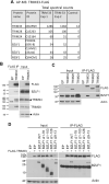
A. TRIM33-FLAG or empty FLAG (negative control) was expressed in AGS-EBV-Z cells followed by reactivation with dox for 24 hrs, and TRIM33 and associated proteins were isolated on anti-FLAG resin. Recovered proteins were identified by tandem MS. Total spectral counts are shown for the five most prevalent interactors seen in two independent experiments. B. TRIM33-FLAG was expressed in AGS-EBV followed by reactivation by sodium butyrate/TPA treatment and FLAG IPs. Western blots on IP samples and 10% of input samples are shown using the indicated antibodies. C. AGS cells were transfected with plasmids expressing BZLF1 and FLAG-tagged TRIM33, TRIM28, TRIM24 or empty FLAG plasmid (EP). 36 hours later, IPs were performed using FLAG antibody, followed by Western blotting with BZLF1 and FLAG antibodies. Input lysates were also probed with actin antibody. D. AGS cells were transfected with plasmids expressing FLAG-TRIM33 and BZLF1 WT or mutants (or empty plasmid) as indicated. IPs were performed using anti-FLAG M2 beads, followed by Western blotting as in C. The lower band in the BZLF1 blot of IP samples is from the IgG light chain.
Fig 9
Fig 9. BZLF1 disrupts the TRIM24/TRIM33/TRIM28 complex.
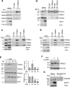
A. AGS-BZLF1 cells before (-) or 24 hrs after dox-induction of BZLF1 were used to perform IPs with TRIM33 or IgG control antibodies, followed by Western blotting with antibodies against TRIM24, TRIM28 and TRIM33. Input lysates were also probed with BZLF1 and tubulin antibodies. B. 293T cells were transfected with a BZLF1 expression plasmid (+) or empty plasmid (-), then IPs were performed with TRIM33 or IgG negative-control antibodies, followed by Western blot on inputs and IPs with antibodies against TRIM24, TRIM28 and TRIM33. Input lysates were also probed with BZLF1 and actin antibodies. C. NPC43-Z cells before (-) or 24 hrs after dox-induction of BZLF1 were used to perform IPs with TRIM33 or IgG control antibodies, followed by Western blotting with antibodies against TRIM24, TRIM28 and TRIM33. Input lysates were also probed with BZLF1 and actin antibodies. D Akata-Z cells before (-) or 24 hrs after dox-induction of BZLF1 were used to perform IPs with TRIM33 or IgG control antibodies, followed by Western blotting as in C. E. AGS-BZLF1 cells before (-) or the indicated hours after dox-induction of BZLF1 were analyzed by Western blotting with antibodies against TRIM33, TRIM28, TRIM24, BZLF1 and actin. TRIM bands were quantified from three independent experiments and average values with standard deviation were plotted. * = 0.01
Fig 10
Fig 10. Model of the repression of the BZLF1 promoter by TRIM24/TRIM28/TRIM33 complexes in latent infection and their disruption by BZLF1 in lytic infection.
The interaction of BZLF1 with TRIM24 and TRIM33 and/or the disruption of TRIM24/TRIM28/TRIM33 complexes by BZLF1 leads to degradation of TRIM24, and phosphorylation and SUMOylation of TRIM33 followed later by its degradation. Created with BioRender.com.

Similar articles

References

    1. Cohen JI, Fauci AS, Varmus H, Nabel GJ. Epstein-Barr virus: an important vaccine target for cancer prevention. Sci Transl Med. 2011;3. doi: 10.1126/scitranslmed.3002878 - DOI - PMC - PubMed
    1. Young LS, Yap LF, Murray PG. Epstein-Barr virus: more than 50 years old and still providing surprises. Nat Rev Cancer. 2016;16: 789–802. doi: 10.1038/nrc.2016.92 - DOI - PubMed
    1. Lanz T v., Brewer RC, Ho PP, Moon JS, Jude KM, Fernandez D, et al.. Clonally expanded B cells in multiple sclerosis bind EBV EBNA1 and GlialCAM. Nature. 2022;603: 321–327. doi: 10.1038/s41586-022-04432-7 - DOI - PMC - PubMed
    1. Bjornevik K, Cortese M, Healy BC, Kuhle J, Mina MJ, Leng Y, et al.. Longitudinal analysis reveals high prevalence of Epstein-Barr virus associated with multiple sclerosis. Science. 2022;375: 296–301. doi: 10.1126/science.abj8222 - DOI - PubMed
    1. Murata T, Sugimoto A, Inagaki T, Yanagi Y, Watanabe T, Sato Y, et al.. Molecular Basis of Epstein-Barr Virus Latency Establishment and Lytic Reactivation. Viruses. 2021;13. doi: 10.3390/V13122344 - DOI - PMC - PubMed