. 2023 Jun 20:11:1165853.
doi: 10.3389/fbioe.2023.1165853. eCollection 2023.

Improving dental epithelial junction on dental implants with bioengineered peptides

Affiliations

Improving dental epithelial junction on dental implants with bioengineered peptides

Ivan V Panayotov et al. Front Bioeng Biotechnol. .

Abstract

Introduction: The functionalization of titanium (Ti) and titanium alloys (Ti6Al4V) implant surfaces via material-specific peptides influence host/biomaterial interaction. The impact of using peptides as molecular linkers between cells and implant material to improve keratinocyte adhesion is reported. Results: The metal binding peptides (MBP-1, MBP-2) SVSVGMKPSPRP and WDPPTLKRPVSP were selected via phage display and combined with laminin-5 or E-cadherin epithelial cell specific peptides (CSP-1, CSP-2) to engineer four metal-cell specific peptides (MCSPs). Single-cell force spectroscopy and cell adhesion experiments were performed to select the most promising candidate. In vivo tests using the dental implant for rats showed that the selected bi functional peptide not only enabled stable cell adhesion on the trans-gingival part of the dental implant but also arrested the unwanted apical migration of epithelial cells. Conclusion: The results demonstrated the outstanding performance of the bioengineered peptide in improving epithelial adhesion to Ti based implants and pointed towards promising new opportunities for applications in clinical practice.

Keywords: bioengineered peptide; epithelial adhesion; implants; phage display; titanium surface functionalization.


Conflict of interest statement

The authors declare that the research was conducted in the absence of any commercial or financial relationships that could be construed as a potential conflict of interest.

Figures

FIGURE 1
FIGURE 1
Maldi-TOF/TOF spectra of MBP-1 (A,B) and MBP-2 (C,D) on Titanium and Ti-alloy (Ti6Al4V) surfaces, respectively, after acetonitrile rinsing.
FIGURE 2
FIGURE 2
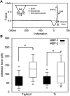
Force spectroscopy of metal binding peptides on Ti and Ti6Al4V. (A) Typical adhesion force curve measured with AFM. The inset shows a schematic representation of the peptide-functionalized tip. (B) Adhesion forces of MBP-1 and MBP-2. The MBP-1 adhesion forces on Ti6Al4V and Ti are 67.07 ± 1.34 pN and 65.42 ± 2.48 pN, respectively. The MBP-2 adhesion forces on the two surfaces are 109.76 ± 2.62 pN and 134.61 ± 2.65 pN, respectively.
FIGURE 3
FIGURE 3
Combinational design of metal-cell specific peptides (MCSP). (A) Metal-binding peptides (MBPs) with high affinity to Ti and Ti6Al4V. (B) Cell specific peptides (CSPs) with affinity to laminin-332 (CSP-1) and to E-cadherin’s ectodomains (CSP-2). (C) The engineered metal-cell specific peptides (MCSPs). Three glycines (G) residues are used as a spacer between the two peptide sequences.
FIGURE 4
FIGURE 4
Strength of keratinocyte cell binding to bare and modified Ti and Ti6Al4V surfaces. Adhesion forces between living keratinocytes and metal surfaces were evaluated using AFM in force mode. (A) Akeratinocyte-decorated cantilever was used to measure a set of forces on bare and functionalized Ti and (B) Ti6Al4V surfaces, respectively. Ti, Ti6Al4V, MCSP-1, MCSP-2, MCSP-3, and MCSP-4 correspond respectively to the bare surfaces and those functionalized with MCSP-1, MCSP-2, MCSP-3, and MCSP-4. Error bars for (A,B) represent the standard deviation of multiple experiments. *: Significant differences correspond to p = 0.0001.
FIGURE 5
FIGURE 5
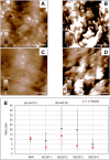
Evaluation of surface modification using AFM height images and surface roughness analysis. AFM height images (1 μm × 1 µm) were recorded in contact mode in liquid at 1 Hz. Height images for the bare and the MCSP-2 modified surfaces are presented: (A) Bare Ti surface, (B) Ti surface modified with MCSP-2, (C) Bare Ti6Al4V surface and (D) Ti6Al4V surface modified with MCSP-2. The scale bar on the height images (A–D) is 200 nm in length. The surface roughness of all measured surfaces is presented in graph (E). The black data corresponds to the bare Ti and its four different coatings with MCSP-1, MCSP-2, MCSP-3, and MCSP-4. The red data correspond to the bare Ti6Al4V and its four different functionalized surfaces. The standard deviations of the surface roughness variations on the scanned surfaces are also shown.
FIGURE 6
FIGURE 6
Assessment of oral keratinocytes adhesion in cell culture against bare and functionalized Ti and Ti6Al4V surfacesusing para-nitrophenil phosphate (pNPP) viability test. White bars correspond to cell adhesion on bare and functionalized (with MCSP-1, MCSP-2, MCSP-3, and MCSP-4, respectively) Ti surfaces, while black bars correspond to cell adhesion on bare and functionalized Ti6Al4V surfaces. * Significant differences (p = 0.0001).
FIGURE 7
FIGURE 7
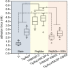
Cell adhesion after bovine serum albumin adsorption on metal-cell specific peptide-2. AFM was used in force mode to evaluate unbinding forces between cell functionalized tip-less cantilevers and three different surfaces of Ti and Ti6Al4V: the blue part corresponds to the bare Ti and Ti6Al4V surfaces, the yellow one shows data for Ti and Ti6Al4V functionalized with the MCSP-2, while the purple one corresponds to the Ti and Ti6Al4V functionalized with MCSP-2 and followed by BSA adsorption. The presence of BSA decreases the adhesion force to values measured for non-functionalized surfaces. Significantly higher cell adhesion was found against Ti/MCSP-2/BSA surfaces compared to bare Ti [*: Significant differences (p = 0.0001)]. Error bars represent the standard deviation of multiple experiments.
FIGURE 8
FIGURE 8
In vivo comparison of the epithelial adhesions on the peptide covered and bare implant surfaces, respectively. Histological slices of peri-implant gingiva in the fourth week after implantation are presented. The immunohistochemical coloration of laminin 332 indicates the epithelial adhesion on the implant surface. The dark arrows indicate cervical and apical points of measurement of the laminin 332 distribution, which is visualized like a dark brown line at the implant-epithelium interface; (A): MCSP-2 covered Ti6Al4Vimplant; (B) bare implant; (C) MBP-1 covered implant; CT: connective tissue; OE: Oral Epithelium; JE: epithelial junction; Bare scale = 10 μm; (D) Statistical analysis of multiple measurements of the length of junctional epithelium for the 6 implanted animals. JE length after 4 weeks of healing demonstrates a statistically significant difference between the bi-functional peptides (MCSP-2) and the bare Ti6Al4V alloy (p ˂ 0.05). The length of JE indicating epithelial adhesion was smaller also for the MBP-1 coated implant, but the apical migration of the epithelial cells was not as limited as on the surfaces functionalized with MCSP-2.

Similar articles

References

    1. Álvarez-López A., Colchero L., Elices M., Guinea G. V., Pérez-Rigueiro J., González-Nieto D. (2022). Improved cell adhesion to activated vapor silanization-biofunctionalized Ti-6Al-4V surfaces with ECM-derived oligopeptides. Biomater. Adv. 133, 112614. 10.1016/j.msec.2021.112614 - DOI - PubMed
    1. Atieh M. A., Ibrahim H. M., Atieh A. H. (2010). Platform switching for marginal bone preservation around dental implants: A systematic review and meta‐analysis. J. Periodontol. 81, 1350–1366. 10.1902/jop.2010.100232 - DOI - PubMed
    1. Atsuta I., Ayukawa Y., Ogino Y., Moriyama Y., Jinno Y., Koyan K. (2012). Evaluations of epithelial sealing and peri‐implant epithelial down‐growth around “step‐type” implants. Oral Impl Res. 23, 459–466. 10.1111/j.1600-0501.2011.02163.x - DOI - PubMed
    1. Baffone G. M., Botticelli D., Canullo L., Scala A., Beolchini M., Lang N. P. (2012). Effect of mismatching abutments on implants with wider platforms – An experimental study in dogs. Clin. Oral Implants Res. 23, 334–339. 10.1111/j.1600-0501.2011.02320.x - DOI - PubMed
    1. Baharloo B., Textor M., Brunette D. M. (2005). Substratum roughness alters the growth, area, and focal adhesions of epithelial cells, and their proximity to titanium surfaces. J. Biomed. Mater Res. A 74, 12–22. 10.1002/jbm.a.30321 - DOI - PubMed