SFRP1 Expression is Inversely Associated With Metastasis Formation in Canine Mammary Tumours
- PMID: 37402051
- PMCID: PMC10319705
- DOI: 10.1007/s10911-023-09543-z
SFRP1 Expression is Inversely Associated With Metastasis Formation in Canine Mammary Tumours
Abstract
Background: Canine mammary tumours (CMTs) are the most frequent tumours in intact female dogs and show strong similarities with human breast cancer. In contrast to the human disease there are no standardised diagnostic or prognostic biomarkers available to guide treatment. We recently identified a prognostic 18-gene RNA signature that could stratify human breast cancer patients into groups with significantly different risk of distant metastasis formation. Here, we assessed whether expression patterns of these RNAs were also associated with canine tumour progression.
Method: A sequential forward feature selection process was performed on a previously published microarray dataset of 27 CMTs with and without lymph node (LN) metastases to identify RNAs with significantly differential expression to identify prognostic genes within the 18-gene signature. Using an independent set of 33 newly identified archival CMTs, we compared expression of the identified prognostic subset on RNA and protein basis using RT-qPCR and immunohistochemistry on FFPE-tissue sections.
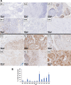
Results: While the 18-gene signature as a whole did not have any prognostic power, a subset of three RNAs: Col13a1, Spock2, and Sfrp1, together completely separated CMTs with and without LN metastasis in the microarray set. However, in the new independent set assessed by RT-qPCR, only the Wnt-antagonist Sfrp1 showed significantly increased mRNA abundance in CMTs without LN metastases on its own (p = 0.013) in logistic regression analysis. This correlated with stronger SFRP1 protein staining intensity of the myoepithelium and/or stroma (p < 0.001). SFRP1 staining, as well as β-catenin membrane staining, was significantly associated with negative LN status (p = 0.010 and 0.014 respectively). However, SFRP1 did not correlate with β-catenin membrane staining (p = 0.14).
Conclusion: The study identified SFRP1 as a potential biomarker for metastasis formation in CMTs, but lack of SFRP1 was not associated with reduced membrane-localisation of β-catenin in CMTs.
Keywords: Canine Mammary Tumours; Metastasis; RNA Signature; SFRP1.
© 2023. The Author(s).
Conflict of interest statement
The authors declare no competing interests.
Figures


Similar articles
-
Evaluation of expression of the Wnt signaling components in canine mammary tumors via RT2 Profiler PCR Array and immunochemistry assays.J Vet Sci. 2017 Sep 30;18(3):359-367. doi: 10.4142/jvs.2017.18.3.359. J Vet Sci. 2017. PMID: 27586466 Free PMC article.
-
Lymphangiogenesis in canine mammary tumours: a morphometric and prognostic study.J Comp Pathol. 2014 Feb-Apr;150(2-3):184-93. doi: 10.1016/j.jcpa.2013.09.006. Epub 2013 Nov 11. J Comp Pathol. 2014. PMID: 24225342
-
Expression and prognostic value of c-met in canine mammary tumours.Vet Comp Oncol. 2018 Dec;16(4):670-676. doi: 10.1111/vco.12439. Epub 2018 Sep 23. Vet Comp Oncol. 2018. PMID: 30129270
-
Exploring the role of microRNAs as diagnostic and prognostic biomarkers in canine mammary tumors.Geroscience. 2024 Dec;46(6):6641-6657. doi: 10.1007/s11357-024-01260-7. Epub 2024 Jul 2. Geroscience. 2024. PMID: 38954129 Free PMC article. Review.
-
Canine mammary tumours, an overview.Reprod Domest Anim. 2011 Dec;46(6):1112-31. doi: 10.1111/j.1439-0531.2011.01816.x. Epub 2011 Jun 6. Reprod Domest Anim. 2011. PMID: 21645126 Review.
Cited by
-
Studying Mammary Physiology and Pathology in Domestic Species Benefits Both Humans and Animals.J Mammary Gland Biol Neoplasia. 2023 Jul 14;28(1):18. doi: 10.1007/s10911-023-09547-9. J Mammary Gland Biol Neoplasia. 2023. PMID: 37450225 Free PMC article. No abstract available.
-
Review of Molecular Technologies for Investigating Canine Cancer.Animals (Basel). 2024 Feb 29;14(5):769. doi: 10.3390/ani14050769. Animals (Basel). 2024. PMID: 38473154 Free PMC article. Review.
References
Publication types
MeSH terms
Substances
LinkOut - more resources
Full Text Sources
Medical

