. 2023 Jul 1;136(13):jcs261121.
doi: 10.1242/jcs.261121. Epub 2023 Jul 4.

How host ER membrane chaperones and morphogenic proteins support virus infection

Affiliations

How host ER membrane chaperones and morphogenic proteins support virus infection

Tai-Ting Woo et al. J Cell Sci. .

Abstract

The multi-functional endoplasmic reticulum (ER) is exploited by viruses to cause infection. Morphologically, this organelle is a highly interconnected membranous network consisting of sheets and tubules whose levels are dynamic, changing in response to cellular conditions. Functionally, the ER is responsible for protein synthesis, folding, secretion and degradation, as well as Ca2+ homeostasis and lipid biosynthesis, with each event catalyzed by defined ER factors. Strikingly, these ER host factors are hijacked by viruses to support different infection steps, including entry, translation, replication, assembly and egress. Although the full repertoire of these ER factors that are hijacked is unknown, recent studies have uncovered several ER membrane machineries that are exploited by viruses - ranging from polyomavirus to flavivirus and coronavirus - to facilitate different steps of their life cycle. These discoveries should provide better understanding of virus infection mechanisms, potentially leading to the development of more effective anti-viral therapies.

Keywords: Coronavirus; ER membrane complex; ER morphogenesis; Endoplasmic reticulum; Flavivirus; Polyomavirus.


Conflict of interest statement

Competing interests B.T. is collaborating with Via Nova Therapeutics. The other authors declare no competing or financial interests.

Figures
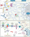
Fig. 1.
Fig. 1.
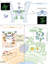
Basic morphology and functions of the ER. (A) Morphologically, the ER is divided into three subdomains: the perinuclear ER, containing sheet-like membranes with associated ribosomes that support protein biosynthesis; the peripheral ER, which harbors tubules that create ER three-way junctions, giving rise to the web-like ER appearance; and the nuclear envelope, which is studded with nuclear pore complexes (NPCs) connecting the nucleoplasm to the cytoplasm. The ER transmembrane protein Climp63 acts as a luminal spacer to maintain the ER sheets of the perinuclear ER, while the ER morphogenesis machinery composed of ATL proteins, RTN proteins and LNP at the peripheral ER promotes formation of ER three-way junctions. ATL proteins trigger fusion between a tubular ER terminus and the side of another tubule via a GTP-dependent process to generate three-way junctions. The vast extent of the ER network within a cell enables it to engage other cellular organelles, including endosomes and mitochondria, via MCSs. (B) The biogenesis of luminal and membrane proteins by ER membrane-associated ribosomes is coupled to the co-translational translocation of the nascent polypeptide chain across the ER membrane and into the ER lumen by the Sec61 translocon. Folding of the imported nascent protein is assisted by several proteins that reside in the ER lumen, such as OST, BiP and PDIs. Membrane proteins are further incorporated into the lipid bilayer with the assistance of the EMC. When a client protein misfolds, sensors of the UPR transduce signals to activate the ER stress response. Misfolded proteins are retro-translocated and directed to the cytosolic proteasome for degradation via the ERAD pathway. In some circumstances, misfolded proteins form luminal aggregates that are targeted and engulfed by the cytosolic phagophore and directed further for lysosomal degradation via the ER-phagy pathway.
Fig. 2.
Fig. 2.
EMC-mediated ER–endosome MSCs support virus infection. (A) Upon entry, the non-enveloped DNA polyomavirus SV40 is endocytosed, reaching the early then late endosome. Through membrane-associated Rab7, the endosome membrane is tethered to the ER membrane via binding to the EMC (i.e. EMC4 and EMC7). Syntaxin18-mediated membrane fusion of a late endosome (harboring SV40) with the ER membrane delivers the virus into the ER lumen. Several ER-resident redox enzymes, including PDIs, reduce and isomerize the disulfide bonds of SV40 capsids inside the ER lumen; this results in conformational changes that expose the inner hydrophobic viral proteins, which support integration into the ER membrane. The membrane-embedded hydrophobic viral particle is stabilized by the chaperone activity of EMC1 of the EMC complex. This prevents premature virus disassembly, thereby enabling the successful penetration of the virus across the lipid bilayer of the ER membrane to reach the cytosol. Upon arrival in the cytosol, the SV40 capsid is disassembled, and the viral genome enters the nucleus via the nuclear pore complex (NPC) for replication and production of new virions. (B) The enveloped RNA flavivirus DENV also ends up in late endosomes after initial receptor-mediated endocytosis. An unknown endosome-associated protein tethers the late endosome to the ER via binding to the EMC. The resulting membrane contact between these two organelles allows for phospholipid transfer from the ER to the endosomal membrane, which facilitates the fusion between endosomal and viral membranes; this leads to release of the nucleocapsid containing the viral genome into the cytosol. The viral genome undergoes co-translational translocation on the ER membrane where the viral polyprotein is synthesized. The partially hydrophobic transmembrane segments of the polyprotein rely on EMC chaperone activity for proper insertion into the ER membrane, which is essential for polyprotein stability and subsequent replication of the viral genome, leading to productive infection.
Fig. 3.
Fig. 3.
Exploiting of the ER morphogenic machinery to promote viral exit from the ER and construction of replication organelles. (A) In order to escape from the ER into the cytosol, polyomavirus SV40 triggers the mobilization of many ER membrane proteins to help establish multi-tubular ER junctions (which are stabilized by LNP proteins). Despite being stabilized, ER junctional sites remain more destabilized as compared to the rest of the ER and thus serve as ideal virus penetration sites. RTN proteins protect the membrane integrity by inducing membrane curvature, promoting membrane flexibility around the protruding virus. This membrane penetration site can be visualized using super-resolution focused ion beam scanning electron microscopy (FIB-SEM) and appears as a flower-like structure. Representative three-dimensional reconstruction images of the multi-tubular ER junctions are shown; images adapted from Bagchi et al. (2021) with permission from Elsevier. The green structures mark the ER, and the red structures depict the SV40 particle. (B) Some (+)RNA viruses, including flaviviruses, induce massive ER membrane rearrangement to form CMs, in which viral structural proteins and NSPs can be detected. The flaviviruses also trigger invagination of the ER membrane to form VPs, which support replication of the viral RNA genome (template RNA is indicated in blue, and the replicated RNA is shown in red). The ER morphogenesis machinery interacts with viral proteins to shape the ER membrane during infection. For example, RTN3 interacts with NS4A and can be recruited to VPs during infection with WNV, DENV and ZIKV. LNP can bind to TEBV NS4B and is recruited to VP-like structures (induced by expression of TBEV NS4A–NS4B fusion protein). Both ATL2 and ATL3 associate with the DENV NS2B–NS3–NS5 replication complex and localize to DENV-induced VPs. However, only ATL2 is required for VP formation, whereas ATL3 colocalizes with DENV envelope protein at assembly sites and might play a role in membrane trafficking or maturation of DENV. (C) Some (+)RNA viruses, including coronaviruses, induce formation of a DMV structure that also supports viral genome replication. The DMV is often found juxtaposed to a lipid droplet (LD) to facilitate lipid transfer to the DMV. It remains unclear how the host ER morphogenesis machinery is co-opted by coronaviruses to form the DMV. In the case of SARS-CoV-2, NSP3 and NSP4 are sufficient to induce DMV formation, but only RTN proteins of the host ER membrane-shaping machinery have been shown to play a functional role in this process. SARS-CoV-2 NSP6 induces ER zippering, forming a connector that allows localization of ER membrane proteins, but not ER luminal proteins, to the DMV.

Similar articles

Cited by

References

    1. Aktepe, T. E., Liebscher, S., Prier, J. E., Simmons, C. P. and Mackenzie, J. M. (2017). The host protein reticulon 3.1A is utilized by flaviviruses to facilitate membrane remodelling. Cell Rep. 21, 1639-1654. 10.1016/j.celrep.2017.10.055 - DOI - PubMed
    1. Bagchi, P., Walczak, C. P. and Tsai, B. (2015). The endoplasmic reticulum membrane J protein C18 executes a distinct role in promoting simian virus 40 membrane penetration. J. Virol. 89, 4058-4068. 10.1128/JVI.03574-14 - DOI - PMC - PubMed
    1. Bagchi, P., Inoue, T. and Tsai, B. (2016). EMC1-dependent stabilization drives membrane penetration of a partially destabilized non-enveloped virus. Elife 5, e21470. 10.7554/eLife.21470 - DOI - PMC - PubMed
    1. Bagchi, P., Torres, M., Qi, L. and Tsai, B. (2020). Selective EMC subunits act as molecular tethers of intracellular organelles exploited during viral entry. Nat. Commun. 11, 1127. 10.1038/s41467-020-14967-w - DOI - PMC - PubMed
    1. Bagchi, P., Liu, X., Cho, W. J. and Tsai, B. (2021). Lunapark-dependent formation of a virus-induced ER exit site contains multi-tubular ER junctions that promote viral ER-to-cytosol escape. Cell Rep. 37, 110077. 10.1016/j.celrep.2021.110077 - DOI - PMC - PubMed

Publication types

MeSH terms

Substances