. 2023 Aug:233:109563.
doi: 10.1016/j.exer.2023.109563. Epub 2023 Jun 29.

Flow cytometric method for the detection and quantification of retinal cell death and oxidative stress

Affiliations

Flow cytometric method for the detection and quantification of retinal cell death and oxidative stress

Shubha Subramanya et al. Exp Eye Res. 2023 Aug.

Abstract

Retinal cell death is the major cause of vision loss in many forms of blinding retinal disease. A plethora of research is focused on understanding the mechanisms of retinal cell death to identify potential neuroprotective strategies that prevent vision loss in these diseases. Traditionally, histological techniques have been used to determine the type and extent of cell death in the retina. These techniques, such as TUNEL labeling and immunohistochemistry, are laborious and time consuming, resulting in low throughput and variable results depending on the experimenter. To increase throughput and reduce variability, we developed several flow cytometry-based assays to detect and quantify retinal cell death. The methods and accompanying data presented demonstrate that flow cytometry can readily detect both retinal cell death and oxidative stress and importantly, the efficacy of neuroprotective agents. These methods will be of interest to investigators looking to increase throughput and efficiency without compromising sensitivity as the methods herein reduce analysis time from several months to less than a week. As such, the flow cytometry methods presented have the potential to expedite research efforts focused on developing novel strategies for retinal cell neuroprotection.

Keywords: Apoptosis; Caspase; Flow cytometry; Reactive oxygen species; Retina; Retinal detachment.


Figures

Figure 1 –
Figure 1 –. Tissue processing and flow cytometry gating
(A) Flow diagram showing steps involved in dissociating whole neural retina into a single cell suspension. Mouse retina is dissected using the cut and pick method and digested in a trypsin solution for 20 minutes. The tissue is then passed repeatedly though an 18G needle before 2 mL of FACS buffer is added to inactivate the trypsin solution. The tissue homogenate is then centrifuged, supernatant removed, and the cells are resuspended in 1 mL of PBS. The single cell suspension is now ready for staining. (B) Gating strategy for flow cytometric analysis. Cells are initially gated using forward scatter and side scatter to remove any broken cells or debris from further analysis (red gate). The cells within the red gate are then further refined to only examine singlet cells (blue gate). Cells within the blue gate are then analyzed for a change in fluorescent intensity for each individual stain (green gate). The fold change of the percentage of cells within the green gate are used to calculate changes in marker expression.
Figure 2 –
Figure 2 –. Flow cytometry can detect early and late markers of retinal cell death after noxious stimuli.
Contour plots showing an increased percentage of (A) Annexin V, (B) 7AAD (7-Aminoactinomycin D), and (C) TUNEL (terminal deoxynucleotidyl transferase dUTP nick end labeling) positive cells compared to their isotype or negative controls. Animals received indicated amount of ferrous sulfate intravitreal (final vitreous concentration), and retinas were harvested 24 hours later.
Figure 3 –
Figure 3 –. Flow cytometry can detect and quantitate retinal cell death secondary to experimental retinal detachment.
Mouse retinas were harvested 3 days after experimental retinal detachment. Dissociated cells were stained to assess for cell death. Contour plots depicting increased (A) TUNEL (terminal deoxynucleotidyl transferase dUTP nick end labeling), (B) cleaved caspase-8, and (C) caspase-8 cleaved at Asp387 staining. Graphs showing corresponding fold change increase in stained cells are displayed to the right of their respective contour plots. There was a significant increase in the percentage of TUNEL positive and cleaved caspase-8 positive cells compared to negative control or isotype control. There was a trend towards increased caspase 8 cleaved at Aspartate-387. N=2–3 for control and attached samples; N=2–11 for detached samples from 4 independent experiments. Alexa Fluor 594 (AF594). **p<0.01, *p<0.05
Figure 4 –
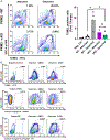
Figure 4 –. Flow cytometry detects and quantitates changes in retinal cell death markers with neuroprotective strategies.
(A) Experimental retinal detachment was induced in either wildtype (PKM2-WT) or Pkm2fl/fl;Rho-Cre+ (PKM2-cKO) and retinas were harvested 3 days later. Contour plots depict a decrease in the percent of TUNEL (terminal deoxynucleotidyl transferase dUTP nick end labeling) positive cells in PKM2-cKO mice compared to PKM2-WT animals. The graph shows a statistically significant fold change in TUNEL positive cells after detachment in PKM2-cKO animals compared to PKM2-WT animals. N=2–5 per group (B) In a separate experiment, experimental retinal detachment was induced in WT animals and either DMSO or ZVAD-FMK (200 μM) was administered sub-retinally at the time of detachment. Contour plots showing decreased TUNEL, cleaved caspase-8, and cleaved caspase-8 at Asp387 staining 3 days status post experimental retinal detachment after treatment with ZVAD. Alexa Fluor 594 (AF594). * p<0.05, ns – not significant
Figure 5 –
Figure 5 –. Assessment of oxidative stress markers in dissociated mouse retina.
200 μM tBH (final vitreous concentration, tert-butyl hydroperoxide) was injected intravitreally, and retinas were harvested 24 hours later. Contour plots showing a significant increase in (A) CM-H2DCFDA (6-chloromethyl-2’,7’-dichlorodihydrofluorescein diacetate) staining indicating increased levels of reactive oxygen species, and (B) an increase in C11-BODIPY (4,4-difluoro-4-bora-3a,4a-diaza-s-indacene) positive cells indicating lipid peroxidation occurs after treatment with tBH. (C) Calcein-AM fluorescence is shifted to a lower intensity after treatment with tBH, indicating an increase in the labile iron pool retina. The histogram shows a shift in the peak intensity (dotted line) to a lower fluorescent intensity in tBH treated retinas. The graphs to the right of respective contour plots quantify the difference in positive cells between vehicle and tBH treated retinas. N=4. **p<0.01, *p<0.05.
Figure 6 –
Figure 6 –. Demonstration of increased retinal cell death after treatment with tBH using flow cytometric methods.
200 μM tBH (final vitreous concentration, tert-butyl hydroperoxide) was injected intravitreally. Retinas were harvested 24 hours later and processed for flow cytometry. Contour plots depicting (A) increased TUNEL (terminal deoxynucleotidyl transferase dUTP nick end labeling), (b) cleaved caspase-8, and (C) caspase-8 cleaved at Asp387 positive cells after tBH treatment. Corresponding bar graphs displayed to the right of the respective contour plots quantify the fold change in positive cells. There was a significant increase in the percentage of TUNEL positive, cleaved caspase-8 positive and caspase-8 cleaved at Asp387 positive cells compared to negative control or isotype control N=4. Alexa Fluor 594 (AF594). **p<0.01, *p<0.05.

Similar articles

Cited by

References

    1. Awwad S, Henein C, Ibeanu N, Khaw PT, Brocchini S, 2020. Preclinical challenges for developing long acting intravitreal medicines. Eur. J. Pharm. Biopharm 153, 130–149. - PubMed
    1. Besirli CG, Chinskey ND, Zheng QD, Zacks DN, 2010. Inhibition of retinal detachment-induced apoptosis in photoreceptors by a small peptide inhibitor of the fas receptor. Invest. Ophthalmol. Vis. Sci 51, 2177–2184. - PMC - PubMed
    1. Campochiaro PA, Iftikhar M, Hafiz G, Akhlaq A, Tsai G, Wehling D, Lu L, Wall GM, Singh MS, Kong X, 2020. Oral N-acetylcysteine improves cone function in retinitis pigmentosa patients in phase I trial. J. Clin. Invest 130, 1527–1541. - PMC - PubMed
    1. Carmody RJ, McGowan AJ, Cotter TG, 1998. Rapid detection of rod photoreceptor apoptosis by flow cytometry. Cytometry 33, 89–92. - PubMed
    1. Chan TC, Wilkinson Berka JL, Deliyanti D, Hunter D, Fung A, Liew G, White A, 2020. The role of reactive oxygen species in the pathogenesis and treatment of retinal diseases. Exp. Eye Res 201, 108255. - PubMed

Substances