. 2023 Aug 25;51(15):7936-7950.
doi: 10.1093/nar/gkad543.

Alteration of replication protein A binding mode on single-stranded DNA by NSMF potentiates RPA phosphorylation by ATR kinase

Affiliations

Alteration of replication protein A binding mode on single-stranded DNA by NSMF potentiates RPA phosphorylation by ATR kinase

Yujin Kang et al. Nucleic Acids Res. .

Abstract

Replication protein A (RPA), a eukaryotic single-stranded DNA (ssDNA) binding protein, dynamically interacts with ssDNA in different binding modes and plays essential roles in DNA metabolism such as replication, repair, and recombination. RPA accumulation on ssDNA due to replication stress triggers the DNA damage response (DDR) by activating the ataxia telangiectasia and RAD3-related (ATR) kinase, which phosphorylates itself and downstream DDR factors, including RPA. We recently reported that the N-methyl-D-aspartate receptor synaptonuclear signaling and neuronal migration factor (NSMF), a neuronal protein associated with Kallmann syndrome, promotes RPA32 phosphorylation via ATR upon replication stress. However, how NSMF enhances ATR-mediated RPA32 phosphorylation remains elusive. Here, we demonstrate that NSMF colocalizes and physically interacts with RPA at DNA damage sites in vivo and in vitro. Using purified RPA and NSMF in biochemical and single-molecule assays, we find that NSMF selectively displaces RPA in the more weakly bound 8- and 20-nucleotide binding modes from ssDNA, allowing the retention of more stable RPA molecules in the 30-nt binding mode. The 30-nt binding mode of RPA enhances RPA32 phosphorylation by ATR, and phosphorylated RPA becomes stabilized on ssDNA. Our findings provide new mechanistic insight into how NSMF facilitates the role of RPA in the ATR pathway.


Figures

Graphical Abstract
Graphical Abstract
Figure 1.
Figure 1.
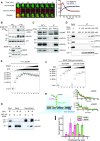
NSMF interacts with RPA at DNA damage sites and dissociates RPA from ssDNA. (A) Laser microirradiation experiments were performed on HeLa cells transfected with eGFP-NSMF and mCherry-RPA32. The kinetics of NSMF and RPA32 localization at DNA damage sites were examined. Left: fluorescence images of a laser-irradiated cell as a function of time. The red lines indicate the laser-irradiated sites. Right: quantification of relative fluorescence intensity of eGFP and mCherry. The initial fluorescent intensity at the damage site was set as 100 for each cell, and the recruitment kinetics were plotted. The intensity was averaged for more than 10 cells under each condition, and the data represent the mean + SEM for three repeated experiments. (B) (Top) Chromatin fraction analysis for the NSMF effect on RPA32 phosphorylation under replication stress. (Bottom) NSMF expression in WT and KO cells. The asterisks (*) represent nonspecific bands. (C) Endogenous immunoprecipitation (IP) assay for NSMF and the RPA trimer in HeLa cells. WCL: whole cell lysates. (D) In vitro IP assay for NSMF and RPA binding. Purified NSMF and RPA trimer were immunoprecipitated with anti-GFP at different concentrations. Each RPA subunit was analyzed by western blotting. (E) EMSA for NSMF and the RPA–ssDNA complex. RPA (0, 25, 75, 100, 125, and 150 nM) was titrated with 10 nM 91-nt ssDNA. NSMF (0, 10, 20, 40, 80, 140, and 200 nM) was titrated with the RPA (150 nM)–ssDNA complex. (F) The effect of NSMF on the formation of RPA–ssDNA complexes. Left: only RPA (0, 25, 75, 125, and 150 nM) was titrated with the 91-nt ssDNA without NSMF. Right: NSMF (200 nM) was pre-incubated with 10 nM ssDNA, and then RPA was titrated. (G) Magnetic bead pulldown assay for RPA dissociation by NSMF. The supernatant and bound fractions were analyzed by western blotting using an anti-FLAG antibody (αFLAG) for NSMF and an RPA32 antibody (αRPA32). (H) Single-molecule photobleaching assay for NSMF-mediated RPA dissociation from ssDNA. Left: schematic of the single-molecule photobleaching assay. The 91-nt ssDNA was anchored on a slide surface passivated by a lipid bilayer via a biotin-streptavidin linkage. eGFP molecules tagged on RPA proteins were bleached step-wise under continuous laser illumination, and the fluorescence signal was detected by total internal reflection fluorescence microscopy. Right: time traces of the fluorescence intensities of RPA-eGFP bound to ssDNA molecules in the absence (top) or presence (bottom) of NSMF. The steps of fluorescence intensity indicate the photobleaching events of individual eGFP molecules. (I) Histogram for the photobleaching steps of RPA-eGFPs bound to ssDNA in the presence (magenta) or absence (green) of NSMF. Error bars represent the standard deviation in triplicate. The number of analyzed molecules was >100.
Figure 2.
Figure 2.
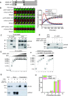
RPA binding–defective mutant NSMF-ΔD2 does not destabilize RPA from ssDNA. (A) Domain constructs for NSMF-WT and NSMF-ΔD2, a mutant in which RPA interaction domain D2 (74–239 amino acids) was deleted. (B) The laser microirradiation experiments were performed with NSMF KO HeLa cells transfected with mCherry-RPA32 and eGFP-NSMF WT, ΔD2, or empty vector. Left: fluorescent images of laser-irradiated cells as a function of time. The white lines indicate the laser-irradiated sites. Right: quantification of the relative fluorescence intensities of eGFP and mCherry. The initial fluorescent intensity at the damage site was set as 100 for each cell, and the recruitment kinetics were plotted. The average intensities for >10 cells for each condition are presented. Data represent the mean + SEM for three repeated experiments. (C) Interaction between RPA32 and either NSMF-WT or NSMF-ΔD2 was determined using immunoprecipitation from HeLa cells transfected with FLAG-NSMF WT or ΔD2. The asterisks denoted bands for NSMF WT and ΔD2. (D) In vitro binding assay using purified RPA32 and either purified NSMF-eGFP WT or ΔD2. (E) EMSA for the NSMF-ΔD2 and RPA–ssDNA complex. RPA (0, 25, 75, 125, and 150 nM) was titrated with 10 nM 91-nt ssDNA, and NSMF-ΔD2 (0, 10, 20, 40, 80, 140, and 200 nM) was titrated with the RPA–ssDNA complex at 150 nM RPA. (F) The effect of NSMF-ΔD2 on the formation of the RPA–ssDNA complex. Left: only RPA was titrated with 91-nt ssDNA without NSMF. Right: NSMF-ΔD2 (200 nM) was pre-incubated with 10 nM ssDNA and then titrated with RPA (0, 25, 75, 125, and 150 nM). (G) Magnetic bead pulldown assay for RPA dissociation by NSMF-ΔD2. NSMF-ΔD2 was incubated with RPA–ssDNA complexes coated on magnetic beads. The supernatant and bound fractions were analyzed by western blotting using an anti-FLAG antibody (αFLAG) for NSMF and an anti-RPA32 antibody (αRPA32). (H) Histogram for the photobleaching steps of RPA-eGFP bound to ssDNA in the presence (magenta) or absence (green) of NSMF-ΔD2. Error bars represent the standard deviation in triplicate. The number of analyzed molecules was >100.
Figure 3.
Figure 3.
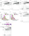
NSMF modulates the RPA binding mode. (A–C) EMSA for NSMF with RPA bound to (A) 60-nt, (B) 30-nt, and (C) 14-nt ssDNA. RPA (0, 25, 75, 100, 125, and 150 nM) was titrated with 10 nM of each ssDNA. RPA (150 nM) was titrated with NSMF (0, 10, 20, 40, 80, 140, and 200 nM). (D, E) Histograms for photobleaching events of RPA-eGFP bound to (D) 60-nt and (E) 30-nt ssDNA in the presence (magenta) or absence (green) of NSMF. The addition of NSMF reduced the number of bound RPA molecules. More than 100 molecules were analyzed for each dataset. Error bars represent the standard deviation in triplicate. (F) NSMF (0, 10, 20, 40, 80, 140, and 200 nM) was titrated with 25 nM RPA bound to 30-nt ssDNA. At 25 nM, RPA had a single binding mode (30-nt mode) without a band shift. (G) Top: the domain structure for the DNA binding defective mutant of RPA (DBM-RPA), in which Trp107 and Phe135 were both replaced by Ala. Bottom: EMSA for NSMF with DBM-RPA and 30-nt ssDNA. DBM-RPA (25 nM) was bound to 10 nM 30-nt ssDNA and then titrated with NSMF (0, 10, 20, 40, 80, 140, and 200 nM).
Figure 4.
Figure 4.
RPA destabilization mechanism by NSMF. (A) Domain constructs for the RPA70 deletion mutants. (B) IP assays between NSMF and the RPA70 deletion mutants in the presence of benzonase. NSMF interacted with the DBD-C domain (D5: 436–610 amino acids). (C) Domain constructs for the RPA32 deletion mutants. (D) IP assays between NSMF and the RPA32 deletion mutants in the presence of benzonase. NSMF interacted with the C-terminal domain (D3-2: 200–270 amino acids). (E) Quantification of the EMSA data for NSMF (WT or ΔD2) and ssDNA in Figure S3A and S3C. Filled square: NSMF-WT and blank circle: NSMF-ΔD2. The error bars represent the standard deviation in triplicate. The quantified data were fitted using the hyperbola function, formula image. The Kd values for NSMF-WT and NSMF-ΔD2 on ssDNA were estimated to be 200 ± 22 nM and 540 ± 59 nM, respectively. (F) Endogenous RPA70 and RPA14 in NSMF-WT or KO HeLa cell lysates were immunoprecipitated with an anti-RPA32 antibody, and western blotting was performed using the indicated antibodies. NSMF did not disrupt the RPA trimer in the absence of DNA in vivo.
Figure 5.
Figure 5.
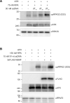
RPA 30-nt binding mode induced by NSMF enhances RPA32 phosphorylation by ATR invitro. (A) In vitro phosphorylation of RPA32 by ATR depends on the ssDNA length (10-nt [dT10], 20-nt [dT20], or 30-nt [dT30] ssDNA consisting of only thymines). ATR was pulled down from HeLa cell extracts treated with 2 mM hydroxyurea using anti-ATR antibody-coated beads. The samples were analyzed by western blotting using the indicated antibodies. (B) NSMF-mediated enhancement of RPA32 phosphorylation by ATR in vitro. Excess RPA was incubated with 91-nt ssDNA followed by 80 nM NSMF and pulled-down ATR. The proteins were analyzed by western blotting using the indicated antibodies.
Figure 6.
Figure 6.
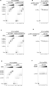
NSMF does not destabilize phosphorylated RPA. (A) EMSA for the binding of WT RPA and pmRPA-S33D (0, 25, 75, 100, 125, and 150 nM) to 91-nt ssDNA. (B) EMSA for the NSMF effect on pmRPA-S33D. NSMF (0, 20, 40, 80, 140, and 200 nM) was titrated with 200 nM pmRPA-S33D pre-incubated with 10 nM 91-nt ssDNA. (C) EMSA for WT RPA and pmRPA-S4/8D&S33D (0, 25, 75, 100, 125, and 150 nM) to compare their binding to 91-nt ssDNA. (D) EMSA for the NSMF effect on pmRPA-S4/8D&S33D. NSMF (0, 20, 40, 80, 140, and 200 nM) was titrated with 200 nM pmRPA-S4/8D&S33D pre-incubated with 10 nM 91-nt ssDNA. (E) EMSA for WT RPA and pRPA (0, 25, 75, 100, 125, and 150 nM) to compare their binding to 91-nt ssDNA. (F) EMSA for NSMF effect on pRPA. NSMF (0, 10, 20, 40, 80, 140, and 200 nM) was titrated with 200 nM pRPA pre-incubated with 10 nM 91-nt ssDNA.
Figure 7.
Figure 7.
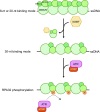
Model for the effect of NSMF on RPA phosphorylation. Immediately after ssDNA is exposed due to replication stress, RPA compactly binds to ssDNA in less stable mode(s). NSMF partially dissociates RPA from the ssDNA, and the remaining RPA molecules form a stable 30-nt binding mode, which promotes RPA phosphorylation via ATR and ATRIP.

Similar articles

Cited by

References

    1. Wold M.S. Replication protein A: a heterotrimeric, single-stranded DNA-binding protein required for eukaryotic DNA metabolism. Annu. Rev. Biochem. 1997; 66:61–92. - PubMed
    1. Iftode C., Daniely Y., Borowiec J.A.. Replication protein A (RPA): the eukaryotic SSB. Crit. Rev. Biochem. Mol. 1999; 34:141–180. - PubMed
    1. Ciccia A., Elledge S.J.. The DNA damage response: making it safe to play with knives. Molecular Cell. 2010; 40:179–204. - PMC - PubMed
    1. Marechal A., Zou L.. RPA-coated single-stranded DNA as a platform for post-translational modifications in the DNA damage response. Cell Res. 2015; 25:9–23. - PMC - PubMed
    1. Marechal A., Zou L.. DNA damage sensing by the ATM and ATR kinases. Cold Spring Harb. Perspect. Biol. 2013; 5. - PMC - PubMed

Publication types