Beneficial Effects of Zoledronic Acid on Tendons of the Osteogenesis Imperfecta Mouse (Oim)
- PMID: 37375779
- PMCID: PMC10303798
- DOI: 10.3390/ph16060832
Beneficial Effects of Zoledronic Acid on Tendons of the Osteogenesis Imperfecta Mouse (Oim)
Abstract
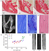
Osteogenesis imperfecta (OI) is a genetic disorder of connective tissue characterized by spontaneous fractures, bone deformities, impaired growth and posture, as well as extra-skeletal manifestations. Recent studies have underlined an impairment of the osteotendinous complex in mice models of OI. The first objective of the present work was to further investigate the properties of tendons in the osteogenesis imperfecta mouse (oim), a model characterized by a mutation in the COL1A2 gene. The second objective was to identify the possible beneficial effects of zoledronic acid on tendons. Oim received a single intravenous injection of zoledronic acid (ZA group) at 5 weeks and were euthanized at 14 weeks. Their tendons were compared with those of untreated oim (oim group) and control mice (WT group) by histology, mechanical tests, western blotting and Raman spectroscopy. The ulnar epiphysis had a significantly lower relative bone surface (BV/TV) in oim than WT mice. The tendon of the triceps brachii was also significantly less birefringent and displayed numerous chondrocytes aligned along the fibers. ZA mice showed an increase in BV/TV of the ulnar epiphysis and in tendon birefringence. The tendon of the flexor digitorum longus was significantly less viscous in oim than WT mice; in ZA-treated mice, there was an improvement of viscoelastic properties, especially in the toe region of stress-strain curve, which corresponds to collagen crimp. The tendons of both oim and ZA groups did not show any significant change in the expression of decorin or tenomodulin. Finally, Raman spectroscopy highlighted differences in material properties between ZA and WT tendons. There was also a significant increase in the rate of hydroxyproline in the tendons of ZA mice compared with oim ones. This study highlighted changes in matrix organization and an alteration of mechanical properties in oim tendons; zoledronic acid treatment had beneficial effects on these parameters. In the future, it will be interesting to better understand the underlying mechanisms which are possibly linked to a greater solicitation of the musculoskeletal system.
Keywords: bone–tendon unit; oim; osteogenesis imperfecta; tendon; zoledronic acid.
Conflict of interest statement
The authors declare no conflict of interest.
Figures


Similar articles
-
Biomechanical, Microstructural and Material Properties of Tendon and Bone in the Young Oim Mice Model of Osteogenesis Imperfecta.Int J Mol Sci. 2022 Sep 1;23(17):9928. doi: 10.3390/ijms23179928. Int J Mol Sci. 2022. PMID: 36077325 Free PMC article.
-
Are Changes in Composition in Response to Treatment of a Mouse Model of Osteogenesis Imperfecta Sex-dependent?Clin Orthop Relat Res. 2015 Aug;473(8):2587-98. doi: 10.1007/s11999-015-4268-z. Clin Orthop Relat Res. 2015. PMID: 25903941 Free PMC article.
-
Collagen from the osteogenesis imperfecta mouse model (oim) shows reduced resistance against tensile stress.J Clin Invest. 1997 Jul 1;100(1):40-5. doi: 10.1172/JCI119519. J Clin Invest. 1997. PMID: 9202055 Free PMC article.
-
Raloxifene reduces skeletal fractures in an animal model of osteogenesis imperfecta.Matrix Biol. 2016 May-Jul;52-54:19-28. doi: 10.1016/j.matbio.2015.12.008. Epub 2015 Dec 18. Matrix Biol. 2016. PMID: 26707242
-
OIM and related animal models of osteogenesis imperfecta.Connect Tissue Res. 1995;31(4):265-8. doi: 10.3109/03008209509010820. Connect Tissue Res. 1995. PMID: 15612365 Review.
Cited by
-
Extra-Skeletal Manifestations in Osteogenesis Imperfecta Mouse Models.Calcif Tissue Int. 2024 Dec;115(6):847-862. doi: 10.1007/s00223-024-01213-4. Epub 2024 Apr 19. Calcif Tissue Int. 2024. PMID: 38641703 Review.
References
-
- Muñoz-Garcia J., Heymann D., Giurgea I., Legendre M., Amselem S., Castañeda B., Lézot F., William Vargas-Franco J. Pharmacological options in the treatment of osteogenesis imperfecta: A comprehensive review of clinical and potential alternatives. Biochem. Pharmacol. 2023;213:115584. doi: 10.1016/j.bcp.2023.115584. - DOI - PubMed
Grants and funding
LinkOut - more resources
Full Text Sources
Miscellaneous

