. 2023 May 30;12(6):1176.
doi: 10.3390/antiox12061176.

ARAG, an Antioxidant-Rich Gel, Shows Superiority to Mepilex Ag in the Treatment of Deep Partial Thickness Burns without Sacrificing Antimicrobial Efficiency

Affiliations

ARAG, an Antioxidant-Rich Gel, Shows Superiority to Mepilex Ag in the Treatment of Deep Partial Thickness Burns without Sacrificing Antimicrobial Efficiency

Brian Michael Cartwright et al. Antioxidants (Basel). .

Abstract

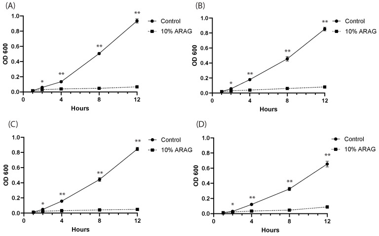
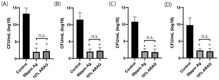
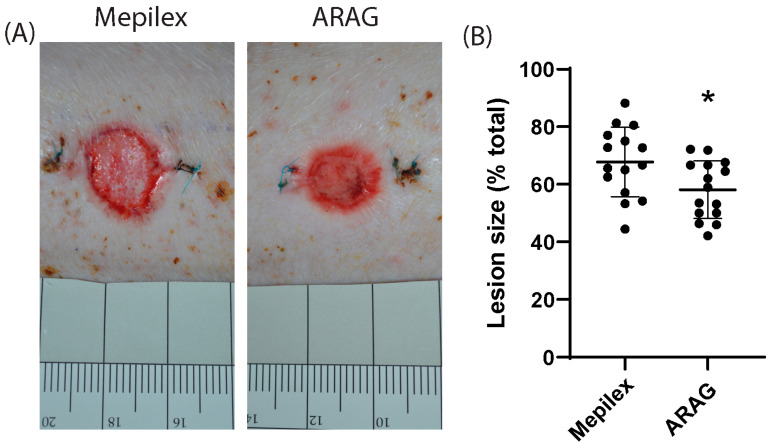
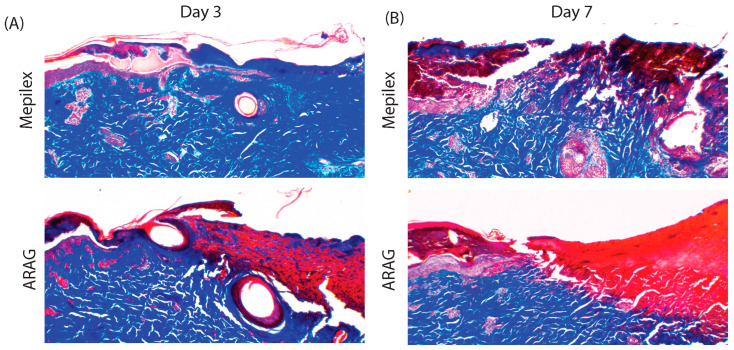
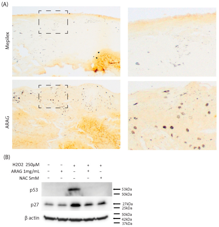
Current treatments for deep tissue burns are limited, and most serve only to enhance hydration or prevent bacterial growth. This leaves burn healing dependent on slow natural processes to debride the wound and reestablish the epidermal and dermal layers of the skin. Infections are well known to destabilize this process through a variety of mechanisms, most notably through increased inflammation and the resulting oxidative stress. In this study, we show that ARAG (an antioxidant-rich antimicrobial gel) can suppress the growth of multiple bacteria commonly found to infect burns (Klebsiella pneumoniae, Proteus vulgaris, Pseudomonas aeruginosa, and Staphylococcus aureus). This inhibition is comparable to that conferred by silver ion release from burn dressings such as Mepilex-Ag. We further show, using a porcine model for deep partial-thickness burns, that ARAG allows for enhanced wound healing over Mepilex-Ag, the current standard of care. Histological findings indicate this is likely due to increased wound debridement and dampening of late inflammatory processes, leading to more balanced physiologic healing. Taken together, these findings show promise for ARAG as a superior alternative to the current standard of care.

Keywords: TPGS; antimicrobial; antioxidant; burns; deep partial thickness burns; wound healing.


Conflict of interest statement

William A. Clark is a stakeholder in AOXbio and the holder of the patent for Lavengel, a product derived from ARAG. All other researchers declare no conflict of interest.

Figures

Figure 1
Figure 1
ARAG is a potent antioxidant capable of preventing oxidative damage induced cell death. (A) The antioxidant activity of ARAG was tested by a DPPH free radical scavenging assay. When compared to ascorbic acid, a known antioxidant, it was found that ARAG possessed good free radical scavenging ability. While this was lower than ascorbic acid at lower concentrations, it became near equivalent with an increased concentration of ARAG (98.3 ± 0.577% ascorbic acid vs. 93.6 ± 2.51% ARAG at 1 mg/mL). To test the ability of ARAG to prevent oxidative damage-induced loss of cell viability and cell death, BJ cells were treated with either 1 mg/mL ARAG or 5 mM NAC for one hour prior to a 12-hour treatment with 250 µM H2O2. (B) Results of MTT cell viability assays revealed that ARAG was highly effective at preventing hydrogen peroxide loss of viability and that this protective effect is near equivalent to that of the glutathione precursor NAC. n = 3, * p < 0.01 (C) Analysis of cell lysates by Western blot for the apoptotic marker c-PARP1 supported MTT findings showing a reduction in apoptosis in ARAG-treated cells exposed to hydrogen peroxide. This reduction was similar but less than that of NAC.
Figure 2
Figure 2
ARAG inhibits the planktonic growth of common burn bacteria. Overnight stock cultures of (A) P. aeruginosa, (B) S. aureus, (C) K. pneumoniae, and (D) P. vulgaris were inoculated into LB and grown at 37 °C with shaking for 12 h either in the presence or absence of 10% ARAG. OD600 measurements were taken at the time points indicated and normalized to the starting OD measurement (n = 4 individual cultures per group). It was found that 10% ARAG rapidly inhibited the growth of all four bacteria tested based on the culture density determined by OD600 measurement. Control cultures (mock inoculated LB) were plated on LB agar without dilution following 12 h of incubation and showed no sign of growth after 24 h (data not shown). * p < 0.01; ** p < 0.001.
Figure 3
Figure 3
ARAG is non-inferior to ionized silver in inhibiting planktonic growth. Stock cultures of (A) P. aeruginosa, (B) S. aureus, (C) K. pneumoniae, and (D) P. vulgaris were normalized to 1 × 10⁶ and inoculated into 2 mL LB containing either saline, 50 ppm ionized silver, or 10% ARAG. Bacteria were then grown at 37 °C with shaking for 24 h before being diluted 10-fold and spread onto LB-agar plates. Plates were incubated for 24 h before being counted to determine CFU/mL. It was found that 10% ARAG significantly reduced the viability of all four bacteria tested, as indicated by the low number of CFUs. Ionized silver, the active anti-microbial component of Mepilex, showed similar results to the 10% ARAG group regarding comparison to the control; however, no significant difference was found between the 50 ppm ionized silver and 10% ARAG groups. Control cultures (mock-inoculated LB) were plated without dilution on LB agar following 24 h of incubation and showed no sign of growth after 24 h (data not shown). n = 4 individual cultures per group; * p < 0.05; n.s. = not significant; CFU = colony-forming units.
Figure 4
Figure 4
ARAG is superior to Mepilex for burn wound healing. (A) Representative images of burns at 21 days are shown. Wounds were assessed by digital photography, followed by quantification of area and comparison to the initial lesion size. (B) A comparison of paired burns showed a significant average reduction in lesion size of 9.65% in ARAG-treated burns over Mepilex-treated burns (*, p = 0.0031).
Figure 5
Figure 5
ARAG decreases the late inflammatory response. Representative images of paired skin biopsies stained with H&E at (A) day three and (B) day seven (4×). Zoomed images (10×) of the wound front (epidermal-dermal junction, black dashed box) are shown to the right. At day three, there is little difference between the levels of leukocytic infiltrate (hematoxylin, dark blue, stained cells) [white arrow] within the wound front. At day seven, there is a marked increase in leukocytic infiltration in both Mepilex and ARAG samples in comparison to day three; however, that infiltration is denser and present over a wider area within the tissue of Mepilex-treated animals.
Figure 6
Figure 6
ARAG increases debridement at the burn healing front. Representative images (4×) of paired skin biopsies stained with Masson’s trichrome at (A) day three and (B) day seven. ARAG-treated burns display enhanced debridement and tissue remodeling, beginning at day three and increasing by day seven. This is demonstrated by the decreased or absence of collagen staining (blue) at the wound front (epidermal-dermal junction) at days three and seven, respectively. Unlike ARAG-treated samples, little to no collagen breakdown is seen at day three in the Mepilex group. At day seven, there is increased collagen breakdown within the Mepilex group; however, it is incomplete in comparison to ARAG.
Figure 7
Figure 7
ARAG maintains cellular proliferation in vivo and decreases markers of oxidative stress-induced senescence in vitro. (A) Representative immunohistochemical staining (10×) of Ki-67, a nuclear marker for cell proliferation, of paired skin biopsies on day seven. Zoomed images of portions of the epidermal-dermal interface (black dashed box) are shown to the right. An increased number of Ki-67-labeled nuclei can be seen in the ARAG-treated group, indicating a higher rate of proliferating cells. To determine if this is due to the prevention of oxidative stress-induced cell cycle arrest and senescence, BJ cells were pretreated with either 1 mg/mL ARAG or 5 mM NAC for one hour before being subjected to a 12-hour treatment with 250 µM H2O2. (B) It was seen that ARAG and NAC both prevented the induction of p27 and p53 following hydrogen peroxide treatment. This suggests that blockage of oxidative stress by ARAG can prevent the upregulation of senescence-associated factors.

Similar articles

References

    1. Barillo D.J., Goode R. Fire fatality study: Demographics of fire victims. Burns. 1996;22:85–88. doi: 10.1016/0305-4179(95)00095-X. - DOI - PubMed
    1. Snell J.A., Loh N.H., Mahambrey T., Shokrollahi K. Clinical review: The critical care management of the burn patient. Crit. Care. 2013;17:241. doi: 10.1186/cc12706. - DOI - PMC - PubMed
    1. White C.E., Renz E.M. Advances in surgical care: Management of severe burn injury. Crit. Care Med. 2008;36:S318–S324. doi: 10.1097/CCM.0b013e31817e2d64. - DOI - PubMed
    1. Cancio L.C. Topical Antimicrobial Agents for Burn Wound Care: History and Current Status. Surg. Infect. 2021;22:3–11. doi: 10.1089/sur.2020.368. - DOI - PubMed
    1. Liu H.F., Zhang F., Lineaweaver W.C. History and Advancement of Burn Treatments. Ann. Plast. Surg. 2017;78:S2–S8. doi: 10.1097/SAP.0000000000000896. - DOI - PubMed

Grants and funding

Funding for portions of this study was supported by a CCRHS Research Enhancement Grant and an ETSU Student-Faculty Collaborative Research Grant awarded to W. Andrew Clark.

LinkOut - more resources