. 2023 Jul 13;66(13):8510-8525.
doi: 10.1021/acs.jmedchem.3c00144. Epub 2023 Jun 26.

Synthesis of a Novel Boronic Acid Transition State Inhibitor, MB076: A Heterocyclic Triazole Effectively Inhibits Acinetobacter-Derived Cephalosporinase Variants with an Expanded-Substrate Spectrum

Affiliations

Synthesis of a Novel Boronic Acid Transition State Inhibitor, MB076: A Heterocyclic Triazole Effectively Inhibits Acinetobacter-Derived Cephalosporinase Variants with an Expanded-Substrate Spectrum

Rachel A Powers et al. J Med Chem. .

Abstract

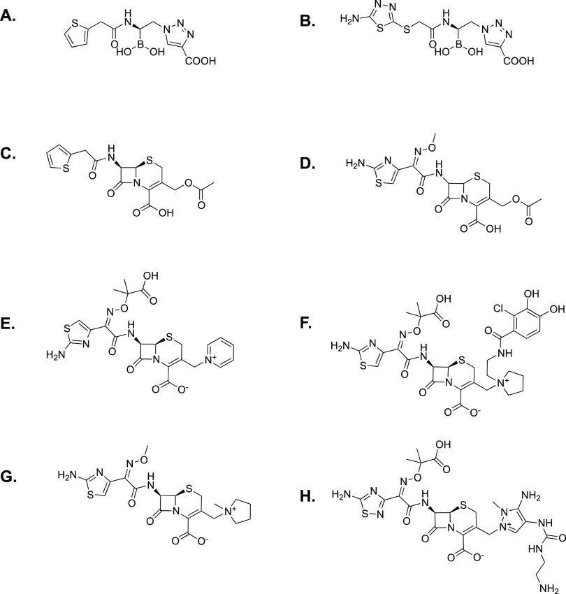
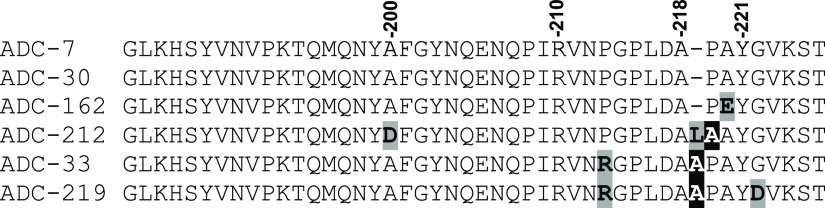
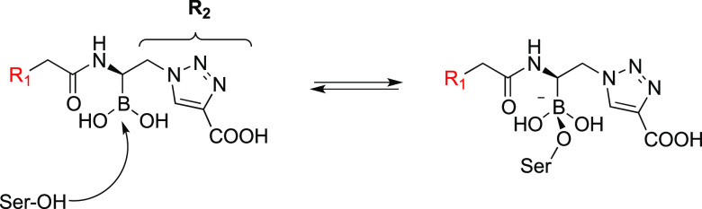
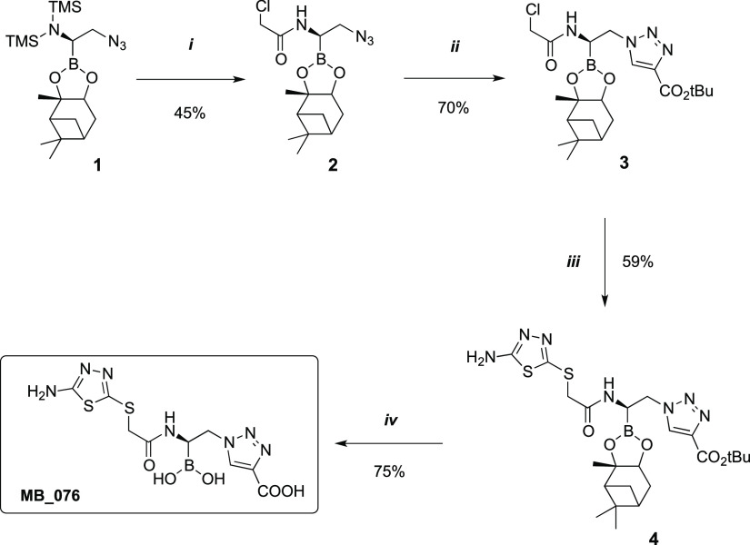
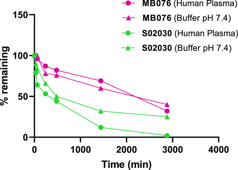
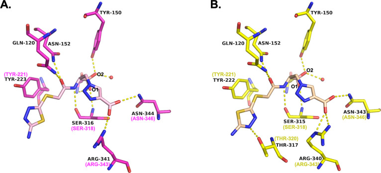
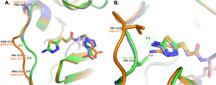
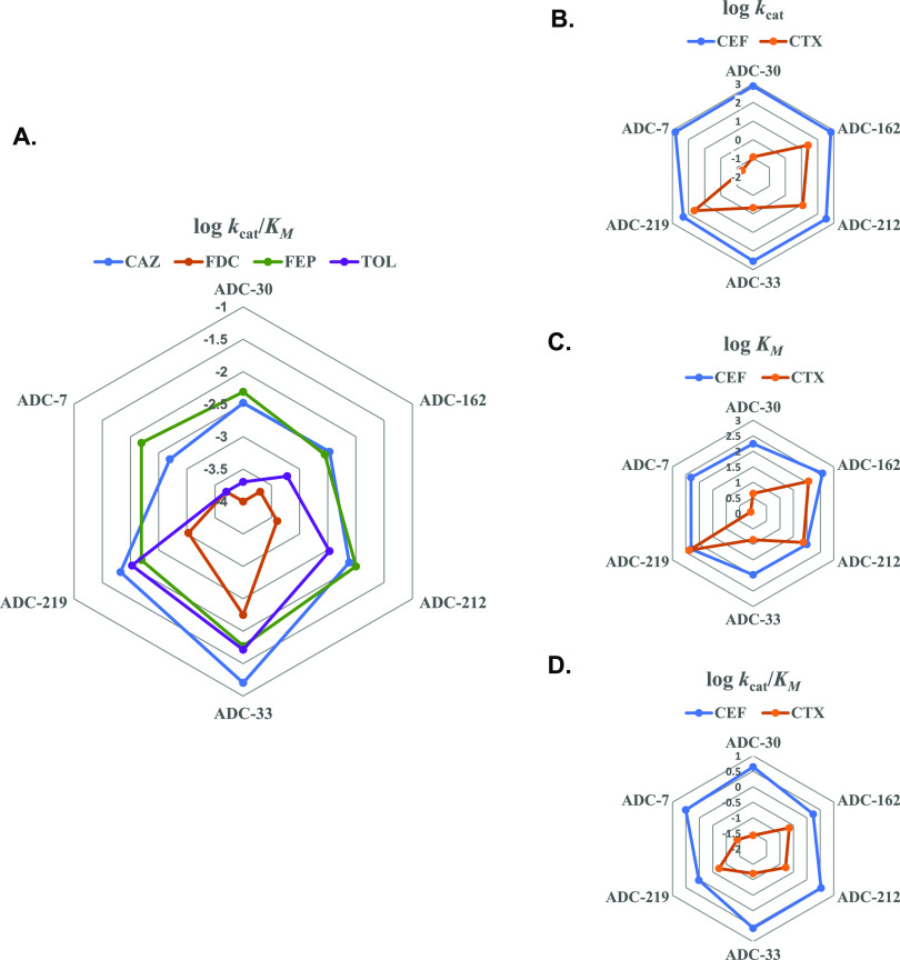
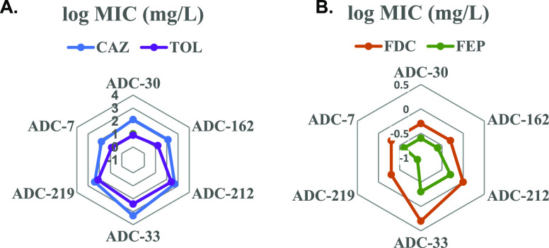
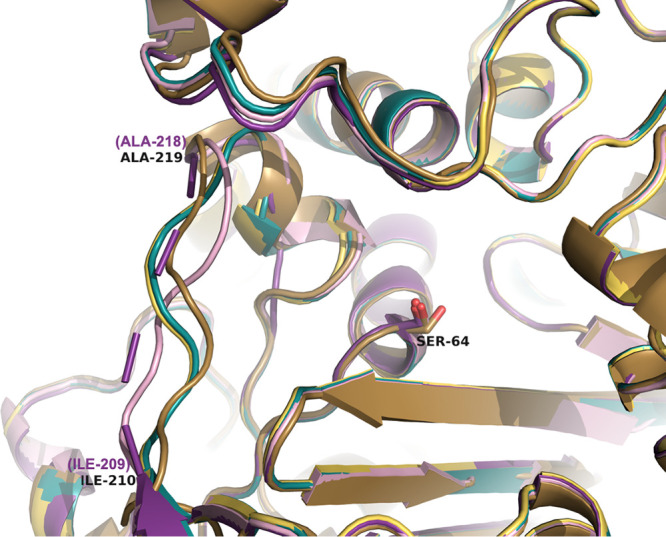
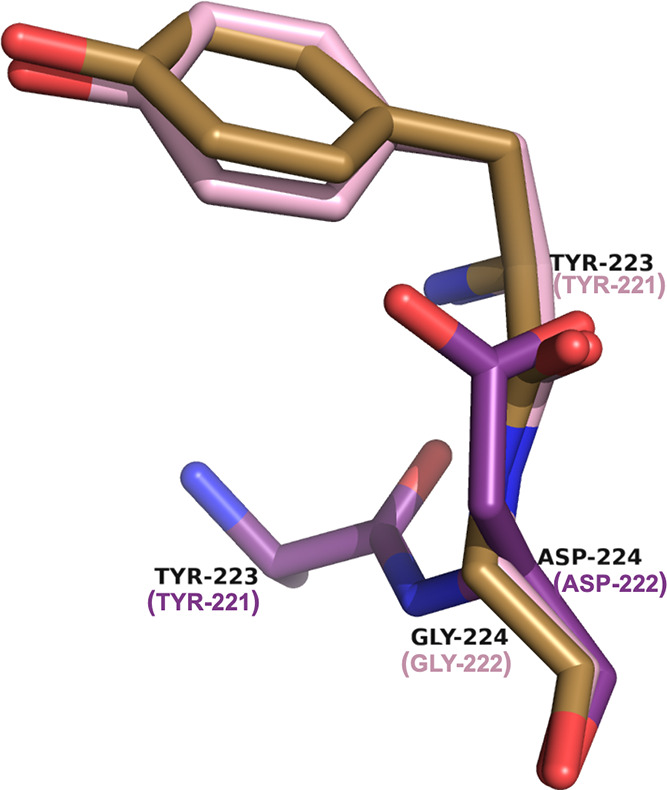
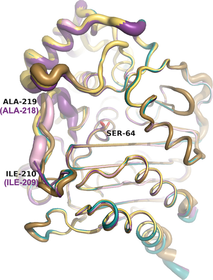
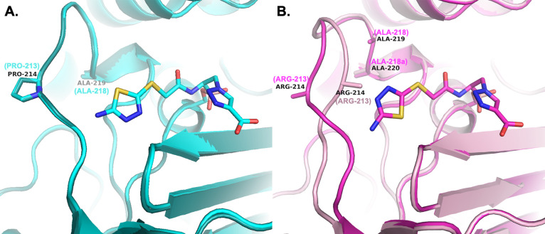
Class C Acinetobacter-derived cephalosporinases (ADCs) represent an important target for inhibition in the multidrug-resistant pathogen Acinetobacter baumannii. Many ADC variants have emerged, and characterization of their structural and functional differences is essential. Equally as important is the development of compounds that inhibit all prevalent ADCs despite these differences. The boronic acid transition state inhibitor, MB076, a novel heterocyclic triazole with improved plasma stability, was synthesized and inhibits seven different ADC β-lactamase variants with Ki values <1 μM. MB076 acted synergistically in combination with multiple cephalosporins to restore susceptibility. ADC variants containing an alanine duplication in the Ω-loop, specifically ADC-33, exhibited increased activity for larger cephalosporins, such as ceftazidime, cefiderocol, and ceftolozane. X-ray crystal structures of ADC variants in this study provide a structural context for substrate profile differences and show that the inhibitor adopts a similar conformation in all ADC variants, despite small changes near their active sites.


Conflict of interest statement

The authors declare no competing financial interest.

Figures

Figure 1
Figure 1
Structures of the antibiotic substrates and BATSI inhibitors used in this study: (A) S02030; (B) MB076; (C) cephalothin; (D) cefotaxime; (E) ceftazidime; (F) cefiderocol; (G) cefepime; (H) ceftolozane.
Figure 2
Figure 2
Multiple alignment of the Ω-loop region of ADCs-7, -30, -162, -212, -33, -219 using Clustal Omega. The SANC numbering scheme positions are listed above the sequences.
Figure 3
Figure 3
Schematic representation of the BATSIs binding to the active site of a serine β-lactamase resembling the tetrahedral transition state of the β-lactam hydrolysis reaction leading to inhibition. In the case of BATSIs, this is a reversible competitive process.
Scheme 1
Scheme 1. (i) (1) MeOH, THF, 0 °C to RT, (2) ClCH2COCl, THF, −30 °C; (ii) t-Butylpropiolate, Sodium Ascorbate, CuSO4, t-ButOH/H2O 1:1, 60 °C; (iii) 5-Amino-1,3,4-thiadiazole-2-thiol, TEA, MeCN, RT; (vi) (1) TFA, Et3SH, DCM, 0 °C to RT, (2) Isobutylboronic Acid, HCl 3 M, MeCN/n-Hexane 1:1, RT
Figure 4
Figure 4
Comparison of the in vitro stability of MB076 and S02030 in plasma and in buffer (pH 7.4). Percentage of compound remaining over time is displayed.
Figure 5
Figure 5
(A) Superposition of ADCs in complex with the BATSI MB076. Carbons for each enzyme are colored as follows: ADC-7 (green, 8FQM), ADC-30 (cyan, 8FQW), ADC-33 (magenta, 8FQO), ADC-162 (yellow, 8FQQ), ADC-212 (orange, 8FQS), and ADC-219 (purple, 8FQU). Nitrogen atoms are blue, oxygens red, and sulfurs yellow. The Ω-loop is shown on the left-hand side of the image, near the distal aminothiadiazole ring of the R1 group of MB076. (B) Superposition of ADC-7 bound to lead compound S02030 (carbons colored white) with its complex with MB076. This figure and all subsequent crystallographic structure images were created with PyMOL (Schrodinger, LLC).
Figure 6
Figure 6
Conserved interactions between representative ADCs and MB076. The active sites of (A) ADC-33 (carbons magenta, 8FQO) and (B) ADC-162 (carbons yellow, 8FQQ). Carbon atoms of the inhibitor are colored light pink (ADC-33) and light peach (ADC-162) for contrast. Where SANC numbering differs from the PDB residue numbering, the SANC number is indicated in parentheses and magenta or yellow. Hydrogen bonding interactions are indicated with yellow dashed lines for distances between 2.5 and 3.2 Å. Water molecules are drawn as red spheres.
Figure 7
Figure 7
(A) Changes in the trajectory of the Ω-loop in ADC-212 (orange, 8FQS) and ADC-219 (purple, 8FQU). The conformation of the Ω-loop in ADC-7 (green, 8FQM) is shown for comparison, representing the predominant loop structure in the other variants. ADC-212 (B monomer) and ADC-219 (A monomer) were superposed with ADC-7 (B monomer) with RMSDs in Cα positions of 0.294 Å and 0.288 Å, respectively. (B) Presumed clash with Ω-loop residue Val211 of ADC-212 results in a slightly different conformation for MB076. Where SANC numbering differs from the PDB residue numbering, the SANC number is indicated in parentheses and orange or green.
Figure 8
Figure 8
Radar plots of kinetic parameters for the ADC variants with various cephalosporin substrates. (A) kcat/KM for ADC variants with ceftazidime (CAZ), cefiderocol (FDC), ceftolozane (TOL), and cefepime (FEP). (B) kcat, (C) KM, and(D) kcat/KM for ADC variants with cephalothin (CEF) and cefotaxime (CTX). Values from Table 4 are plotted as logarithms.
Figure 9
Figure 9
Radar plots of AST results for ADC variants. (A) MICs for ADC variants with ceftazidime (CAZ) and ceftolozane (TOL); (B) MICs for ADC variants with cefiderocol (FDC) and cefepime (FEP). Values from Table 2 are plotted as logarithms.
Figure 10
Figure 10
Comparison of the Ω-loop region in the apo ADC variant structures. The active site serine residue (Ser64) is labeled. Ile209 and Ala218 are labeled to indicate the area of the Ω-loop. Carbons of ADC-30 are colored turquoise (8FQV), ADC-33 are light pink (8FQN), ADC-162 light yellow (8FQP), ADC-212 gold (8FQR), and ADC-219 violet (8FQT). Where SANC numbering differs from the PDB residue numbering, the SANC number is indicated in parentheses and violet.
Figure 11
Figure 11
Alternate location of Tyr221 in the apo ADC-219 (violet, 8FQT) structure (side chain not visible in the electron density map). The mutation of Gly222Asp in ADC-219 repositions Asp222 into the position usually occupied by Tyr221. The standard position of Tyr221 is indicated in the apo ADC-33 (light pink, 8FQN) and apo ADC-212 (gold, 8FQR) structures for comparison. SANC numbering for residues is provided in parentheses.
Figure 12
Figure 12
Analysis of B-factors for the variants in their apo forms. The Ω-loop contains higher B-factors as indicated by the larger tubes, suggesting increased mobility. Carbons of ADC-30 are colored turquoise (8FQV), ADC-33 are light pink (8FQN), ADC-162 light yellow (8FQP), ADC-212 gold (8FQR), and ADC-219 violet (8FQT). Where SANC numbering differs from the PDB residue numbering, the SANC number is indicated in parentheses and violet.
Figure 13
Figure 13
Comparison of the Ω-loops upon MB076 inhibitor binding. (A) Variants that do not contain an Ala insertion; ADC-30 (apo and complex) is shown as a representative example. (B) Ala insertion variants (Adup); ADC-33 (apo-light pink and complex-magenta) as a representative example. The side chain of Arg213 was not visible in the electron density maps of either the apo or complexed structures of ADC-33 and is indicated only by the Cβ atom of the side chain. Where SANC numbering differs from the PDB residue numbering, the SANC number is indicated in parentheses and cyan (ADC-30, 8FQV, 8FQW), light pink (ADC-33 apo, 8FQN), or magenta (ADC-33 complex, 8FQO).

Similar articles

Cited by

References

    1. Centers for Disease Control . US Department of Health and Human Services, COVID-19: U.S. Impact on Antimicrobial Resistance, Special Report 2022; Atlanta, GA, 2022.
    1. Beesley T.; Gascoyne N.; Knott-Hunziker V.; Petursson S.; Waley S. G.; Jaurin B.; Grundstrom T. The inhibition of class C β-lactamases by boronic acids. Biochem. J. 1983, 209, 229–233. 10.1042/bj2090229. - DOI - PMC - PubMed
    1. Cartwright S. J.; Waley S. G. β-Lactamase inhibitors. Med. Res. Rev. 1983, 3, 341–382. 10.1002/med.2610030402. - DOI - PubMed
    1. Crompton I. E.; Cuthbert B. K.; Lowe G.; Waley S. G. β-lactamase inhibitors. The inhibition of serine β-lactamases by specific boronic acids. Biochem. J. 1988, 251, 453–459. 10.1042/bj2510453. - DOI - PMC - PubMed
    1. Powers R. A.; Blazquez J.; Weston G. S.; Morosini M. I.; Baquero F.; Shoichet B. K. The complexed structure and antimicrobial activity of a non-β-lactam inhibitor of AmpC β-lactamase. Protein Sci. 1999, 8, 2330–2337. 10.1110/ps.8.11.2330. - DOI - PMC - PubMed

Publication types

MeSH terms