. 2023 Jul 27;97(7):e0025523.
doi: 10.1128/jvi.00255-23. Epub 2023 Jun 26.

KIF16B Mediates Anterograde Transport and Modulates Lysosomal Degradation of the HIV-1 Envelope Glycoprotein

Affiliations

KIF16B Mediates Anterograde Transport and Modulates Lysosomal Degradation of the HIV-1 Envelope Glycoprotein

Nicholas Weaver et al. J Virol. .

Abstract

The human immunodeficiency virus type 1 (HIV-1) envelope glycoprotein (Env) is incorporated into virions at the site of particle assembly on the plasma membrane (PM). The route taken by Env to reach the site of assembly and particle incorporation remains incompletely understood. Following initial delivery to the PM through the secretory pathway, Env is rapidly endocytosed, suggesting that recycling is required for particle incorporation. Endosomes marked by the small GTPase Rab14 have been previously shown to play a role in Env trafficking. Here, we examined the role of KIF16B, the molecular motor protein that directs outward movement of Rab14-dependent cargo, in Env trafficking. Env colocalized extensively with KIF16B+ endosomes at the cellular periphery, while expression of a motor-deficient mutant of KIF16B redistributed Env to a perinuclear location. The half-life of Env labeled at the cell surface was markedly reduced in the absence of KIF16B, while a normal half-life was restored through inhibition of lysosomal degradation. In the absence of KIF16B, Env expression on the surface of cells was reduced, leading to a reduction in Env incorporation into particles and a corresponding reduction in particle infectivity. HIV-1 replication in KIF16B knockout cells was substantially reduced compared to that in wild-type cells. These results indicated that KIF16B regulates an outward sorting step involved in Env trafficking, thereby limiting lysosomal degradation and enhancing particle incorporation. IMPORTANCE The HIV-1 envelope glycoprotein is an essential component of HIV-1 particles. The cellular pathways that contribute to incorporation of envelope into particles are not fully understood. Here, we have identified KIF16B, a motor protein that directs movement from internal compartments toward the plasma membrane, as a host factor that prevents envelope degradation and enhances particle incorporation. This is the first host motor protein identified that contributes to HIV-1 envelope incorporation and replication.

Keywords: HIV-1; KIF16B; envelope glycoprotein; kinesin; recycling; trafficking.


Conflict of interest statement

The authors declare no conflict of interest.

Figures
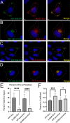
FIG 1
FIG 1
Env localizes to KIF16B-positive compartments and is redistributed by dominant-negative KIF16B. Approximate cell periphery outlines are shown as dashed lines. (A) Subcellular distribution of NL4-3 Env when coexpressed with KIF16B-WT-YFP in HeLa cells. Cells were fixed and immunolabeled with human neutralizing antibody 2G12 to stain HIV-1 gp120. Green, KIF16B; red, Env; rightmost image, overlay. (B) Distribution of NL4-3 Env when coexpressed with dominant KIF16B-S109A-YFP. Staining is as described for panel A. (C and D) Coexpression of proviral NL4-3 with WT and dominant-negative KIF16B, respectively. Staining for Env as described for panels A and B. (E) Percent Env signal at periphery of cells observed for each combination of coexpression shown in panels A to D. Signal at the periphery was the sum all Env signal within 30% of the distance from edge of the cell to the nucleus. Results are shown as means ± standard deviations from a total of 10 representative images. ns, not significant; ***, P < 0.0008; ****, P < 0.0001. P values were calculated using Student's unpaired t test using GraphPad Prism 9. (F) The degree of colocalization between KIF16B constructs and Env was measured by Pearson's correlation coefficient equation using Volocity 6.5.1 software after thresholding. Bars, 5 μm.
FIG 2
FIG 2
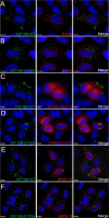
KIF16B and Env colocalization is not generalized to other viral envelope glycoproteins. Dashed lines indicate cell periphery. (A and B) Influenza HA glycoprotein was coexpressed with KIF16B-WT-YFP and KIF16B-S109A-YFP, respectively, in HeLa cells. Cells were fixed and immunolabeled with 11692-T54 antibody to stain HA. Green, KIF16B; red, HA; rightmost image, overlay. (C and D) SARS-Cov2-S protein was coexpressed with WT (C) or dominant-negative KIF16B (D). Cells were fixed and immunolabeled with anti-spike antibody 1A9 to stain S. Green, KIF16B; red, SARS-Cov2-S; rightmost image, overlay. (E and F) Coexpression of KIF16B-WT-YFP or KIF16B-S109A-YFP with Ebola GP. Cells were fixed and immunostained with 4F3 antibody specific to EBOV GP. Green, KIF16B; red, EBOV GP; rightmost image, overlay. Bars, 10 μm.
FIG 3
FIG 3
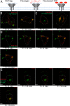
Env on the plasma membrane is endocytosed into KIF16B-positive compartments. (A) FAP-Env construct shown as a cartoon. The FAP tag was placed on the V2 loop of the Env ectodomain. This tag is inherently nonfluorescent on the far left. In the middle, the addition of a nonpermeable dye is shown. On the far right, the FAP tag becomes fluorescent, allowing visualization of a pulsed population of surface Env. (B) FAP-Env coexpressed with KIF16B-WT-YFP in HeLa cells. Left image, FAP-Env was pulse-labeled for 10 min on ice. Cells were then fixed and imaged immediately to show population of Env on plasma membrane surface. Right image, cells were pulsed as before and then were returned to a 37°C incubator for 15 min to allow internalization, and then fixed and imaged to show FAP-Env endocytosed into KIF16B-positive compartments. (C) FAP-Env construct coexpressed with KIF16B-S109A-YFP in HeLa cells. Left image is FAP-Env collecting on surface as before. Right image is the cell shown after 15 min of internalization as before. (D and E) Frames from live-cell imaging video of FAP-Env coexpressed with KIF16B-WT-YFP (D) or KIF16B-S109A-YFP (E). Time at T =-0 min shows the cell before pulse. Subsequent time points show FAP-Env collecting in KIF16B-positive compartments. Dotted lines are a visual aid to represent periphery of the cell body. Bars, 10 μm.
FIG 4
FIG 4
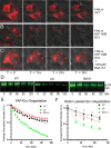
KIF16B KO leads to enhanced degradation of endocytosed Env in the lysosome. (A, B, and C) Frames from live-cell movies for Fap-Env expressed in WT HeLa cells with vehicle DMSO (A), KIF16B KO HeLa cells with DMSO (B), or KIF16B KO HeLa cells with 100 μM Baf-A1 (C). Representative frames are shown for each hour over the time course. One cell in each series of images is outlined with a dashed line. (B) Western blots show biotinylated Env from surface-labeled cells, with lysates chased over the time course, pulled down with streptavidin beads, and blotted for Env. Labels indicate WT, KIF16B KO, and KIF16B KO plus Baf-A1 HeLa cells. (C) Quantification of signal intensity of each time point over the FAP-Env imaging experiment normalized to the first frame. (D) Quantification of the Western blot signal for each time point for the biotin-streptavidin pulse-chase experiment. Error bars represent SD from 5 independent experiments. Fit curves for both panels C and D were generated using the Prism 9 nonlinear regression one-phase decay algorithm. Bars, 60 μm.
FIG 5
FIG 5
KIF16B KO shunts Env to lysosomal compartments. (A) HIV-1 NL4-3 Env expressed in WT HeLa cells. Magnification, ×60. Bar, 10 μm. (B) NL4-3 Env expressed in KIF16B KO HeLa cells. Magnification, ×60. Bar = 10 μm. (C) NL4-3 Env expressed in KIF16B KO HeLa cells with Baf-A1 added for 3 h prior to imaging. Magnification, ×60. Bar, 10 μm. (D) HIV-1 NL4-3 Env expressed in WT HeLa cells. Magnification, ×20. Bar, 35 μm. (E) NL4-3 Env expressed in KIF16B KO HeLa cells. Magnification, ×20. Bar, 35 μm. (F) NL4-3 Env expressed in KIF16B KO HeLa cells with Baf-A1 added 3 h prior to imaging. Magnification, ×20. Bar, 35 μm. For all imaging, cells were fixed and immunolabeled for Env with 2G12 antibody and for endogenous LAMP1 with H4A3 antibody. (G) Pearson’s correlation coefficient showing the degree of colocalization between Env and LAMP1 positive compartments between WT, KIF16B KO cells, and WT cells with Baf-A1 in panels A, B, and C, respectively. Pearson's correlation coefficient equation used Volocity 6.5.1 software after manual thresholding. One-way analysis of variance performed using Prism 9 to obtain significance values for difference in colocalization. ****, P < 0.0001. (H) Comparison of LAMP1 fluorescence intensity in WT and KIF16B KO cells. Ten individual cells for each cell type were selected to quantify total signal (per micrometer squared), and mean values ± SD. An unpaired t test was utilized to analyze significance (P = 0.182; nonsignificant [ns]).
FIG 6
FIG 6
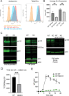
KIF16B depletion leads to reduction of Env on the cell surface, particle incorporation, infectivity, and viral replication. (A) Flow plots for cell surface staining (nonpermeabilized) and total Env content (permeabilized) in WT H9 and KIF16B KO H9 cells. Cells were fixed and stained for Env using anti-gp120 2G12 antibody. (B) Quantification of flow cytometry mean fluorescent intensity of Env expression on surface and total cell, averaged across 5 independent experiments. Unpaired t test was performed on Prism 9 to for significance values between surface versus total. **, P < 0.005. (C) WT and KIF16B H9 cells were infected with VSV-G-pseudotyped NL4-3. Shown are Western blots of Env and Gag content for particles in viral supernatant (left) and cell lysates (middle) collected on day 5 following infection. Note that loading was normalized for p24 content (supernatants) or for total cellular protein (cell lysates). Rightmost blot, single-round infection of WT and KIF16B KO H9 cells infected with VSV-G-pseudotyped NL4-3 at an MOI of 0.5 and harvested for cell lysate analysis after 24 h. Uninfected WT and KO cell lysates were run on the same blot as controls. Blotting was performed with 2G12 antibody against HIV-1 gp160 and ACTN05 (Invitrogen) against actin as a loading control. (D) Infectivity of viral particles released from WT or KIF16B KO H9 cells was determined from 3 independent experiments evaluated using TZM-bl indicator cells. (E) WT (black line) and KIF16B KO (green line) cells were infected with VSV-G-pseudotyped NL4-3 virus. Viral growth was monitored over time by detection of p24 in the culture supernatant.
FIG 7
FIG 7
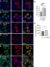
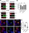
Semipermissive HeLa cells demonstrated reduced dependence on KIF16B. (A) HeLa cells and KIF16B KO HeLa cells were transfected with NL4-3 provirus (top) or NL4-3 CT144 provirus (bottom), and cell lysates and viral pellets were analyed by Western blotting. Molecular mass markers are shown on the left. (B) Fluorescence signals for gp41 and p24 in cells and supernatants from three independent experiments were measured, with the gp41/p24 ratio normalized to a setting of 1.0 for NL4-3 in WT HeLa cells. Differences between Env particle content in particles derived from WT or KIF16B KO cells were analyzed by an unpaired t test and found to be nonsignificant. Shown are mean values + standard deviations. (C) WT and CT144 versions of JR-FL Env were coexpressed in HeLa cells with GFP-KIF16B-S109A. Bars: top = 7 μm; bottom = 10 μm. (D) Pearson’s correlation coefficient values and mean representing colocalizaton of Env and YFP-KIF16B-S109A. Analysis was by unpaired t test; ***, P < 0.001.
FIG 8
FIG 8
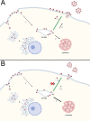
(A and B) Model of Env Trafficking with (A) and without (B) KIF16B. (A) In a wild-type cell, Env is produced in the rough endoplasmic reticulum and follows the secretory pathway to the cell surface. Env is then endocytosed into early endosomes (EEs; path 1 in the figure), which later may sort to the endosomal recycling compartment (ERC) for recycling. This compartment is depicted as “EE/ERC.” A fraction of Env in EEs can progress to fuse with lysosomes, leading to lysosomal degradation in path 2. In path 3, Env is directed in an anterograde manner to the PM, where it is available for incorporation into viral particles. Path 3 is the relevant pathway that is partially dependent upon KIF16B. (B) In a cell with KIF16B KO, a diminished fraction of Env is delivered to the PM from the EE/ERC via path 3. This results in an increase in the amount of Env shunted to the lysosome (path 2, illustrated as a broader arrow) and enhanced degradation of Env. The change in the distribution of endocytosed Env is shown graphically as fewer trimers on the budding and mature virion, as well as fewer trimers on the PM and more trimers in the lysosome. The figure was created with BioRender.

Similar articles

Cited by

References

    1. Egan MA, Carruth LM, Rowell JF, Yu X, Siliciano RF. 1996. Human immunodeficiency virus type 1 envelope protein endocytosis mediated by a highly conserved intrinsic internalization signal in the cytoplasmic domain of gp41 is suppressed in the presence of the Pr55gag precursor protein. J Virol 70:6547–6556. PMC190695. doi:10.1128/JVI.70.10.6547-6556.1996. - DOI - PMC - PubMed
    1. Ohno H, Aguilar RC, Fournier MC, Hennecke S, Cosson P, Bonifacino JS. 1997. Interaction of endocytic signals from the HIV-1 envelope glycoprotein complex with members of the adaptor medium chain family. Virology 238:305–315. doi:10.1006/viro.1997.8839. - DOI - PubMed
    1. Rowell JF, Stanhope PE, Siliciano RF. 1995. Endocytosis of endogenously synthesized HIV-1 envelope protein. Mechanism and role in processing for association with class II MHC. J Immunol 155:473–488. doi:10.4049/jimmunol.155.1.473. - DOI - PubMed
    1. Anokhin B, Spearman P. 2022. Viral and host factors regulating HIV-1 envelope protein trafficking and particle incorporation. Viruses 14:1729. doi:10.3390/v14081729. - DOI - PMC - PubMed
    1. Checkley MA, Luttge BG, Freed EO. 2011. HIV-1 envelope glycoprotein biosynthesis, trafficking, and incorporation. J Mol Biol 410:582–608. PMC3139147. doi:10.1016/j.jmb.2011.04.042. - DOI - PMC - PubMed

Publication types

LinkOut - more resources