

Recent trends in macromolecule-conjugated hybrid quantum dots for cancer theranostic applications
- PMID: 37346950
- PMCID: PMC10281231
- DOI: 10.1039/d3ra02673f
Recent trends in macromolecule-conjugated hybrid quantum dots for cancer theranostic applications
Abstract
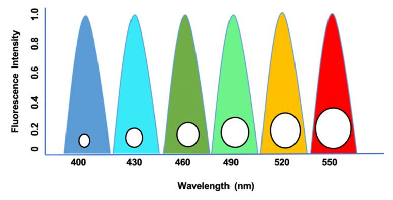
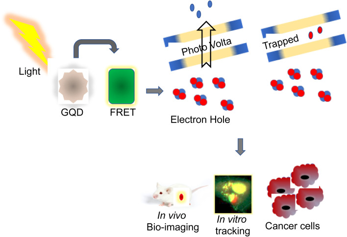
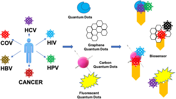
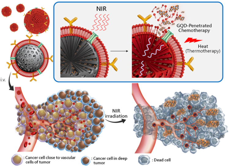
Quantum dots (QDs) are small nanoparticles with semiconductor properties ranging from 2 to 10 nanometers comprising 10-50 atoms. The single wavelength excitation character of QDs makes it more significant, as it can excite multiple particles in a confined surface simultaneously by narrow emission. QDs are more photostable than traditional organic dyes; however, when injected into tissues, whole animals, or ionic solutions, there is a significant loss of fluorescence. HQD-based probes conjugated with cancer-specific ligands, antibodies, or peptides are used in clinical diagnosis. It is more precise and reliable than standard immunohistochemistry (IHC) at minimal protein expression levels. Advanced clinical studies use photodynamic therapy (PDT) with fluorescence imaging to effectively identify and treat cancer. Recent studies revealed that a combination of unique characteristics of QDs, including their fluorescence capacity and abnormal expression of miRNA in cancer cells, were used for the detection and monitoring progression of cancer. In this review, we have highlighted the unique properties of QDs and the theranostic behavior of various macromolecule-conjugated HQDs leading to cancer treatment.
This journal is © The Royal Society of Chemistry.
Conflict of interest statement
The authors declare no conflict of interest.
Figures


Similar articles
-
64Cu-1,4,7,10-Tetraazacyclododecane-1,4,7,10-tetraacetic acid-quantum dot-vascular endothelial growth factor.2008 Jul 1 [updated 2008 Aug 12]. In: Molecular Imaging and Contrast Agent Database (MICAD) [Internet]. Bethesda (MD): National Center for Biotechnology Information (US); 2004–2013. 2008 Jul 1 [updated 2008 Aug 12]. In: Molecular Imaging and Contrast Agent Database (MICAD) [Internet]. Bethesda (MD): National Center for Biotechnology Information (US); 2004–2013. PMID: 20641777 Free Books & Documents. Review.
-
Quantum dots-deoxycholic acid (DOCA)-conjugated low molecular weight heparin (LMWH) nanoparticles.2012 Nov 28 [updated 2013 Feb 28]. In: Molecular Imaging and Contrast Agent Database (MICAD) [Internet]. Bethesda (MD): National Center for Biotechnology Information (US); 2004–2013. 2012 Nov 28 [updated 2013 Feb 28]. In: Molecular Imaging and Contrast Agent Database (MICAD) [Internet]. Bethesda (MD): National Center for Biotechnology Information (US); 2004–2013. PMID: 23469382 Free Books & Documents. Review.
-
Quantum dot 800–prostate-specific membrane antigen antibody J591.2011 Feb 28 [updated 2011 May 18]. In: Molecular Imaging and Contrast Agent Database (MICAD) [Internet]. Bethesda (MD): National Center for Biotechnology Information (US); 2004–2013. 2011 Feb 28 [updated 2011 May 18]. In: Molecular Imaging and Contrast Agent Database (MICAD) [Internet]. Bethesda (MD): National Center for Biotechnology Information (US); 2004–2013. PMID: 21634075 Free Books & Documents. Review.
-
Aggregation-Induced Emission (AIE) Dots: Emerging Theranostic Nanolights.Acc Chem Res. 2018 Jun 19;51(6):1404-1414. doi: 10.1021/acs.accounts.8b00060. Epub 2018 May 7. Acc Chem Res. 2018. PMID: 29733571
-
Semiconductor quantum dots for bioimaging and biodiagnostic applications.Annu Rev Anal Chem (Palo Alto Calif). 2013;6(1):143-62. doi: 10.1146/annurev-anchem-060908-155136. Epub 2013 Mar 20. Annu Rev Anal Chem (Palo Alto Calif). 2013. PMID: 23527547 Free PMC article. Review.
Cited by
-
Applicability of Quantum Dots in Breast Cancer Diagnostic and Therapeutic Modalities-A State-of-the-Art Review.Nanomaterials (Basel). 2024 Aug 31;14(17):1424. doi: 10.3390/nano14171424. Nanomaterials (Basel). 2024. PMID: 39269086 Free PMC article. Review.
References
-
- Bagher A. M. Sens. Transducers. 2016;198:37–43.
-
- Chatterjee D. K. Gnanasammandhan M. K. Zhang Y. Small. 2010;6:2781–2795. - PubMed
-
- Florescu M. Lee H. Puscasu I. Pralle M. Florescu L. Ting D. Z. Dowling J. P. Sol. Energy Mater. Sol. Cells. 2007;91:1599–1610.
-
- Niemeyer C. M. Curr. Opin. Chem. Biol. 2000;4:609–618. - PubMed
Publication types
LinkOut - more resources
Full Text Sources