Review
. 2023 Jun 2:10:1113827.
doi: 10.3389/fcvm.2023.1113827. eCollection 2023.

The role of monocytes in thrombotic diseases: a review

Affiliations
Review

The role of monocytes in thrombotic diseases: a review

Zhongyu Han et al. Front Cardiovasc Med. .

Abstract

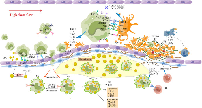
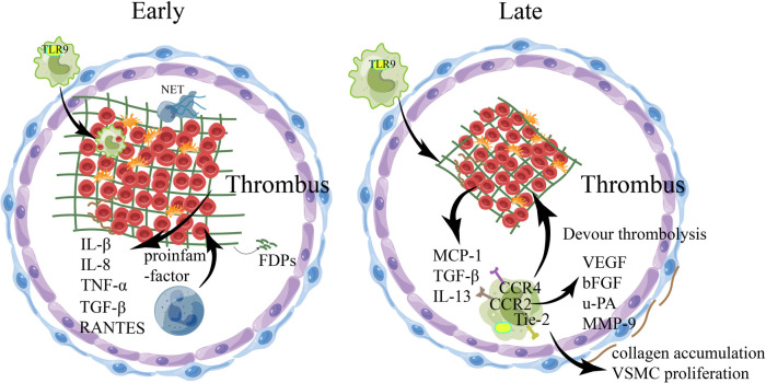
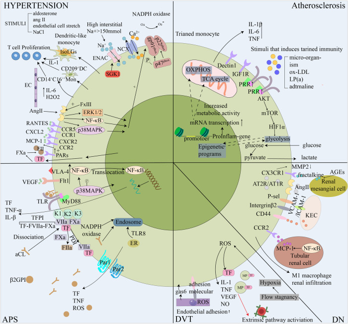
Cardiovascular and cerebrovascular diseases are the number one killer threatening people's life and health, among which cardiovascular thrombotic events are the most common. As the cause of particularly serious cardiovascular events, thrombosis can trigger fatal crises such as acute coronary syndrome (myocardial infarction and unstable angina), cerebral infarction and so on. Circulating monocytes are an important part of innate immunity. Their main physiological functions are phagocytosis, removal of injured and senescent cells and their debris, and development into macrophages and dendritic cells. At the same time, they also participate in the pathophysiological processes of pro-coagulation and anticoagulation. According to recent studies, monocytes have been found to play a significant role in thrombosis and thrombotic diseases of the immune system. In this manuscript, we review the relationship between monocyte subsets and cardiovascular thrombotic events and analyze the role of monocytes in arterial thrombosis and their involvement in intravenous thrombolysis. Finally, we summarize the mechanism and therapeutic regimen of monocyte and thrombosis in hypertension, antiphospholipid syndrome, atherosclerosis, rheumatic heart disease, lower extremity deep venous thrombosis, and diabetic nephropathy.

Keywords: arterial thrombosis; intravenous thrombolysis; macrophage; monocyte; thrombotic diseases.


Conflict of interest statement

The authors declare that the research was conducted in the absence of any commercial or financial relationships that could be construed as a potential conflict of interest.

Figures

Figure 1
Figure 1
The involvement of monocytes in atherosclerosis-thrombosis. The release of pro-inflammatory factors from cells in the plaque and the damage of ECs caused circulating monocytes to roll, roll slowly, adhere firmly, and travel through the tissue to differentiate. Macrophages phagocytose excessive Ox-LDL and other immune substances, causing apoptosis and necrosis of cells, lipid deposition, the generation of a large number of inflammatory factors and tissue factors, and promoting the transport of more circulating monocytes. Activated Th1 cells produce interferon γ, which inhibits smooth muscle cell synthesis of interstitial collagen. The interaction between Th1 cells and M1 macrophages leads to the overproduction of MMP-1, MMP-8 and MMP-13, and the interstitial collagenase can promote the breakdown of interstitial collagen. At the same time, it can induce M1 macrophages to overexpress tissue factors. These processes make the fibrous cap more vulnerable to rupture, and inflammatory activation leads to the increased potential for thrombosis. Platelet-rich thrombosis forms around atherosclerotic plaque rupture and endothelial damage when endosubcutaneous collagen is exposed to circulation under high shear flow. Platelets, through their glycoproteins, interact with collagen and collagen-deposited vWF to change their shape and adhere to the injured site. Attachment results in the secretion of ADP, serotonin, and thromboxane (TxA2), which recruit and activate more platelets. The platelet-monocyte complex (PMC) is activated by the interaction of various receptor ligands between platelets and monocytes. Platelets and monocytes also shed extracellular vesicles, release cytokines and chemokines, and tissue factors are increased to promote thrombosis. 5-HT, 5-hydroxytryptamine; E-sel, E-selection; FXIII, Factor XIII; FIB, fibrinogen; GM-CSF, granulocyte-macrophage colony stimulating factor; IL-8, interleukin-8; LDL, low-density lipoproteins; L-sel, L-selection; MCP-1, monocyte chemotactic protein-1; oxidized LDL; M-CSF, macrophage colony-stimulating factor; MMP, matrix metalloproteinases; Ox-LDL, oxidized LDL; PEV, platelet-derived extracellular vesicles; PMC, platelet-monocyte complex; P-sel, P-selection; ROS, reactive oxygen species; SRs, scavenger receptors; TF, tissue factor; TNF-α, tumor necrosis factor-α; TxA2, thromboxane; VCAM-1, vascular cell adhesion molecule-1; vWF, von Willebrand factor.
Figure 2
Figure 2
The involvement of relevant immune cells during venous thrombosis. During venous thrombus regression, numerous pro-inflammatory factors are released into the local environment, with an early influx of Neutrophils and macrophages, followed by monocytes, which regulate the production and activity of plasminase and MMPs. In the early stage of thrombolysis, fibrinolysis occurs at a high rate under the action of neutrophils, resulting in the production of fibrin degradation products, the emergence of collagen fibrils within the thrombolus, and the induction of inflammatory cytokines and various proteases by thrombolt-associated immune cells. With the structuring of the thrombus, the dissolution rate of fibrin slows down, collagen proliferation in the thrombus increases, monocytes infiltrate phagocytosis and mobilize EPC, and matrix remodeling of mmp secreted by macrophages occurs, which may eventually recover through the blood flow of the thrombus. bFGF, basic fibroblast growth factor; EPC, endothelial progenitor cells; FDPs, fibrin/fibrinogen degradation products;IL-8, interleukin-8; MCP-1, monocyte chemotactic protein-1; MMP, matrix metalloproteinases; NET, Neutrophil extracellular traps; TNF-α, tumor necrosis factor-α; TGF-β, transforming growth factor-β; TLR-9, toll-like receptor 9;uPA, uridylyl phosphate adenosine; VEGF, vascular endothelial growth factor; VSMC, Vascular smooth muscle cell.
Figure 3
Figure 3
Schematic diagram of the role of monocytes in five thrombotic diseases. In response to the hypertensive stimulant, monocytes transform and activate, release hypertensive cytokines and differentiate into dendritic cells to promote T cell proliferation through the effects of the vascular system, kidney and sympathetic nervous system, elevateing blood pressure, thus increasing the risk of blood vessel rupture. Monocytes are involved in atherosclerosis by training immune processes. This is mediated by epigenetic and metabolic reprogramming. By enhancing cell-related endometabolic pathways, monocytes can develop a long-lasting proinflammatory phenotype. In APS, the anti-β2gpi antibody binds to phospholipid-binding protein β2GPI and acts on monocytes to trigger the Toll-like receptor 4-myeloid differentiation primary response 88 (TLR-4-MyD88) signaling pathway. In addition, aPL triggers coagulation and inflammatory signals by dissociating suppressed TF cell surface complexes. TFPI expressed in myeloid cells specifically supports aPL-induced thrombosis. The hypoxic environment of DVT stimulates the expression of TF and some pro-inflammatory cytokines in monocytes, initiates the exogenous coagulation pathway, activates ECs and promotes the recruitment of monocytes. In the diabetic environment, the accumulation of advanced glycation end products (AGEs) increases the expression of fractalkine in human renal mesangial cells, and the interaction between monocytes and human renal mesangial cells promotes DN inflammation through MMP2 and fractalkine. Ang-II is over-produced and stimulated in the diabetic kidney, the adhesion of monocytes to ECs is enhanced, and M1 macrophages infiltrate the kidney under the regulation of the RAS system. AGEs, Advanced glycation end products; Ang II, Angiotensin II; aPL, antiphospholipid antibody; APS, Antiphospholipid syndrome; AT1R, angiotensin type 1 receptor; β2GPI, beta2glycoprotein I; DC, dendritic cells; DN, Diabetic nephropathy; DVT, Deep vein thrombosis; ENAC, epithelial sodium channel; ER, Endoplasmic reticulum; gas6, growth arrest specific 6; KEC, Kidney endothelial cell; LP(a), Lipoprotein a; IGF1R, insulin like growth factor 1 receptor;MCP-1, monocyte chemotactic protein-1; MMP, matrix metalloproteinases; MP, microparticles; NCX, Sodium-calcium exchangers; NF-κB, nuclear factor κB; NOX-2, NADPH oxidase-2; OXPHOS, oxidative phosphorylation; PARs, protease activated receptors; PRR, pattern recognition receptor; RAS, renin-angiotensin system; ROS, reactive oxygen species; TCA, tricarboxylic acid; TF, tissue factor; TFPI, TF pathway inhibitor; TLR, toll-like receptors; VEGF, vascular endothelial growth facto;VLA-4,very late antigen 4.

Similar articles

Cited by

References

    1. Schafer AI, Levine MN, Konkle BA, Kearon C. Thrombotic disorders: diagnosis and treatment. Hematology Am Soc Hematol Educ Program. (2003) 1:520–39. 10.1182/asheducation-2003.1.520 - DOI - PubMed
    1. Muñoz Tovar RA, Alvarez Perdomo LC, Rojas Molina SM, Salazar SJ. Submassive pulmonary thromboembolism in a patient with thrombocytopenia: therapeutic challenge. Case Rep Crit Care. (2019) 2019:1919401. 10.1155/2019/1919401 - DOI - PMC - PubMed
    1. GBD 2017 SDG Collaborators: Lozano R, Fullman N, Abate D, Abay SM, Abbafati C, Abbasi N, et al. Measuring progress from 19 90 to 2017 and projecting attainment to 2030 of the health-related sustainable development goals for 195 countries and territories: a systematic analysis for the global burden of disease study 2017. Lancet. (2018) 392(10159):2091–138. 10.1016/s0140-6736(18)32281-5 - DOI - PMC - PubMed
    1. Boyd AN. An inflammatory basis for coronary thrombosis. Am J Pathol. (1928) 4(2):159–66.5. - PMC - PubMed
    1. Wolf AA, Yáñez A, Barman PK, Goodridge HS. The ontogeny of monocyte subsets. Front Immunol. (2019) 10:1642. 10.3389/fimmu.2019.01642 - DOI - PMC - PubMed

Grants and funding

The work was supported by Chengdu University of Traditional Chinese Medicine undergraduate research practice innovation project (ky-2023015; ky-2023081; ky-2023083).