. 2023 Jun 17;13(1):9825.
doi: 10.1038/s41598-023-36642-y.

Tozorakimab (MEDI3506): an anti-IL-33 antibody that inhibits IL-33 signalling via ST2 and RAGE/EGFR to reduce inflammation and epithelial dysfunction

Affiliations

Tozorakimab (MEDI3506): an anti-IL-33 antibody that inhibits IL-33 signalling via ST2 and RAGE/EGFR to reduce inflammation and epithelial dysfunction

Elizabeth England et al. Sci Rep. .

Abstract

Interleukin (IL)-33 is a broad-acting alarmin cytokine that can drive inflammatory responses following tissue damage or infection and is a promising target for treatment of inflammatory disease. Here, we describe the identification of tozorakimab (MEDI3506), a potent, human anti-IL-33 monoclonal antibody, which can inhibit reduced IL-33 (IL-33red) and oxidized IL-33 (IL-33ox) activities through distinct serum-stimulated 2 (ST2) and receptor for advanced glycation end products/epidermal growth factor receptor (RAGE/EGFR complex) signalling pathways. We hypothesized that a therapeutic antibody would require an affinity higher than that of ST2 for IL-33, with an association rate greater than 107 M-1 s-1, to effectively neutralize IL-33 following rapid release from damaged tissue. An innovative antibody generation campaign identified tozorakimab, an antibody with a femtomolar affinity for IL-33red and a fast association rate (8.5 × 10⁷ M-1 s-1), which was comparable to soluble ST2. Tozorakimab potently inhibited ST2-dependent inflammatory responses driven by IL-33 in primary human cells and in a murine model of lung epithelial injury. Additionally, tozorakimab prevented the oxidation of IL-33 and its activity via the RAGE/EGFR signalling pathway, thus increasing in vitro epithelial cell migration and repair. Tozorakimab is a novel therapeutic agent with a dual mechanism of action that blocks IL-33red and IL-33ox signalling, offering potential to reduce inflammation and epithelial dysfunction in human disease.


Conflict of interest statement

AstraZeneca funded this study and participated in the study design, data collection, data analysis and data interpretation. AstraZeneca reviewed the publication, without influencing the opinions of the authors, to ensure medical and scientific accuracy and the protection of intellectual property. The corresponding author had access to all data in the study and was responsible for submitting the manuscript for publication. E.E., D.G.R., I.C.S., S.C., C.C., D.J.C., K.F.H., C.C.E.H., D.A.S., C.H., E.C.H., M.D.S., E.S.C., T.J.V., T.E. and M.P. are employees of AstraZeneca and may hold stock or stock options. D.T.Y.C., B.P.K., D.C.L., J.B.M., R.D.M., L.R., K.A.V., R.J.B. and T.M. are former employees of AstraZeneca and may hold stock or stock options.

Figures
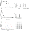
Figure 1
Figure 1
IL-33 binds sST2 with a high affinity and a fast association rate. (a) The affinity (KD) of IL-33red (residues 112–270) interacting with sST2 was obtained after a 1:1 binding model was fitted to two KinExA datasets simultaneously; the black solid line with filled squares are the 30 pM fixed [sST2] data (■) and the grey line with filled diamonds are the 2.0 pM fixed [sST2] data (◆). (b) A plot of 95% CIs (indicated by the grey rectangle) around the affinity estimate (dashed line) shown in panel a. (c) A plot of 95% CIs around the active receptor CBP parameter shown in panel a. (d) The association rate constant (ka) for IL-33 interacting with sST2 using KinExA. (e) The 95% CI around the association rate constant estimate shown in panel d. (f) In silico modelling of the suppression profile expected from antibodies with varying kinetic and affinity profiles, during acute IL-33 spike profiles parameterized using measured IL-33 spike data from ALT challenged mice, following the establishment of steady state suppression. The spike suppression was modelled assuming prior dosing of 150 mg neutralizing antibody every 4 weeks with an IL-33 degradation half-life of 60 min and an IL-33 spike half-life of 15 min (spike occurring at 2,160 h; refer to Supplementary Fig. 1a). Antibody affinity (KD) was varied over a 0.1–10 pM range (solid versus dashed lines, respectively) with the association rate (ka), which was varied in tenfold steps over the 105–108 M−1 s−1 range (red, blue, grey and green, respectively). ALT, Alternaria alternata; CBP, constant-binding partner; CI, confidence interval; IL, interleukin; KinExA, Kinetic Exclusion Assays; sST2, soluble serum stimulated 2.
Figure 2
Figure 2
Characterization of the lead antibody (33v20064) in biochemical and in vitro assays. (a) IL-33 binding sST2 biochemical competition assay (mean ± SD of duplicate determinations). (b) Inhibition of NF-κB signalling in HUVECs (mean ± SD, n = 3). (c) Inhibition of IL-8 release in HUVECs (mean ± SD, n = 2). (d) Affinity of lead antibody for human IL-33 at concentrations of 1–256 nM, which was determined by surface plasmon resonance. For each sensorgram trace 1:1 fit lines are shown in black. Figure shows a representative dataset from four separate experiments. HUVEC, human umbilical vein endothelial cell; IL, interleukin; mAb, monoclonal antibody; NF-κB, nuclear factor kappa-light-chain-enhancer of activated B cells; RU, response units; SD, standard deviation.
Figure 3
Figure 3
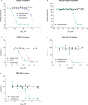
In vitro pharmacology of tozorakimab. (a) Epitope competition assay. (b) Biochemical receptor–ligand competition assay. (c) Inhibition of recombinant IL-33red (residues 112–270)-driven IL-8 release in HUVECs. (d) Inhibition of recombinant IL-33C>S (residues 112–270)-driven IL-13 release in human blood-derived mast cells (three individual donors shown, data points are the mean of duplicate tests). (e) Inhibition of recombinant IL-33C>S (residues 112–270)-driven IFN-γ release from human PBMCs (six individual donors, data points are the mean of duplicate tests). (ac) The data shown are a representative example from at least two independent experiments. Data points are plotted as mean ± SD of duplicate determinations. HUVEC, human umbilical vein endothelial cell; IFN-γ, interferon-gamma; IL, interleukin; PBMC, peripheral blood mononuclear cell; red, reduced; SD, standard deviation; sST2, soluble serum stimulated 2.
Figure 4
Figure 4
Affinity and association rate of tozorakimab for IL-33red measured using KinExA. (a) The affinity (KD) of tozorakimab for IL-33red (residues 112–270) was obtained after a 1:1 binding model was fitted to two KinExA datasets simultaneously; the black solid line and filled squares are the data from IL-33red titrated into 5 pM fixed [IgG] (■) and the grey solid line and filled diamonds are from a 0.20 pM fixed [IgG] data titration (◆). (b) Error curve indicating the extent (grey shaded rectangle) of 95% CIs around the affinity value (indicated by dashed line) from the fit to the data in panel a. (c) Fitting error plot of the 95% CIs of the active receptor CBP parameter that arise from the fit in panel a. (d) The association rate constant for the tozorakimab Fab fragment binding IL-33red (residues 112–270) measured using KinExA. (e) Error plot of the 95% CI around the association rate (ka) estimate from the fit in panel d. CBP, constant-binding partner; CI, confidence interval; Fab, antigen-binding fragment; IgG, immunoglobulin G; KinExA, kinetic exclusion assays; IL, interleukin; red, reduced.
Figure 5
Figure 5
Tozorakimab can inhibit endogenous IL-33red-driven inflammatory responses in vitro and in vivo. (a) Western blot of human IL-33 in lung tissue lysate derived from a healthy individual who was an ex-smoker (representative of n = 3). For the full western blot, please see Supplementary figure S4. (b) Inhibition of IL-8 release in HUVECs treated with human lung lysate (mean ± SD, n = 3). (c) Study to determine inhibition of endogenous IL-33-dependent IL-5 in BALF following an ALT challenge. Tozorakimab administered i.p. 24 h before ALT challenge. The study was performed in female humanized-IL-33 KI, mouse-IL-33 KO mice (huIL-33KI). (d) Inhibition of ALT driven IL-5 release in BALF. PBS, human-isotype mAb and mouse-isotype mAb controls were used (n = 6). A Browne–Forsyth ANOVA was used to test the impact of dose on log10 transformed IL-5 levels to confirm that the samples were of similar variance. The post hoc Dunnett’s multiple comparison test was then used to generate p values (as stated) for the comparisons of interest (Prism 8). ALT, Alternaria alternata; ANOVA, analysis of variance; BALF, bronchoalveolar lavage fluid; ELISA, enzyme-linked immunosorbent assay; FL, full length; HUVEC, human umbilical vein endothelial cell; IL, interleukin; i.p., intraperitoneal; KI, knock in; KO, knockout; mAb, monoclonal antibody; PBS, phosphate buffered saline; red, reduced; SD, standard deviation; sST2, soluble serum stimulated 2.
Figure 6
Figure 6
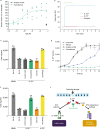
Tozorakimab potently inhibits the formation of IL-33ox. (a) The A549 epithelial cell scratch wound healing assay was normalized to the untreated control (data points are a mean of six replicates, n = 4 separate experiments). (b) Surface plasmon resonance binding profiles for tozorakimab Fab fragment binding to chip-immobilized N-terminally tagged wild-type IL-33red or wild-type IL-33ox. Both IL-33 constructs were based on the 112–270 residue form. Amine biotinylated ubiquitin served as a protein control. The bar indicates the timing of the 2 min injection of 10 nM monomerized Fab. (c) Impact of recombinant IL-33ox in the presence of test antibodies on human lung epithelial cell migration. The data shown (mean ± SD of triplicate determinations) are a representative example from two independent experiments. (d) Time course of recombinant IL-33red oxidation in the presence of antibodies. IL-33ox levels were detected using IL-33ox ELISA (mean ± SD of duplicate determinations). (e) Impact of recombinant IL-33red, oxidized for 24 h in the presence of test antibodies, on human lung epithelial cell migration. The data shown (mean ± SD of triplicate determinations) are a representative example from two independent experiments. (f) A schematic of the proposed mode of action of tozorakimab. IL-33red, that is rapidly released following epithelial damage, binds tozorakimab with a high affinity and a fast association rate. Tozorakimab directly neutralizes IL-33red inflammatory activities at the ST2 receptor. The IL-33red–tozorakimab complex prevents the oxidation of IL-33, IL-33ox–RAGE/EGFR signalling and epithelial dysfunction. EGFR, epidermal growth factor receptor; ELISA, enzyme-linked immunosorbent assay; IL, interleukin; IL-1RAP, interleukin-1 receptor accessory protein; mAb, monoclonal antibody; ox, oxidized; RAGE, receptor for advanced glycation end products; red, reduced; RU, response units; SD, standard deviation.

Similar articles

Cited by

References

    1. Cayrol C, Girard JP. Interleukin-33 (IL-33): A nuclear cytokine from the IL-1 family. Immunol. Rev. 2018;281:154–168. doi: 10.1111/imr.12619. - DOI - PubMed
    1. Gungor O, et al. IL-33 and ST2 levels in chronic kidney disease: Associations with inflammation, vascular abnormalities, cardiovascular events, and survival. PloS One. 2017;12:e0178939. doi: 10.1371/journal.pone.0178939. - DOI - PMC - PubMed
    1. Drake LY, Kita H. IL-33: Biological properties, functions, and roles in airway disease. Immunol. Rev. 2017;278:173–184. doi: 10.1111/imr.12552. - DOI - PMC - PubMed
    1. Sundnes O, et al. Rapid systemic surge of IL-33 after severe human trauma: A prospective observational study. Mol Med. 2021;27:29. doi: 10.1186/s10020-021-00288-1. - DOI - PMC - PubMed
    1. Zizzo G, Cohen PL. Imperfect storm: Is interleukin-33 the Achilles heel of COVID-19? Lancet Rheumatol. 2020;2:e779–e790. doi: 10.1016/s2665-9913(20)30340-4. - DOI - PMC - PubMed