. 2022 Jul 13;29(2):87-103.
doi: 10.32604/or.2022.03529. eCollection 2021.

cGAS regulates the DNA damage response to maintain proliferative signaling in gastric cancer cells

Affiliations

cGAS regulates the DNA damage response to maintain proliferative signaling in gastric cancer cells

Bin Liu et al. Oncol Res. .

Abstract

The activation of some oncogenes promote cancer cell proliferation and growth, facilitate cancer progression and metastasis by induce DNA replication stress, even genome instability. Activation of the cyclic GMP-AMP synthase (cGAS) mediates classical DNA sensing, is involved in genome instability, and is linked to various tumor development or therapy. However, the function of cGAS in gastric cancer remains elusive. In this study, the TCGA database and retrospective immunohistochemical analyses revealed substantially high cGAS expression in gastric cancer tissues and cell lines. By employing cGAS high-expression gastric cancer cell lines, including AGS and MKN45, ectopic silencing of cGAS caused a significant reduction in the proliferation of the cells, tumor growth, and mass in xenograft mice. Mechanistically, database analysis predicted a possible involvement of cGAS in the DNA damage response (DDR), further data through cells revealed protein interactions of the cGAS and MRE11-RAD50-NBN (MRN) complex, which activated cell cycle checkpoints, even increased genome instability in gastric cancer cells, thereby contributing to gastric cancer progression and sensitivity to treatment with DNA damaging agents. Furthermore, the upregulation of cGAS significantly exacerbated the prognosis of gastric cancer patients while improving radiotherapeutic outcomes. Therefore, we concluded that cGAS is involved in gastric cancer progression by fueling genome instability, implying that intervening in the cGAS pathway could be a practicable therapeutic approach for gastric cancer.

Keywords: Cell proliferation; DNA damage response; Gastric cancer; MRN complex; cGAS.


Conflict of interest statement

The authors have declared that they have no conflict of interest.

Figures
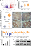
Figure 1
Figure 1. High expression of cGAS in gastric cancer tissues and gastric cancer cell lines. A. Volcano map of gastric cancer and normal tissue showing differentially expressed genes in TCGA database (cGAS and several vital genes of HR repair were specially labelled). B. The expression data of cGAS in gastric cancer tissues and normal human gastric mucosa in the TCGA database. C. The level of cGAS expression in different T stages of gastric cancer. D–G. Representative immunohistochemical results: D. adjacent tissue, score 2 (scale bar:20 μm); E. Cancer tissue, score 4; F. Cancer tissue, score 8; G. Cancer tissue, score 12. H. Retrospective immunohistochemical analysis score of cGAS in gastric cancer tissues and adjacent tissues. I. cGAS expression data for 37 gastric cancer cell lines in the CCLE database. J. RT-qPCR for cGAS relative expression of mRNA in 3 gastric cancer cell lines and GES-1 cell line (n = 3). K. Western blotting of cGAS in three gastric cancer cell lines and GES-1 cell line (* p < 0.05, ** p < 0.01, *** p < 0.001, **** p < 0.0001).
Figure 2
Figure 2. cGAS knockdown inhibits GC cells proliferation in vitro. A, B. The relative mRNA expression and protein expression of cGAS after siRNA triggered knockdown in AGS cells. C. CCK-8 assay show reduced cell proliferation in si-cGAS AGS cells. D, E. EdU assay of AGS cells in vitro, EdU (red) and hoechst33342 (blue). F, G. The cGAS knockdown efficiency in MKN45 cells. H. cell proliferation curve of si-cGAS MKN45 cells. I, J. EdU assay of MKN45 cells in vitro (**p < 0.01, ***p < 0.001, ****p < 0.0001, n = 3).
Figure 3
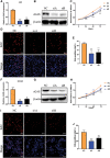
Figure 3. cGAS knockdown inhibits the proliferation of MKN45 cell line in vivo. A. The cGAS knockdown efficiency in MKN45 cells. B. Subcutaneous tumor volume, the growth curve of sh-cGAS MKN45 cells in nude mice. C. Representative images showing the tumors’ sizes were dissected 14 days after subcutaneous injection of sh-cGAS MKN45 and control cells. D. Subcutaneous tumor mass quality. E. H&E staining of MKN45 tumor tissues (**p < 0.01, ***p < 0.001, scale bar:100 μm).
Figure 4
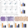
Figure 4. Knockdown of cGAS affects gastric cell function. A–C. Flow cytometry assay detected moderate apoptosis in si-cGAS AGS (up) and MKN45(down) cells. D, E. si-cGAS reduced the migration of AGS (left) and MKN45(right) cells. F, G. Several migrated AGS and MKN45 cells. H–K. Wound healing observed slow migration of si-cGAS AGS (left) and MKN45(right) cells. (*p < 0.05, **p < 0.01, ***p < 0.001, ****p < 0.0001, n = 3).
Figure 5
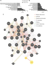
Figure 5. Bioinformatics analysis reveals the role of cGAS in DNA damage repair. A, B. GO and KEGG functional analysis of the top 200 co-expressed genes with cGAS from the TCGA dataset. C. Functional proteins’ interaction networks display potential physical interaction between cGAS and MRN complex.
Figure 6
Figure 6. cGAS were regulating MRN complex formation and cell cycle checkpoint. A–C. The correlation of the MRN complex subunit expression with cGAS. D. Co-IP experiment and follows immunoblotting (IB) assay revealed that cGAS interacted with NBN and MRE11 in AGS cells. E. Western blot results of the MRN complex and cell cycle checkpoint proteins in cGAS knockdown AGS and MKN45 cells.
Figure 7
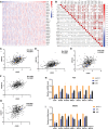
Figure 7. cGAS is involved in DNA HR repair and promote genomic instability. A. Heat maps of cGAS and HR repair genes’ expression; B. The Pearson correlation between cGAS and HR repair genes in the TCGA database represents the correlation coefficient value. C–G. Five HR repair genes with the highest correlation with cGAS in gastric cancer. H, I. RT-qPCR results of HR repair genes co-expressed with cGAS in sh-cGAS AGS and MKN45 cells. J. Western blot results of cGAS overexpression. K. Comet assay determined the genome stability in GES-1 cells with cGAS overexpression. L. Comet tail moment length. (*p < 0.05, **p < 0.01, ***p < 0.001, ****p < 0.0001)
Figure 8
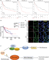
Figure 8. cGAS cause poor prognosis in GC patients. A–C. K-M survival curve demonstrating the overall survival times of GC patients with different cGAS expressions. The datasets were derived from GSE15459, GSE51105, and GSE22377 in the GEO database, respectively (scale bar: 10 μm). D. K-M survival curve presenting the overall survival times of GC patients from the TCGA database stratified based on the treatment (radiotherapy (RT) and non-radiotherapy) and expression levels of cGAS (cutoff: median value). E. Immunofluorescence of endogenously and exogenously localization of cGAS after IR exposure (scale bar: 10 μm). F. The schematic diagram of cGAS in gastric cancer progression and radiotherapy.

Similar articles

Cited by

References

    1. Siegel, R. L., Miller, K. D., Jemal, A. (2020). Cancer statistics, 2020. CA: A Cancer Journal for Clinicians , 70, 7–30. - PubMed
    1. Shimizu, T., Chiba, T., Marusawa, H. (2017). Helicobacter pylori-mediated genetic instability and gastric carcinogenesis. Current Topics in Microbiology and Immunology , 400, 305–323. DOI 10.1007/978-3-319-50520-6. - DOI - PubMed
    1. Koeppel, M., Garcia-Alcalde, F., Glowinski, F., Schlaermann, P., Meyer, T F. (2015). Helicobacter pylori infection causes characteristic DNA damage patterns in human cells. Cell Reports , 11(11), 1703–1713. DOI 10.1016/j.celrep.2015.05.030. - DOI - PubMed
    1. Sun, L., Wu, J., Du, F., Chen, X., Chen, Z. J. (2013). Cyclic GMP-AMP synthase is a cytosolic DNA sensor that activates the type I interferon pathway. Science , 339(6121), 786–791. DOI 10.1126/science.1232458. - DOI - PMC - PubMed
    1. Cai, X., Chiu, Y. H., Chen, Z. J. (2014). The cGAS-cGAMP-STING pathway of cytosolic DNA sensing and signaling. Molecular Cell , 54(2), 289–296. DOI 10.1016/j.molcel.2014.03.040. - DOI - PubMed