

Computational approaches for evaluating morphological changes in the corneal stroma associated with decellularization
- PMID: 37304146
- PMCID: PMC10250676
- DOI: 10.3389/fbioe.2023.1105377
Computational approaches for evaluating morphological changes in the corneal stroma associated with decellularization
Abstract
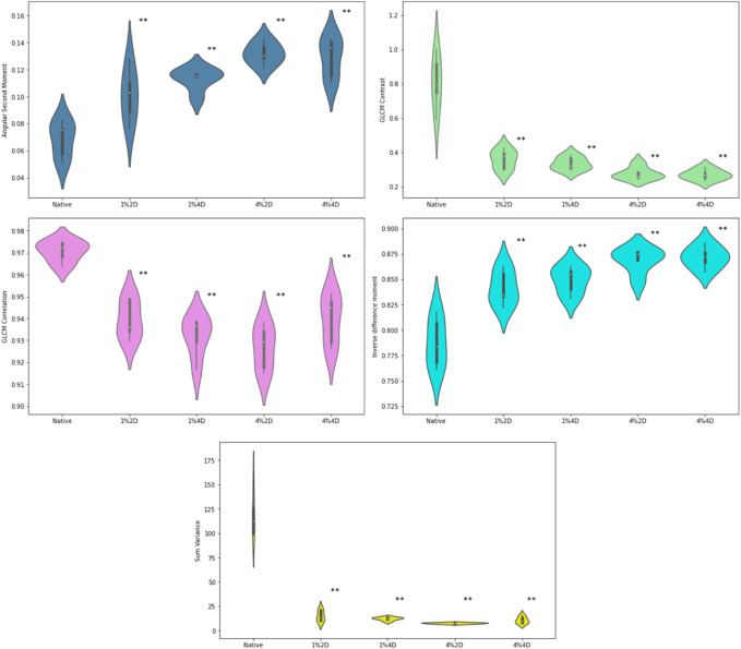
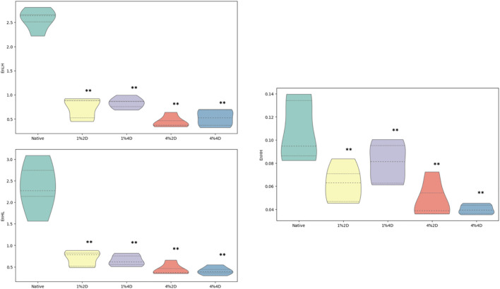
Decellularized corneas offer a promising and sustainable source of replacement grafts, mimicking native tissue and reducing the risk of immune rejection post-transplantation. Despite great success in achieving acellular scaffolds, little consensus exists regarding the quality of the decellularized extracellular matrix. Metrics used to evaluate extracellular matrix performance are study-specific, subjective, and semi-quantitative. Thus, this work focused on developing a computational method to examine the effectiveness of corneal decellularization. We combined conventional semi-quantitative histological assessments and automated scaffold evaluations based on textual image analyses to assess decellularization efficiency. Our study highlights that it is possible to develop contemporary machine learning (ML) models based on random forests and support vector machine algorithms, which can identify regions of interest in acellularized corneal stromal tissue with relatively high accuracy. These results provide a platform for developing machine learning biosensing systems for evaluating subtle morphological changes in decellularized scaffolds, which are crucial for assessing their functionality.
Keywords: GLCM (gray level co-event matrix); cornea; corneal stroma; decellualrization; keratocytes.
Copyright © 2023 Pantic, Cumic, Valjarevic, Shakeel, Wang, Vurivi, Daoud, Chan, Petroianu, Shibru, Ali, Nesic, Salih, Butt and Corridon.
Conflict of interest statement
The authors declare that the research was conducted in the absence of any commercial or financial relationships that could be construed as a potential conflict of interest.
Figures

Similar articles
-
Acellular ostrich corneal stroma used as scaffold for construction of tissue-engineered cornea.Int J Ophthalmol. 2016 Mar 18;9(3):325-31. doi: 10.18240/ijo.2016.03.01. eCollection 2016. Int J Ophthalmol. 2016. PMID: 27158598 Free PMC article.
-
Reconstruction of a tissue-engineered cornea with porcine corneal acellular matrix as the scaffold.Cells Tissues Organs. 2010;191(3):193-202. doi: 10.1159/000235680. Epub 2009 Aug 19. Cells Tissues Organs. 2010. PMID: 19690400
-
An efficient strategy to recellularization of a rat aorta scaffold: an optimized decellularization, detergent removal, and Apelin-13 immobilization.Biomater Res. 2022 Sep 22;26(1):46. doi: 10.1186/s40824-022-00295-1. Biomater Res. 2022. PMID: 36138491 Free PMC article.
-
Strategies for developing decellularized corneal scaffolds.Exp Eye Res. 2013 Mar;108:42-7. doi: 10.1016/j.exer.2012.12.012. Epub 2012 Dec 31. Exp Eye Res. 2013. PMID: 23287438 Review.
-
Human SMILE-Derived Stromal Lenticule Scaffold for Regenerative Therapy: Review and Perspectives.Int J Mol Sci. 2022 Jul 19;23(14):7967. doi: 10.3390/ijms23147967. Int J Mol Sci. 2022. PMID: 35887309 Free PMC article. Review.
Cited by
-
A proposed model of xeno-keratoplasty using 3D printing and decellularization.Front Pharmacol. 2023 Sep 20;14:1193606. doi: 10.3389/fphar.2023.1193606. eCollection 2023. Front Pharmacol. 2023. PMID: 37799970 Free PMC article.
-
Integrated environmental and health economic assessments of novel xeno-keratografts addressing a growing public health crisis.Sci Rep. 2024 Oct 27;14(1):25600. doi: 10.1038/s41598-024-77783-y. Sci Rep. 2024. PMID: 39465317 Free PMC article.
-
Enhancing the expression of a key mitochondrial enzyme at the inception of ischemia-reperfusion injury can boost recovery and halt the progression of acute kidney injury.Front Physiol. 2023 Feb 8;14:1024238. doi: 10.3389/fphys.2023.1024238. eCollection 2023. Front Physiol. 2023. PMID: 36846323 Free PMC article.
-
Capturing effects of blood flow on the transplanted decellularized nephron with intravital microscopy.Sci Rep. 2023 Mar 31;13(1):5289. doi: 10.1038/s41598-023-31747-w. Sci Rep. 2023. PMID: 37002341 Free PMC article.
-
Machine learning approaches to detect hepatocyte chromatin alterations from iron oxide nanoparticle exposure.Sci Rep. 2024 Aug 23;14(1):19595. doi: 10.1038/s41598-024-70559-4. Sci Rep. 2024. PMID: 39179629 Free PMC article.
References
-
- Adankon M.M., Cheriet M. (2015). Support Vector Machine. In: Li, S.Z., Jain, A.K. (eds) Encyclopedia of Biometrics. Springer, Boston, MA. 10.1007/978-1-4899-7488-4_29 - DOI
LinkOut - more resources
Full Text Sources