

Application of magnetic resonance imaging-related techniques in the diagnosis of sepsis-associated encephalopathy: present status and prospect
- PMID: 37304016
- PMCID: PMC10248056
- DOI: 10.3389/fnins.2023.1152630
Application of magnetic resonance imaging-related techniques in the diagnosis of sepsis-associated encephalopathy: present status and prospect
Abstract
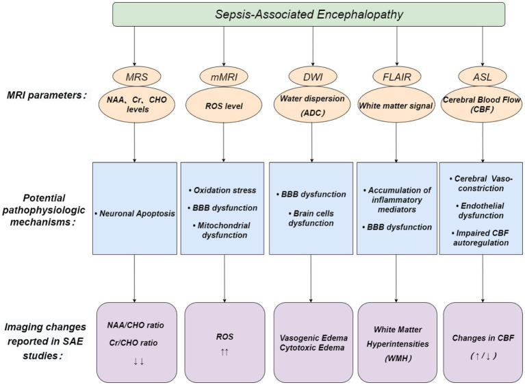
Sepsis-associated encephalopathy (SAE) refers to diffuse brain dysfunction secondary to systemic infection without central nervous system infection. The early diagnosis of SAE remains a major clinical problem, and its diagnosis is still exclusionary. Magnetic resonance imaging (MRI) related techniques, such as magnetic resonance spectroscopy (MRS), molecular MRI (mMRI), arterial spin-labeling (ASL), fluid-attenuated inversion recovery (FLAIR), and diffusion-weighted imaging (DWI), currently provide new options for the early identification of SAE. This review collected clinical and basic research and case reports related to SAE and MRI-related techniques in recent years, summarized and analyzed the basic principles and applications of MRI technology in diagnosing SAE, and provided a basis for diagnosing SAE by MRI-related techniques.
Keywords: cognitive impairment; imaging diagnosis; magnetic resonance imaging; sepsis; sepsis-associated encephalopathy.
Copyright © 2023 Wu, Wang, Song, Hu, Jing and Zhu.
Conflict of interest statement
The authors declare that the research was conducted in the absence of any commercial or financial relationships that could be construed as a potential conflict of interest.
Figures
Similar articles
-
Fulminant encephalopathy in a child with hyperferritinemic sepsis: a case report.BMC Neurol. 2020 Mar 2;20(1):73. doi: 10.1186/s12883-020-01661-z. BMC Neurol. 2020. PMID: 32122316 Free PMC article.
-
Imaging in sepsis-associated encephalopathy--insights and opportunities.Nat Rev Neurol. 2013 Oct;9(10):551-61. doi: 10.1038/nrneurol.2013.177. Epub 2013 Sep 3. Nat Rev Neurol. 2013. PMID: 23999468 Review.
-
Early Diagnosis of Murine Sepsis-Associated Encephalopathy Using Dynamic PET/CT Imaging and Multiparametric MRI.Mol Imaging Biol. 2022 Dec;24(6):928-939. doi: 10.1007/s11307-022-01743-z. Epub 2022 May 25. Mol Imaging Biol. 2022. PMID: 35612771
-
Sepsis-associated encephalopathy.Transl Med UniSa. 2012 Jan 18;2:20-7. Print 2012 Jan. Transl Med UniSa. 2012. PMID: 23905041 Free PMC article.
-
Central role of microglia in sepsis-associated encephalopathy: From mechanism to therapy.Front Immunol. 2022 Jul 26;13:929316. doi: 10.3389/fimmu.2022.929316. eCollection 2022. Front Immunol. 2022. PMID: 35958583 Free PMC article. Review.
References
-
- Bartnik-Olson B. L., Alger J. R., Babikian T., Harris A. D., Holshouser B., Kirov I. I., et al. . (2021). The clinical utility of proton magnetic resonance spectroscopy in traumatic brain injury: recommendations from the ENIGMA MRS working group. Brain Imaging Behav. 15, 504–525. doi: 10.1007/s11682-020-00330-6, PMID: - DOI - PMC - PubMed
Publication types
LinkOut - more resources
Full Text Sources