. 2023 Jun 12;56(1):31.
doi: 10.1186/s40659-023-00441-6.

Acupuncture regulates the apoptosis of ovarian granulosa cells in polycystic ovarian syndrome-related abnormal follicular development through LncMEG3-mediated inhibition of miR-21-3p

Affiliations

Acupuncture regulates the apoptosis of ovarian granulosa cells in polycystic ovarian syndrome-related abnormal follicular development through LncMEG3-mediated inhibition of miR-21-3p

Xiaohong Chen et al. Biol Res. .

Abstract

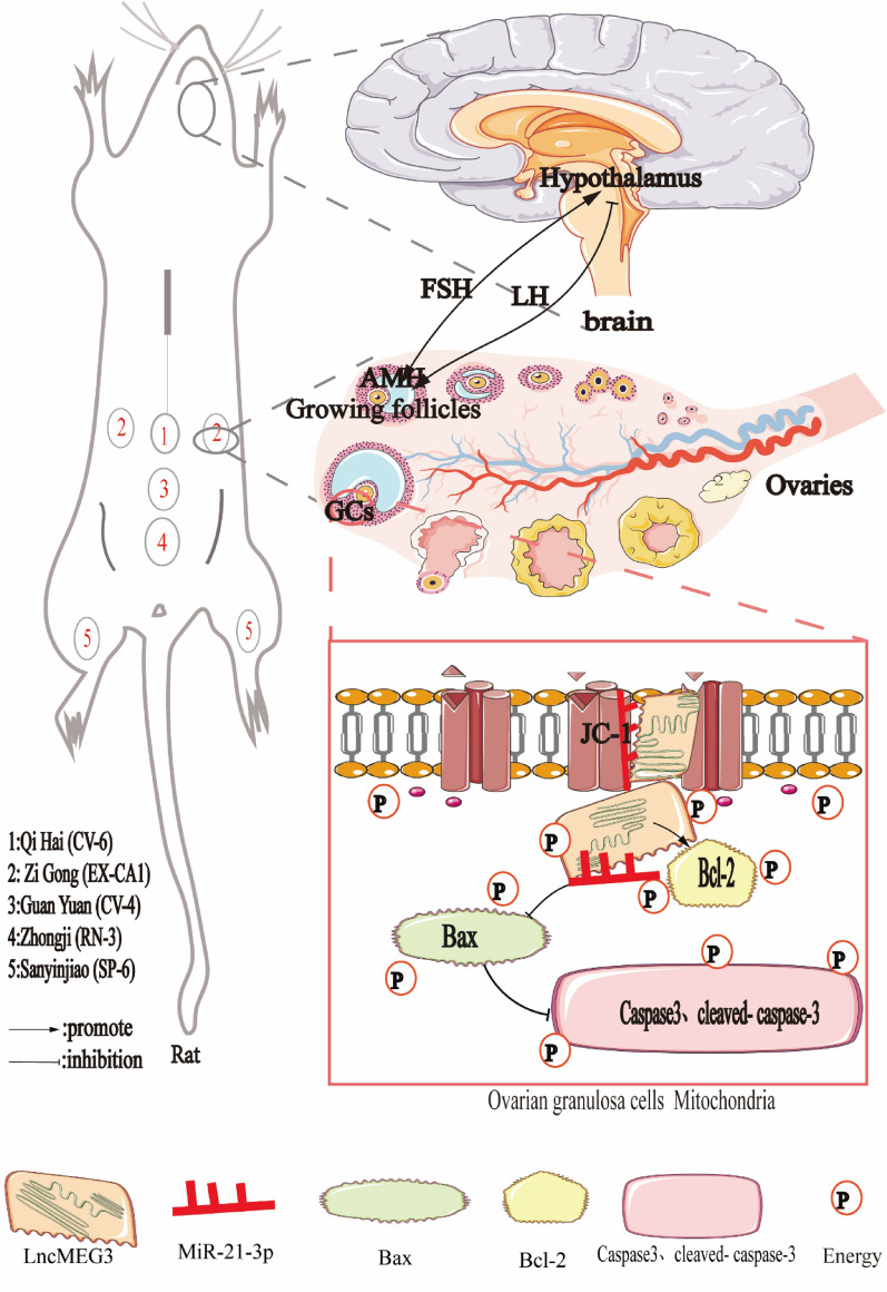
Background: The main features of polycystic ovary syndrome (PCOS) are abnormal follicular development and ovulatory dysfunction, which are caused by excessive apoptosis of ovarian granulosa cells. Acupuncture has been shown to improve follicular development abnormalities in patients with PCOS, but its mechanism is unknown. This study hypothesized that the mechanism of acupuncture on follicular development abnormalities in PCOS patients is the inhibition of granulosa cell apoptosis through LncMEG3-mediated regulation of miR-21-3p.

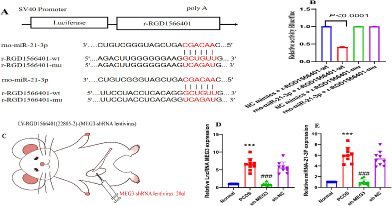
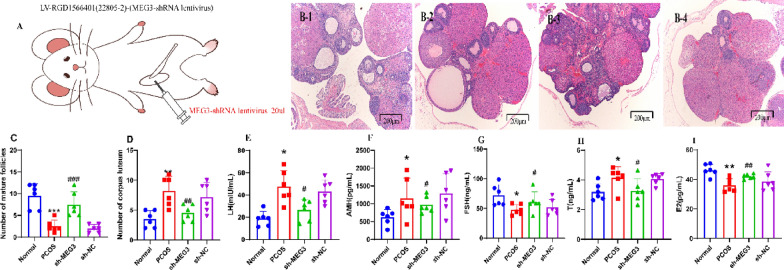
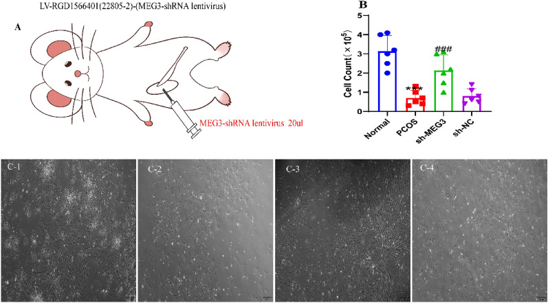
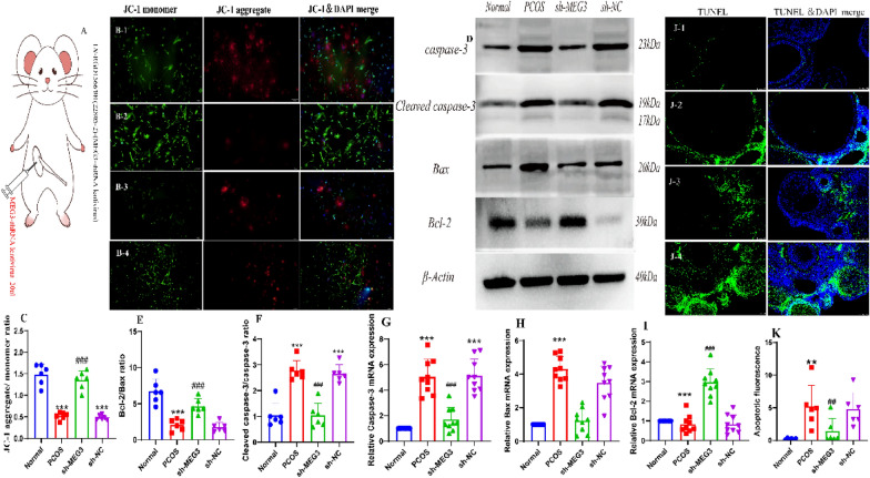
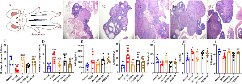
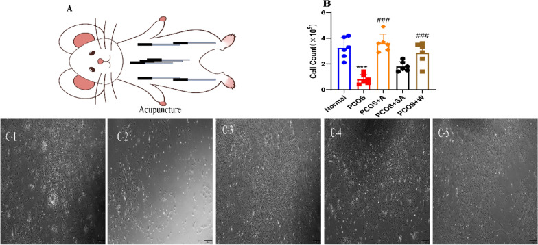
Methods: A PCOS-like rat model was established using subcutaneous injection of dehydroepiandrosterone (DHEA). Acupuncture was performed on rats for 15 d (CV-4, RN-3, CV-6, SP-6 and EX-CA 1). Ovarian morphology was observed by HE staining, and sex hormone and AMH levels were detected by ELISA. Primary granulosa cells were isolated from each group of rats to assess the association of acupuncture treatment, LncMEG3, miR-21-3p, and granulosa cell apoptosis in rats with PCOS.

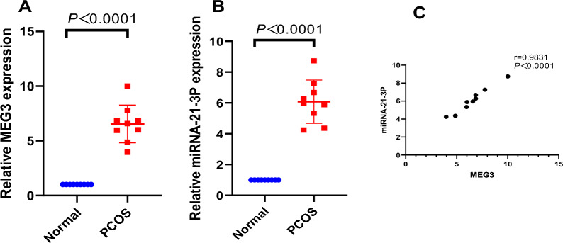
Results: LncMEG3 and miR-21-3p were highly expressed in the ovarian granulosa cells of rats with PCOS, and LncMEG3-mediated regulation of miR-21-3p was involved in the development of PCOS in rats. Silencing of MEG3 attenuated sex hormone dysregulation and ovarian histopathological changes in PCOS rats and promoted follicle cell development and maturation. In addition, silencing MEG3 increased the viability and number of granulosa cells. In addition, silencing MEG3 further inhibited early and late apoptosis of ovarian granulosa cells in PCOS rats. Acupuncture improved polycystic ovarian morphology and sex hormone levels in PCOS rats. Acupuncture intervention increased the viability and number of granulosa cells. Acupuncture intervention inhibited early and late apoptosis of ovarian granulosa cells in PCOS rats by targeting miR-21-3p via LncMEG3.

Conclusion: These results suggest that acupuncture can downregulate LncMEG3, thereby targeting and regulating miR-21-3p to suppress early and late granulosa cell apoptosis and normalize their proliferation. These factors ultimately compensate for abnormal follicular development. These findings shed light on the clinical potential of acupuncture as a safe treatment for follicular developmental abnormalities in PCOS.

Keywords: Acupuncture; Granulosa cell apoptosis; LncMEG3; PCOS; miR-21-3p.


Conflict of interest statement

The authors declare that they have no competing interests.

Figures

Fig. 1
Fig. 1
MEG3 and miR-21-3p expression are high in rats with PCOS. A MEG3 expression in ovarian granulosa cells of rats with PCOS by RT‒qPCR; B miR-21-3p expression in ovarian granulosa cells of rats with PCOS by RT‒qPCR; C Pearson analysis of the relationship between MEG3 and miR-21-3p in ovarian granulosa cells of rats with PCOS
Fig. 2
Fig. 2
MEG3 binds to miR-21-3p, a direct target gene of miR-21-3p in PCOS rats. A Putative complementary binding site of miR-21-3p and MEG3; B Fluorophore activity assay verified the targeting relationship between MEG3 and miR-21-3p; C Experimental strategy for the inhibition of ovarian MEG3 expression by viral injection; D Detection of LncMEG3 by RT‒qPCR in ovarian granulosa cells of PCOS rats; E The expression of miR-21-3p was detected by RT‒qPCR in the ovarian granulosa cells of PCOS rats. A-E, N = 6. Data reflect all measurements and are expressed as the mean ± standard deviation. A t-test was used for statistical analysis between two groups, and one-way ANOVA and Tukey's post hoc test were used for statistical analysis between multiple groups. ***P < 0.0001 compared to the normal group; ### P < 0.0001 compared to the sh-NC group
Fig. 3
Fig. 3
Silencing MEG3 ameliorates abnormal sex hormone levels and ovarian histopathological changes in PCOS rats. A Experimental strategy for the inhibition of ovarian MEG3 expression by viral injection; B HE staining was used to observe pathological changes in the ovaries of PCOS rats after MEG3 downregulation (scale bar = 200 μm); C Number of mature follicles in the ovarian tissue of PCOS rats after MEG3 downregulation; D Number of corpus luteum structures in the ovarian tissue of PCOS rats after MEG3 downregulation; E Serum secretion of LH in PCOS rats after silencing MEG3; F Serum secretion of AMH in PCOS rats after silencing MEG3; G Serum secretion of FSH in PCOS rats after silencing MEG3;H T secretion in the serum of PCOS rats after MEG3 silencing; I E2 secretion in the serum of PCOS rats after MEG3 silencing. A-I, N = 6. These data are expressed as the mean ± standard deviation, *P < 0.05, **P < 0.01, ***P < 0.0001 compared with the normal group; #P < 0.05, ##P < 0.01, ###P < 0.0001 compared with the sh-NC group, and statistically analysis using one-way ANOVA and Tukey's post hoc test. e2 stands for oestradiol, AMH stands for anti-Mullerian hormone, T stands for testosterone, FSH stands for follicle-stimulating hormone, and LH stands for luteinizing hormone. 1: Normal group, 2: PCOS group, 3:sh-MEG3 group, 4:sh-NC group
Fig. 4
Fig. 4
Silencing MEG3 increases the number of granulosa cells. A Experimental strategy for the inhibition of ovarian MEG3 expression by viral injection; B MEG3 downregulation of ovarian granulosa cells in PCOS rats; C Primary granulosa cells were extracted and cultured to observe cell number; The number of granulosa cells in PCOS rat ovaries were significantly increased after MEG3 downregulation (scale bar = 100 μm). A-C, N = 6. These data are expressed as the mean ± standard deviation, ***P < 0.0001 compared to the normal group; ###P < 0.0001 compared to the sh-NC group and were statistically analysis using one-way ANOVA and Tukey's post hoc test. 1: Normal group, 2: PCOS group, 3:sh-MEG3 group, 4:sh-NC group
Fig. 5
Fig. 5
Silencing of MEG3 inhibited apoptosis in PCOS rat ovarian granulosa cells. A Experimental strategy for the inhibition of ovarian MEG3 expression by viral injection; B The JC-1 method was used to detect the early stage of apoptosis in PCOS rat ovarian granulosa cells after MEG3 downregulation (scale bar = 100 μm):at high mitochondrial membrane potential, which can produce red fluorescence, at low mitochondrial membrane potential, which produces green fluorescence, and the blue fluorescent DAPI dye labels the nucleus, and Merge is a superimposed image showing the three fluorescent signals; C The ratio of JC-1 aggregate (red) to monomer (green) intensity in myotubes, Cell experiments were independently repeated three times; D cleaved caspase-3, caspase-3, Bax and Bcl-2 protein bands in PCOS rat ovarian granulosa cells after MEG3 downregulation; E Western blotting to detect the Bcl-2/Bax ratio in PCOS rat granulosa cells after MEG3 downregulation in granulosa cells; F Western blotting to detect cleaved caspase-3/caspase-3 protein expression in PCOS rat granulosa cells after MEG3 down regulation; G Detection of caspase-3 by RT‒qPCR in ovarian granulosa cells of PCOS rats; H Detection of Bax by RT‒qPCR in ovarian granulosa cells of PCOS rats; I Detection of Bcl-2 by RT‒qPCR in ovarian granulosa cells of PCOS rats; J Apoptosis of PCOS rat ovarian granulosa cells after MEG3 downregulation was detected by the TUNEL method (scale bar = 100 μm); green fluorescent FITC dye labelled apoptotic cells, blue fluorescent DAPI dye labelled nuclei, and Merge is a superimposed image showing both fluorescent signals; K fluorescence values of PCOS rat ovarian granulosa cells after MEG3 downregulation indicating apoptosis. D-K, N = 6. These data are expressed as the mean ± standard deviation, **P < 0.01 or ***P < 0.0001 compared to the normal group, ##P < 0.01 or ###P < 0.0001 compared to the sh-NC group and were statistically analysis using one-way ANOVA and Tukey’s post hoc test. 1: Normal group, 2: PCOS group, 3:sh-MEG3 group, 4:sh-NC group
Fig. 6
Fig. 6
Acupuncture ameliorates abnormal sex hormone levels and ovarian histopathological changes in PCOS rats. A Experimental strategy for acupuncture intervention; B HE staining was used to observe pathological changes in the ovaries of PCOS rats after the acupuncture intervention (scale bar = 200 μm); C Number of mature follicles in the ovarian tissue of PCOS rats after the acupuncture intervention; D Number of corpus luteum structures in the ovarian tissue of PCOS rats after acupuncture intervention; E AMH secretion in the serum of PCOS rats after acupuncture intervention; F LH secretion in the serum of PCOS rats after acupuncture intervention; G FSH secretion in the serum of PCOS rats after acupuncture intervention; H T secretion in the serum of PCOS rats after acupuncture intervention; I E2 secretion in the serum of PCOS rats after acupuncture intervention. A-I, N = 6. These data are measured values, expressed as the mean ± standard deviation, * P < 0.05, *** P < 0. 0001 compared to the normal group; #P < 0.05, ###P < 0.0001 compared to the PCOS group, statistically analysis using one-way ANOVA and Tukey’s post hoc test. E2 stands for oestradiol, AMH stands for anti-Mullerian hormone, T stands for testosterone, FSH stands for follicle-stimulating hormone and LH stands for luteinizing hormone. 1: Normal group, 2: PCOS group, 3: PCOS + A group, 4: PCOS + SA group, 5: PCOS + W group
Fig. 7
Fig. 7
Acupuncture intervention in PCOS rats increases the number of granulocytes. A Experimental strategy for acupuncture intervention; B Number of granulosa cells in the ovaries of PCOS rats after acupuncture intervention; C Primary granulosa cells were extracted and cultured to observe cell number. The number of granulosa cells in the ovaries of PCOS rats were significantly increased after acupuncture intervention (scale bar = 100 μm). A-C, N = 6.These data are expressed as the mean ± standard deviation, ***P < 0.0001 compared to the normal group and ####P < 0.0001 compared to the sh-NC and PCOS groups. One-way factorial ANOVA and Tukey’s post hoc test were used for statistical analysis. 1: Normal group, 2: PCOS group, 3: PCOS + A group, 4: PCOS + SA group, 5: PCOS + W group
Fig. 8
Fig. 8
Inhibition of apoptosis of ovarian granulosa cells in PCOS rats through LncMEG3-mediated regulation of miR-21-3p after acupuncture intervention. A Experimental strategy for acupuncture intervention; B Cleaved caspase-3, caspase-3, Bax, and Bcl-2 protein bands in granulosa cells of PCOS rat ovaries after acupuncture intervention. C Protein blotting for cleaved caspase-3/caspase-3 protein expression in granulosa cells of PCOS rats after acupuncture intervention. D Western blotting for Bcl-2/Bax protein expression in granulosa cells of PCOS rats after needle prick intervention. E Expression of LncMEG3 in ovarian granulosa cells of PCOS rats detected by RT‒qPCR after acupuncture intervention. F Expression of miR-21-3p in ovarian granulosa cells of PCOS rats detected by RT‒qPCR after acupuncture intervention; G Detection of caspase-3 by RT‒qPCR in ovarian granulosa cells of PCOS rats; H Detection of Bax by RT‒qPCR in ovarian granulosa cells of PCOS rats; I Detection of Bcl-2 by RT‒qPCR in ovarian granulosa cells of PCOS rats; B The JC-1 method was used to detect the early stage of apoptosis in PCOS rat ovarian granulosa cells after MEG3 downregulation (scale bar = 100 μm): at high mitochondrial membrane potential, which can produce red fluorescence, at low mitochondrial membrane potential, which produces green fluorescence, and the blue fluorescent DAPI dye labels the nucleus, and Merge is a superimposed image showing the three fluorescent signals; C The ratio of JC-1 aggregate (red) to monomer (green) intensity in myotubes, Cell experiments were independently repeated three times; L Apoptosis of ovarian granulosa cells in PCOS rats after acupuncture intervention was detected by the TUNEL method (scale bar = 100 μm), with green fluorescent FITC dye labelling the apoptotic cells and blue fluorescent DAPI dye labelling the nuclei; the merged image is a superimposed image of both fluorescent signals; M Fluorescence values of ovarian granulosa cells in PCOS rats after acupuncture intervention reflecting apoptosis. B-M, N = 6. These data are expressed as the mean ± standard deviation, ***P < 0.0001 compared to the normal group and ####P < 0.0001 compared to the sh-NC group, using one-way ANOVA and Tukey's post hoc test for statistical analysis. 1: Normal group, 2: PCOS group, 3: PCOS + A group, 4: PCOS + SA group, 5: PCOS + W group
Fig. 9
Fig. 9
Acupuncture regulates the apoptosis of ovarian granulosa cells in the context of abnormal follicular development in PCOS through LncMEG3-mediated inhibition of miR-21-3p

Similar articles

Cited by

References

    1. Walter K. What is polycystic ovary syndrome? Jama-J Am Med Assoc. 2022;327(3):294. doi: 10.1001/jama.2021.19776. - DOI - PubMed
    1. Peng G, Yan Z, Liu Y, Li J, Ma J, Tong N, Wang Y. The effects of first-line pharmacological treatments for reproductive outcomes in infertile women with PCOS: a systematic review and network meta-analysis. Reprod Biol Endocrinol. 2023;21(1):24. doi: 10.1186/s12958-023-01075-9. - DOI - PMC - PubMed
    1. Bahri Khomami M, Moran L, Kenny L, Grieger J, Myers J, Poston L, McCowan L, Walker J, Dekker G, Norman R, et al. Lifestyle and pregnancy complications in polycystic ovary syndrome: the SCOPE cohort study. Clin Endocrinol. 2019;90(6):814–821. doi: 10.1111/cen.13954. - DOI - PubMed
    1. Dumesic DA, Oberfield SE, Stener-Victorin E, Marshall JC, Laven JS, Legro RS. Scientific statement on the diagnostic criteria, epidemiology, pathophysiology, and molecular genetics of polycystic ovary syndrome. Endocr rev. 2015;36(5):487–525. doi: 10.1210/er.2015-1018. - DOI - PMC - PubMed
    1. Jayasena CN, Franks S. The management of patients with polycystic ovary syndrome. Nat rev endocrinol. 2014;10(10):624–636. doi: 10.1038/nrendo.2014.102. - DOI - PubMed