Review
. 2023 May 18;13(5):855.
doi: 10.3390/biom13050855.

(Patho)Physiology of Glycosylphosphatidylinositol-Anchored Proteins I: Localization at Plasma Membranes and Extracellular Compartments

Affiliations
Review

(Patho)Physiology of Glycosylphosphatidylinositol-Anchored Proteins I: Localization at Plasma Membranes and Extracellular Compartments

Günter A Müller et al. Biomolecules. .

Abstract

Glycosylphosphatidylinositol (GPI)-anchored proteins (APs) are anchored at the outer leaflet of plasma membranes (PMs) of all eukaryotic organisms studied so far by covalent linkage to a highly conserved glycolipid rather than a transmembrane domain. Since their first description, experimental data have been accumulating for the capability of GPI-APs to be released from PMs into the surrounding milieu. It became evident that this release results in distinct arrangements of GPI-APs which are compatible with the aqueous milieu upon loss of their GPI anchor by (proteolytic or lipolytic) cleavage or in the course of shielding of the full-length GPI anchor by incorporation into extracellular vesicles, lipoprotein-like particles and (lyso)phospholipid- and cholesterol-harboring micelle-like complexes or by association with GPI-binding proteins or/and other full-length GPI-APs. In mammalian organisms, the (patho)physiological roles of the released GPI-APs in the extracellular environment, such as blood and tissue cells, depend on the molecular mechanisms of their release as well as the cell types and tissues involved, and are controlled by their removal from circulation. This is accomplished by endocytic uptake by liver cells and/or degradation by GPI-specific phospholipase D in order to bypass potential unwanted effects of the released GPI-APs or their transfer from the releasing donor to acceptor cells (which will be reviewed in a forthcoming manuscript).

Keywords: (G)PI-specific phospholipase D (GPLD1); adipose cells; extracellular vesicles; glycosylphosphatidylinositol (GPI)-anchored proteins (GPI-APs); metabolic diseases; protein release; sulfonylurea drugs.


Conflict of interest statement

The authors declare no conflict of interest.

Figures

Figure 1
Figure 1
(a) Schematic structures of typical mammalian GPI-APs and of canonical and atypical transmembrane proteins. (right section) GPI-APs consist of a large protein domain (red), often with N-/O-glycosidically linked carbohydrates (blue) and intrachain disulfide bonds (brown), and a GPI anchor which is constituted of phosphatidylinositol (P-Ino) with long saturated fatty acids (dark green) and a glycan core (light green). The carboxy-terminus of the protein domain and the terminal (third) mannose residue of the glycan core are coupled through ethanolamine via amide (H-N-C=O) and phosphodiester (-P-) bonds, respectively. The fatty acids of the GPI anchor are embedded in the outer leaflet of the PM bilayer, often at lipid rafts, which typically harbor cholesterol and caveolin-1. Caveolin-1 represents an atypical monotopic transmembrane protein with both termini facing the cytoplasm. (left section) The transmembrane protein shown is a typical class I representative, with the amino- and carboxy-terminal polypeptide domains protruding into the extracellular space and cytoplasm, respectively, separated by the α-helical transmembrane domain. (b) Molecular structure of the typical GPI-AP human acetylcholinesterase (hAChE). (upper section) Crystal structure of wildtype and mutant AChE generated using the “Protein Repair One-Stop Shop” program. The cartoon representation of the crystal structure of hAChE displays the mutated residues in the designed variant. hAChE is shown as a backbone trace, color-coded from blue at the amino-terminus (labeled N) to red at the carboxy-terminus (labeled C). The residues of the catalytic triad, Ser-203, His-447 and Glu-334 are displayed as black ball and stick models, and labeled S, H and E, respectively. Fifty-one mutated residues distributed throughout the sequence as generated by Goldenzweig and coworkers [15] are highlighted as red spheres (adapted from ref. [16]). (lower section) Structure of its GPI anchor according to Deeg and coworkers [17]. The three constituents of the GPI anchor (enclosed by green lines) are the EtNH2-P bridge (red), the highly conserved glycan core (black) and the phosphatidate lipid portion (blue), as it is cleaved off by GPI-specific phospholipase D (see below). The presence of additional acyl or alkyl chains at the lipid portion as well as EtNH3+-P moieties at the glycan core (in blue) is variable. Removal of the EtNH3+-P side branches by PGA5P phosphodiesterase is required for efficient recruitment into ER-exit sites (ERES) and subsequent trafficking from the endoplasmic reticulum to the Golgi apparatus (see below, Section 2).
Figure 2
Figure 2
Localization of GPI-APs and their endocytosis (via caveolae) at lipid rafts, which are made up of (glyco)sphingolipids (GSL) at the outer leaflet of the PM bilayer, caveolin-1, phospholipids with saturated fatty acyl chains (sPL), and cholesterol at the inner leaflet. In contrast, non-lipid rafts predominantly contain transmembrane proteins and phospholipids with unsaturated fatty acyl chains (uPL). Lipid rafts are thought to act as nucleation sites for the biogenesis of caveolae upon invagination of the PMs into the cell interior. Following incision and fusion, the formed membrane vesicles would harbor the typical components of lipid rafts, among them cholesterol in concert with caveolin-1 (enriched at the cytoplasmic leaflet of the membrane bilayer), glycosphingolipids (enriched at the luminal leaflet) and GPI-APs. Their GPI anchor may become inserted in the luminal leaflet of the membrane bilayer and their protein moieties located in the vesicle lumen. Not only endocytosis, but also transcytosis of GPI-APs may involve caveolae. Experimental data supporting this view are currently intensively disputed (for a review, see [121,122]).
Figure 3
Figure 3
Inhibition of internalization of GPI-APs during transfer from human adipocytes and stimulation of glycogen synthesis in the ELCs. Transwell co-cultures were run (37 °C, absence of serum and BSA, (a,b), 2 weeks; (c,d), transfer times as indicated) with human donor adipocytes (of lipid-loading stage IV) and GPI-deficient acceptor ELCs in the insert and bottom wells, respectively, in the absence (ad, No siRNA) or presence of siRNAs (30 nM) directed against Cdc42 (ad), Rac1 (a,b) or RhoA (a,b). Subsequently, PMs were prepared from the ELCs of the bottom wells, then coupled to chips by ionic/covalent capture and finally analyzed for the expression of membrane proteins by SAW biosensing. (a) Phase shifts Δ in response to injection of anti-TNAP, anti-CD73 and anti-AChE antibodies (1800 to 2700 sec) were measured as summation signals and are indicated by horizontal hatched lines and brackets for each incubation. (bd) The experiment was repeated three to four times (different transwell co-cultures). Relative amounts of total transferred GPI-APs at PMs of ELCs (summation signals) (b,c) as well as relative glycogen synthesis (0.1 mM glucose) (b,d) in the absence (set at 100%) or presence of the siRNAs are given. Significant differences between the presence and absence of siRNAs at identical transfer times (bd, black symbols) as well as between the various transfer times and the 5-min times for presence and absence of Cdc42 siRNA each (c,d, turquoise and green symbols, respectively) are indicated (means ± S.D.; * p ≤ 0.01, # p ≤ 0.02, § p ≤ 0.05).
Figure 4
Figure 4
Different modes of enzymic release of membrane proteins from PMs by cleavage of their transmembrane domain or GPI anchor. Proteases cleave either at the juxtamembrane region of transmembrane proteins or at the extreme carboxy-terminus of GPI-APs and thereby cause the release of the extracellular domains of both the former and the latter (with no residue of the GPI glycan core left) into the extracellular compartment. The site of the proteinaceous cleavage determines whether the released protein moiety is truncated at its carboxy-terminus by a single (C) or a few amino acids (C*) and may therefore affect the functional state of the released GPI-AP. GPI-specific phospholipases (PL) of specificity C or D (GPI-PLC/D) cleave at distinct positions within the phosphodiester bridge of PI, which thereby results in different cleavage products, i.e., the release of the complete protein moiety together with the phosphoinositolglycan (PIG) as PIG-protein (by GPI-PLC) or with the inositolglycan moiety as inositolglycan-protein (by GPI-PLD), leaving behind at the PMs the corresponding diacyl or acylalkylglycerol and phosphatidate moieties, respectively. Certain chemicals, such as hydrogen fluoride (HF) and nitrous acid (NA), which have been used during the initial characterization of GPI anchorage by non-enzymic means, cleave within the glycan core of the GPI anchor of GPI-APs and cause the separation of the protein moiety from the terminal EtN-P residue in the course of dephosphorylation (HF) or from the non-acetylated GlcN in the course of deamination (NA). Importantly, those chemical methods with their unique cleavage specificities and the resulting release of the GPI-AP protein moieties can only be used in vitro rather than in living organisms. Ino, inositol; CC-N-H, ethanolamine.
Figure 5
Figure 5
The different structural entities and assemblies involved in the release of full-length GPI-APs with the complete GPI anchor remaining attached from the PMs of donor cells are represented by specific monomeric GPI-AP carrier- or binding-proteins (GPI-binding proteins) or micelle-like GPI-AP complexes constituted of (lyso)phospholipids and cholesterol (GLEC) or lipoprotein-like particles (LLPs) surrounded by phospholipid bi- and/or monolayers or extracellular vesicles (EVs), such as exosomes and microvesicles, formed by phospholipid bilayers. They are released from the outer leaflet of the PM bilayer of donor tissue or blood cells of mammalian organisms into extracellular compartments, such as blood or interstitial spaces, spontaneously or in response to specific endogenous or exogenous stimuli.
Figure 6
Figure 6
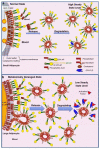
Hypothetical model for the release of full-length GPI-APs from adipocytes and their degradation by serum GPI-PLD (GPLD1), which both determine the steady-state concentration of full-length GPI-APs at micelle-like serum complexes in the normal (Panel a) and metabolically dysregulated state (Panel b). The upregulation of the release of full-length GPI-APs into micelle-like complexes because of destabilization of the PMs of the relevant blood or tissue cells is overcompensated by the increase of the GPLD1 activity. GPLD1 activity is controlled by its interaction with a so-called activating factor (red triangles) and ultimately leads to lower steady-state serum concentrations of the complexes than those in the normal state. The rapid removal of the full-length GPI-APs from the blood stream by the elevated GPLD1 activity is presumably of tremendous importance on the basis of their pronounced amphiphilic character. In fact, micelle-like GPI-AP complexes have been demonstrated to cause lysis of adipocytes upon incubation, as measured as the release of lactate dehydrogenase into the medium [394]. Differential interaction of the activating factor with the GPLD1, which is dependent on the nature of the donor cells and tissues as well as the extent of destabilization of their PMs, may explain the elevated serum GPLD1 activity in the metabolically deranged state.

Similar articles

Cited by

References

    1. Eisenhaber B., Bork P., Eisenhaber F. Prediction of potential GPI-modification sites in proprotein sequences. J. Mol. Biol. 1999;292:741–758. doi: 10.1006/jmbi.1999.3069. - DOI - PubMed
    1. Eisenhaber B., Bork P., Eisenhaber B. Post-translational GPI lipid anchor modification of proteins in kingdoms of life: Analysis of protein sequence data from complete genomes. Protein Eng. 2001;14:17–25. doi: 10.1093/protein/14.1.17. - DOI - PubMed
    1. Poisson G., Chauve C., Chen X., Bergeron A. FragAnchor: A large-scale predictor of glycosylphosphatidylinositol anchors in eukaryote protein sequences by qualitative scoring. Genom. Proteom. Bioinform. 2007;5:121–130. doi: 10.1016/S1672-0229(07)60022-9. - DOI - PMC - PubMed
    1. Ferguson M.A.J., Haldar K., Cross G.A.M. Trypanosoma brucei variant surface glycoprotein has a sn-1,2-dimyristyl glycerol membrane anchor at its COOH terminus. J. Biol. Chem. 1985;260:4963–4968. doi: 10.1016/S0021-9258(18)89166-9. - DOI - PubMed
    1. Haldar K., Ferguson M.A.J., Cross G.A.M. Acylation of a Plasmodium falciparum merozoite surface antigen via sn-1,2-diacyl glycerol. J. Biol. Chem. 1985;260:4969–4974. doi: 10.1016/S0021-9258(18)89167-0. - DOI - PubMed

Publication types

MeSH terms

Grants and funding

T.D.M. received funding from the German Research Foundation (DFG TRR296, TRR152, SFB1123 and GRK 2816/1), the German Center for Diabetes Research (DZD e.V.) and the European Research Council ERC-CoG Trusted (no. 101044445).